Glabella/central brow

Published on 16/03/2015 by admin

Filed under Dermatology

Last modified 22/04/2025

Print this page

rate 1 star rate 2 star rate 3 star rate 4 star rate 5 star
Your rating: none, Average: 0 (0 votes)

This article have been viewed 4743 times

14 Glabella/central brow

Summary and Key Features

Soft tissue augmentation of the glabella and central brow is increasingly recognized as an important part of the pan-facial volumization strategy for many patients

Augmentation of the glabella and central brow with fillers may be performed to correct age-related volume loss, or for the purposes of enhancement

The glabella and central brow are anatomically unforgiving with regard to both safety and aesthetic considerations

An understanding of anatomy and the physicochemical characteristics of fillers (including rheology) drives the selection of appropriate filler products and safe, efficacious injection techniques

For many patients, the combination of fillers with neuromodulators will produce more a natural-looking rejuvenation than treatment with neuromodulators alone

Fillers are also of value for patients with residual rhytides after neuromodulator treatment of the glabella and central brow

A thorough pre-procedural consultation and discussion of realistic objectives are key to optimizing patient satisfaction

Superficial or deep injection approaches may be employed alone or in combination. For both approaches, hyaluronic acid fillers may be the best option owing to their potential reversibility and excellent safety margin

Meticulous sterile technique is essential before and during all filler injection procedures to minimize the risk of infection or biofilm.

When properly performed, filler injections to the glabella and brow have a high rate of patient satisfaction and can profoundly improve the aesthetic appearance of the whole face

Introduction

The glabella and central brow may be considered the last frontier in facial soft tissue augmentation, in that the value of including them in pan-facial volumetry has been recognized only recently.

The recognition of volume loss as a cardinal feature of aging, and the availability of fillers that allowed this volume loss to be more effectively addressed – starting in the US with Food and Drug Administration (FDA) approval of the first hyaluronic acid (HA) filler in 2003 – catalyzed an evolution in our approach to soft tissue augmentation. The emphasis shifted from simply filling in rhytides to the objective of restoring youthful facial contours. For some patients, fillers can be used not just to correct age-related volume loss but also to actually improve upon the contours of their youth and create facial proportions that more closely approximate the ideals of beauty.

Age-related volume loss was first recognized in the lower face and mid-face. Early filler strategies focused on the nasolabial folds and subsequently on correcting volume deficits in the mid-face to secondarily elevate the nasolabial folds. At this time, there was some degree of ‘geographic divide’ between fillers and neuromodulators, with the latter being used more commonly to the upper face. The importance of also restoring volume to the upper face was recognized later, with the realization that age-related volume loss is pan-facial. Filler injection strategies developed for the temples, and now it is understood that true pan-facial volumization for many patients also includes filler implantation in the glabella and forehead, including the central brow.

Advances in our use of fillers have been paralleled by an evolution in our concepts of how best to combine fillers with neuromodulators. The advanced injector now appreciates that fillers can be used synergistically with neuromodulators throughout the face, including the glabella and central brow, rather than being placed differentially. This optimizes results, and potentially also improves their longevity.

The ultimate aim of pan-facial volumization is to restore ideal facial contours. The ideals of facial beauty have been largely preserved across time and cultures. This is probably because these ideals have an evolutionary basis, reflecting youth, good health, and, for women, the presence of sufficient levels of estrogen to confer fertility (Sundaram H). For both sexes, the youthful ideal is a relatively prominent upper and mid-face. In Western cultures, this tapers to a less prominent lower face / jawline in women, and thus a face that is shaped like a heart or inverted triangle. In men, the taper is somewhat less pronounced, with a stronger jawline and more equal proportions of the upper, mid- and lower face resulting in a longer, more rectangular shape.

Asian and other Eastern cultures have historically preferred a more rounded female facial shape, though jawline tapering is still desired. The rounded ideal is seen in images of Indian women dating back to the Indus Valley civilization (3300–1300bc) and Chinese women from the Tang dynasty (ad363–422); in depictions of Queen Maya (the Buddha’s mother); and in Indian poetry from the 5th century ad that exalts the beauty of ‘a face to rival the moon’. Although the traditional Eastern ideal has been somewhat influenced of late by the West, with some Asian women now expressing the desire for a less rounded face and actively seeking the injection of fillers and neuromodulators (e.g. to the masseter muscles) to achieve this, a ‘long face’ is still considered unfeminine. The preference for less angular female facial contours is readily discerned by comparing the movie stars of India’s Bollywood with those of America’s Hollywood. It is important to grasp these cultural differences in perception of beauty and to discuss each patient’s notions of what will enhance her appearance in advance of treatment, since even Asian women who were raised in Western countries may be displeased with soft tissue augmentation that gives them the oval, sculpted look that is currently a hallmark of Caucasian beauty.

In women, a smooth, unfurrowed brow and high, broad, convex forehead contours have been prized since antiquity and enhanced through artificial means. Women of ancient Greece pulled their hair tightly back from their faces to emphasize their foreheads and to lift their brows. Upper face prominence was enhanced in India with forehead decorations such as the bindi, which is still worn in modern times, and the painted forehead designs of the Chinese Tang dynasty served a similar purpose. Renaissance women such as the one depicted in 16th century Italian artist Allori’s Portrait of a Woman and the famous Elizabethan English beauty Lettice Knollys painstakingly plucked their hairlines to achieve the ‘noble brow’. More recently, the 1940s film actress Rita Heyworth had her hairline raised by electrolysis.

Currently, soft tissue fillers are used alone or in combination with neuromodulators for non-surgical improvement of the glabella and forehead, including the central brow. The objective may be corrective, i.e. reduction in rhytides and / or restoration of more youthful contours. Fillers are also used for forehead augmentation – to enhance the convexity and prominence of the forehead in younger patients who have not yet have developed actual volume loss.

Epidemiology and patient selection

Soft tissue augmentation of the glabella and central brow may be performed in the following patients:

1. Patients with residual rhytides following neuromodulator injections. These patients, who generally have some degree of volume loss and decreased skin elasticity, can be identified in advance of neuromodulator injection to the glabella and central brow with a skin stretch test, and counseled at that time regarding the likely need for adjunctive filler. Filler can correct volume loss and compensate to some extent for loss of skin elasticity.

2. Patients who prefer to retain some degree of glabellar and central brow mobility after neuromodulator treatment. Neuromodulator dosing strategies for the glabella and forehead, including the central brow, vary depending on patient preference for a mobile versus a ‘frozen’ look. Patients who wish to retain maximum expressivity and avoid a ‘frozen’ look may be best treated with relatively low doses of neuromodulators to the glabella and forehead, including the central brow, plus adjunctive filler.

3. Patients with volume loss from the glabella and forehead, including the central brow. Glabellar volume loss often assumes the form of two parallel vertical furrows of variable depth. In the forehead including the central brow, volume loss may be focal or more general, respectively resulting in localized or more diffuse, trough-like concavities. Volume loss to the glabella is generally primary and age related. Volume loss to the central brow may also be primary, or secondary to neuromodulator-related atrophy of the frontalis. This is a thin sheet-like muscle and this author has observed anecdotally that it has a propensity to develop atrophic areas in some patients after repeated neuromodulator injections over many years, especially if these injections are of high dosage.

4. Patients desiring augmentation or enhancement of forehead / central brow convexity. There are individual and cultural / ethnic variations in the preference for a convex or even domed forehead. Forehead augmentation has become especially popular in Korea and other Asian countries. It is interesting to note that neuromodulator treatment of the upper face, the most popular non-surgical rejuvenative procedure in the USA, is rarely performed on patients in these countries because they do not tend to frown in the way that Caucasians do.

5. Patients who decline neuromodulator treatment but still wish to improve rhytides in the glabella and central brow regions.

Anatomical considerations

A working knowledge of the anatomy of the glabella and central brow is required in order to optimize both the safety and aesthetic outcome of soft tissue augmentation.

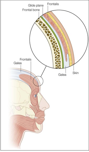
The most superficial tissue plane of the glabella and central brow is the skin, comprising the epidermis and dermis. Beneath this lies the subcutaneous tissue, then the superficial fascia, which covers the underlying muscles. There is loose subgaleal areolar tissue below the muscles, then the periosteum, which represents the deep fascia in this region and rests upon the bone (Fig. 14.1).

Muscles of the glabella and central brow

Medial brow depressors

The procerus is a small, pyramid-shaped muscle that originates from the fascia overlying the inferior aspect of the nasal bone and the superior aspect of the lateral nasal cartilage. It inserts into the skin of the medial forehead between the eyebrows, where its fibers interdigitate with those of the frontalis muscle. The procerus muscle is usually supplied by temporal and lower zygomatic branches of the facial nerve. It contracts vertically, resulting in movement of the eyebrows medially and down, to produce transverse glabellar lines.

The corrugator supercilii muscles lie below the frontalis muscle. They originate bilaterally from the bone at the medial end of the superciliary arches, travel obliquely and superiorly, and insert deeply into the skin above the midpoints of the orbital arches. The corrugator supercilii muscles are innervated by a nerve plexus originating from the temporal branch of the facial nerve. Their contraction moves the eyebrows medially and down, resulting in vertical lines of the glabella and central brow.

The depressor supercilii muscles are regarded by some as separate muscles and by others as part of the orbicularis oculi or corrugator supercilii muscles. They lie bilaterally between the corrugators and procerus, originating from the medial orbital rims in proximity to the lacrimal bones and inserting on the medial aspects of the orbital arches inferiorly to the corrugator supercilii. Innervation is from branches of the facial nerve. Contraction of the depressor supercilii muscles is considered to result in oblique lines of the glabella and central brow.

Important nerves, vessels, and other structures

The supraorbital neurovascular bundle emerges on each side from the supraorbital foramen, which is located 2.7 cm from the midline, at the junction of the medial one-third and the lateral two-thirds of the superior margin of the orbital bone. From here, the supraorbital neurovascular bundle pierces the corrugator supercilii and then pierces the surface of frontalis higher up on the forehead. The supraorbital nerve and vessels ramify into branches that innervate the forehead and scalp back to the vertex.

The supratrochlear neurovascular bundle lies more medially and is smaller. The supratrochlear nerve is a branch of the frontal nerve, which is derived from the ophthalmic division of the trigeminal cranial nerve (V). It is located 1.7 cm from the midline and 0.8 cm anterior to the supraorbital nerve. After exiting the orbit under the medial 1 cm of the brow, between the pulley of the superior oblique muscle and the supraorbital foramen, the supraorbital neurovascular bundle curves up on to the forehead close to the bone and beneath the corrugator supercilii and frontalis. It then divides into branches that pierce these muscles and ramify to supply the skin of the lower mid-forehead including the central brow, the skin of the upper eyelid, and the conjunctiva.

It is also important to be aware of the structure of the eyelids. They are multilayered with the middle layer consisting of a sheet of connective tissue known as the orbital septum, which extends to the rim of the bony orbit. The septum thickens as it inserts onto the orbital rim and this thickening is referred to as the arcus marginalis.

Decision-making: selection and preparation of filler product, selection of injection plane

General considerations

The glabella and central brow are anatomically unforgiving areas in regards to both safety and aesthetic considerations.

To ensure safety, it is necessary to visualize the location of vital structures such as nerves and vessels, and to take measures to avoid them when injecting fillers or neuromodulators.

In regard to aesthetics, the slightest overfilling or misplacement of a fraction of a milliliter of filler within the glabella and central brow can cause suboptimal results. It is therefore recommended that HA fillers be used, as contour correction can be performed if needed by removing them in part or fully by the injection of hyaluronidase. Dilution of fillers with saline and / or lidocaine suspension makes them easier to spread with post-injection tissue molding to achieve smooth contours. This is because dilution reduces filler viscosity. Dilution is best performed with the aid of a sterile two-way female-to-female adaptor to which the syringe of filler and another syringe containing the diluent can be attached.

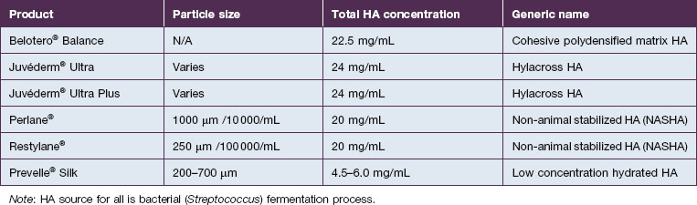
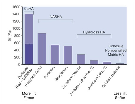
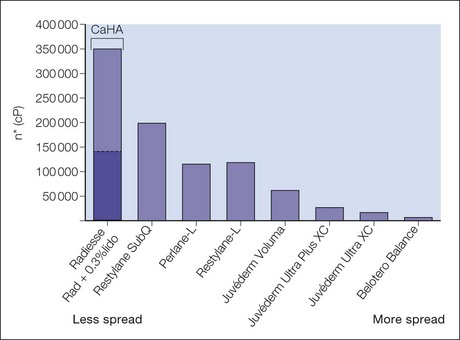
An understanding of structural and functional anatomy and of the physicochemical characteristics of fillers – including the flow-related (rheologic) properties of elasticity (G prime) and viscosity that predict their behavior – informs the selection of appropriate filler products and injection techniques to achieve optimal results (see Box 14.1, Tables 14.1 and 14.2; see also Figs 14.214.6).

Selection of injection plane and filler product

Fillers may be injected into the glabella and the central brow via two approaches: deep and superficial. These approaches may be used alone, or in combination with a multiplane or ‘sandwich’ technique. As discussed below, blunt microcannulas or sharp needles may be employed for filler injection. Filler may be injected with anterograde technique, tracking a path ahead of the injecting cannula or needle, or with retrograde technique, flowing along the path already created by the cannula or needle as it is withdrawn through the tissue (Sundaram H, Monheit G et al).

Deep approach

The path of least resistance, or glide plane, in the forehead including the central brow is the subgaleal (pre-periosteal) plane, which represents the plane between the superficial and deep fascia of this facial zone. This glide plane can be bluntly dissected with ease using the filler product with anterograde flow. The deep approach is well suited to corrective contouring for restoration of volume to the glabella and central brow, and also to augmentation of the forehead including the central brow.

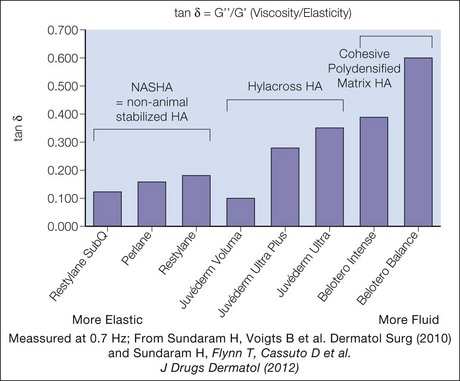
Desirable physical characteristics of a filler product for the deep approach to the glabella and forehead, including the central brow, include medium to low viscosity – for some degree of spread to decrease the risk of contour irregularities – and some lifting capacity, as measured by elastic modulus or G prime. A medium- or low-viscosity product such as Belotero® Balance; Juvéderm® Voluma, Ultra Plus or Ultra; or Prevelle® Silk may be selected. Alternatively, a higher viscosity product such as Perlane® or Restylane® may be diluted to lower its viscosity. Juvéderm® Voluma is currently under study in the US, but is available in Canada, Europe and elsewhere. It is described as having some tissue spread and integration but also significant cohesivity that confers robustness and moldability (Carruthers J, Carruthers A et al). These properties, together with the presence of some particles of varying shape and size (Stocks D, Sundaram H et al), make it appropriate for deep volumization in the pre-periosteal tissue plane. Other products available outside the USA that are appropriate for deep implantation into the glabella and brow include a cohesive polydensified matrix HA (Belotero® Intense, Merz) and a HA with “optimal balance” technology that has the largest gel calibration (particle size) and heaviest crosslinking within its product family (Emervel® Volume, Galderma). In addition to the absolute values of elasticity (firmness) and viscosity (spread) for each filler product, it is also useful to consider the balance of elasticity versus fluidity. This can be done with a rheologic value known as tan delta, which is calculated as the viscosity modulus (G double prime or G”) divided by the elasticity modulus (G prime or G’). [FIG. 14.4] Juvéderm Voluma, Perlane and Restylane have a low tan delta, and thus are more elastic than fluid and suited to deep implantation for contouring. Belotero Balance has a high tan delta, indicating a preponderence of fluidity over elasticity that makes it suitable for flowing through the superficial tissue planes. (Sundaram H, Flynn T et al.)

Filler migration along the glide plane is a theoretical problem. If migration were to occur down to the level of the arcus marginalis, this could result in filler drifting into the thin upper eyelid skin. The risk of migration could be higher with lower viscosity fillers since they have a tendency to spread more. In practice, the glide plane is not so low in tissue resistance that migration is a significant risk. To reduce or eliminate migration risk, it is recommended that the filler should be injected in small aliquots of no more than 0.4 mL each, rather than as a large bolus, and that tissue molding should be restricted to the immediate post-injection period and performed only by the clinician, not by the patient.

Superficial approach

Implantation of fillers into tissue planes that are superficial to the frontalis (high subcutaneous or intradermal) carries no risk of nerve damage at the level of the brow, since the supraorbital and supratrochlear nerves run and ramify within frontalis. There is also no risk of encountering the vessels associated with these nerves, since they follow the same course. The superficial approach may be used for contouring to restore or augment volume, and also for filling rhytides.

Desirable physical characteristics of a filler for the superficial approach include low viscosity so that the filler spreads significantly, decreasing the risk of contour irregularities in these very anatomically unforgiving tissue planes. The filler must also not cause the Tyndall effect (more accurately known as Rayleigh scattering), which is the phenomenon whereby the particles within a bolus of superficially implanted HA filler scatter light back to the observer’s eye. Scattering is inversely proportional to the 4th power of the light wavelength; therefore blue light, which has a shorter wavelength, is scattered more and this imparts a bluish discoloration to the filler bolus. It has been demonstrated that both the Perlane® / Restylane® and Juvéderm® families of HA products have a significant particulate component and are therefore best suited to subdermal implantation when undiluted (Stocks D, Sundaram H et al).

Appropriate filler products for the superficial approach that are currently approved for use in the USA include Belotero® Balance (aka Belotero® Basic outside the USA), which has been shown to have a homogeneous intradermal distribution after implantation that correlates with the fact that it has not been reported to cause the Tyndall effect, since it does not provide any particulate boluses to scatter light. Prevelle® Silk, which has low HA concentration and thus not enough particles per mL to cause the Tyndall effect, may also be used. Alternatively, Juvéderm® Ultra or Restylane® may be diluted 1 : 1 or more with saline and / or lidocaine to reduce its concentration sufficiently to minimize risk of the Tyndall effect; dilution also decreases filler viscosity and thus increases spread, which is desirable. HA products available for use outside the USA that may be injected superficially without causing the Tyndall effect include a cohesive polydensified matrix HA (Belotero® Soft, Merz) and an “optimal balance” HA with small gel calibration (Emervel® Touch, Galderma).

Pre-procedural consultation, assessing and preparing the patient

During the pre-treatment consultation, it is important to determine the patient’s objectives and to discuss realistic outcomes. For example, one cannot obliterate all rhytides of the forehead, including the central brow, owing to the tension the skin in this zone is under from the frontalis. Patients should also understand that, although filler implantation can help offset mild to moderate loss of skin elasticity, significant skin laxity is not fully ameliorable with fillers.

Clinical evaluation of the patient in repose and in animation (frowning and raising the eyebrows) from different angles – particularly from below and from an oblique viewpoint – may be especially helpful to detect volume loss in the central brow, even when it is focal and manifests as a localized concavity. It may be helpful to outline focal concavities with white pencil. If these concavities are subtle, it may be preferable to avoid topical anesthetic cream as the resultant mild skin tumescence due to hydration from the topical cream may obscure them, and instead to apply an ice pack for patient comfort. A simple manual stretch test and snap test can be performed to gauge volume deficit and skin elasticity, and predict the likely response to fillers.

Standardized digital photography should be employed for documentation and three-dimensional imaging can also be helpful. Some clinicians like to present patients with their pre- and post-treatment images as documentation of their results.

Patients should be counseled that avoidance of aspirin, non-steroidal anti-inflammatory drugs, vitamin E and fish oil supplements for 5–7 days prior to filler injections may decrease the risk of post-injection ecchymosis.

In this era of increased concern over filler-related infection or biofilms, it is vital to perform scrupulous sterile skin preparation prior to injection; 4% chlorhexidine gluconate (Hibiclens®) is a good cleansing agent, although it should not be used near the eyes.

Injection technique

The superficial technique is indicated for fine glabellar or central brow lines, while a superficial or deep approach may be employed for treatment of glabellar troughs or for central brow volumization. A sharp needle is preferred for superficial injection, as the intradermal or high subdermal plane has some degree of resistance (especially in the superficial dermal plane). With a non-Tyndall HA filler, a multiplane approach may be employed, with filler implantation both subcutaneously and intradermally (Sundaram H and Kiripolsky M).

Anterograde serial puncture technique with a 30-gauge image-
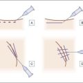
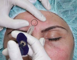
inch (12 mm) needle inserted perpendicular to the skin surface is effective for glabellar or brow lines. Skin blanching may be observed as microaliquots of filler are implanted intradermally. Retrograde serial threading with a 30 G 12 mm or longer needle may be preferred for more trough-like volume deficits, allowing the needle to dissect tunnels in the desired plane into which filler is then placed. Fanning injection at an angle to the trough may be more effective than injecting parallel to it. A longer needle, such as 30 G image inch (38 mm), permits more efficient fanning within the superficial plane. When the needle is fanning within the dermis, the angle of the needle hub to the skin is quite acute (30° or less) and skin tenting is seen. In the superficial dermis, the needle hub grazes the skin surface and the outline of the needle is clearly seen through the surface. Threading may confer a greater risk of ecchymosis than serial puncture, and patients should be apprised of this prior to treatment.

With the deep approach, a blunt microcannula may be preferred for the forehead, including the central brow, for reasons of safety. A 22 or 25G 50 mm microcannula is inserted through an entry point made with a sharp “pilot” needle of equal or greater diameter and passed through and down to lie on the periosteum. Injection into the forehead, including the central brow, is best approached from lateral to medial using a fanning technique while palpating the supraorbital notch. Serial threading with anterograde flow allows the filler product to gently flow through the glide plane (Sundaram H and Weinkle S). Alternatively, a sharp 30 G 12 mm or longer needle may be selected; this is the easier technique for a deep approach to the glabella owing to its limited size.

If treating with filler and neuromodulator in the same session to the forehead including the central brow, where the tissue is relatively thin, these authors’ preference is to inject filler first and then neuromodulator, in order to achieve precise placement of filler within the desired tissue plane prior to injection of fluid boluses of toxin that could distort or change the position of the planes. For the glabella, where the tissue is thicker, these authors find that comparable results are achieved with injection of either filler or neuromodulator first. Cryo-neuromodulation (Myoscience), a recently available non-chemical neuromodulator option, is currently under study in the US and approved in Europe for temporary improvement of dynamic rhytides of the forehead including the central brow. The handheld percutaneous device targets the temporal branch of the facial nerve to prevent signal transduction. The results are reported to be comparable in longevity to those of injected botulinum toxin neuromodulators.

Case Study 1

image

A 42-year-old East Indian woman presents complaining of the skin being ‘uneven’ between and above her eyebrows. She previously received injections of NASHA filler (Perlane® and Restylane®, Medicis) to the mid-face, lower face and temples and was pleased with these results. Examination in repose reveals moderate focal volume loss, resulting in localized trough-like concavities of the central brow and glabella that are more pronounced in animation (frowning and raising the eyebrows). She has Fitzpatrick skin phototype IV. There is good improvement in the concavities with a manual stretch test, and a snap test shows good skin elasticity. Examination in animation (frowning and raising the eyebrows) shows medium glabellar muscle mass and hyperdynamic activity of the muscles of the glabella (procerus, corrugators and depressor supercilii) and forehead (frontalis).

Based on this evaluation, you recommend combination treatment with a HA filler and botulinum neurotoxin. The patient is viewed from the oblique angle and the areas of volume deficit are marked with white pencil.

Botulinum neurotoxin (incobotulinumtoxin A, Xeomin®) is injected into the glabellar region, with 4 units being injected at each of five sites (procerus, corrugator supercilii and depressor supercilii muscles) for a total of 20 units.

You select a low concentration (5.5 mg/mL) non-Tyndall hydrated HA filler (Prevelle® Silk) containing 0.3% lidocaine for superficial injection. The product is injected with a lateral to medial oblique fanning approach and retrograde serial threading into the dermis of the glabella, while retracting the skin with the non-dominant fingers. The patient is periodically viewed during the injection procedure from above, below, and obliquely, to gauge the efficacy of volume correction. Tissue molding is performed after each injection. A total of 1.5 mL of the HA product is injected. You inject the patient’s lower eyelids in the pre-periosteal plane and her upper eyelids in the high subcutaneous plane with the HA filler using a 27 G 38 mm blunt microcannula. You also inject the lower eyelids intradermally with a 30 G 12 mm sharp needle.

At follow-up 3 weeks later, the patient is very pleased with her results (Figure 14.7A and B).

Case Study 2

image

A 57-year-old German woman presents complaining that she looks tired and prematurely aged. She has had no previous filler or neuromodulator injections. Examination in repose reveals moderate volume loss from the glabella and central brow, resulting in localized trough-like concavities, and fine rhytides that are more pronounced in animation (frowning and raising the eyebrows). She has Fitzpatrick skin phototype I. There is good improvement in the concavities and the rhytides with a manual stretch test, and a snap test shows moderate skin elasticity. Examination in animation (frowning and raising eyebrows) shows medium glabellar muscle mass and mild to moderate hyperdynamic activity of the muscles of the glabella (procerus, corrugators, and depressor supercilii) and forehead (frontalis). The patient also has moderate volume loss in the lower eyelid (nasojugal) region and the mid-face.

You recommend treatment with soft tissue fillers and select a high concentration (22.5 mg/mL) non-Tyndall cohesive polydensified matrix hyaluronic acid (HA) (Belotero® Balance), to which you add 0.45% lidocaine, for superficial injection into the glabella and central brow. The product is injected via a sharp 27 G image-
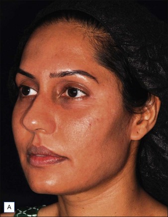
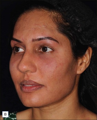
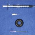
inch (12 mm) needle with retrograde serial threading and medial to lateral and lateral to medial oblique fanning approach into the dermis and subdermis of the glabella and central brow, while retracting the skin with the non-dominant fingers. The patient is periodically viewed during injection from above, below, and obliquely, to gauge the efficacy of volume correction. Tissue molding is performed after each injection. A total of 1 mL of the HA product is injected. You also inject the patient’s lower eyelids with 2 mL of a high concentration (20 mg/mL) cohesive polydensified HA filler (Belotero® Soft) to which you have added 0.3% lidocaine, using a 27 G 40 mm blunt microcannula for pre-periosteal and subdermal injection and a 30 G 12 mm (image-inch) sharp needle for intradermal injection. You inject her mid-face pre-periosteally and subcutaneously with 3 mL of calcium hydroxylapatite (Radiesse® ) to which you have added 0.3% lidocaine. The patient is pleased with her results and remarks that she looks younger and her nose now appears smaller.

Case Study 3

image

A 42-year-old German woman presents complaining that she looks angry even when she is not, exhausted, and older than her actual age. She has had no previous filler or neuromodulator injections.

Examination in repose shows fine vertical rhytides of the glabella and central brow, also some fine horizontal rhytides in the inferior portion of the glabella. A manual skin stretch test shows mild to moderate improvement in the vertical rhytides and good improvement in the horizontal rhytides. A skin snap test shows moderate elasticity. Animation (frowning) causes deepening of the fine rhytides, and reveals medium glabellar muscle mass with moderate hyperdynamic activity of the muscles of the glabella (procerus, corrugators, and depressor supercilii) and of the nasalis muscle. She has Fitzpatrick skin phototype I. The patient has moderate volume loss in the lower eyelid (nasojugal) region and mid-face. She has hyperdynamic rhytides of the lateral periocular and perioral regions, and cobblestoning of the chin on animation.

You recommend treatment with combined soft tissue fillers and neuromodulators. You inject the fine horizontal and vertical rhytides of the glabella and central brow with 1 mL of a high-concentration (20 mg/mL) cohesive polydensified hyaluronic acid (HA) filler (Belotero® Soft) to which you have added 0.3% lidocaine, using a 30 G 12 mm sharp needle with superficial serial puncture “blanch” technique and tissue molding. You also inject the patient’s lower eyelids with 1 mL of this HA product using a 27 G 40 mm blunt microcannula for pre-periosteal and subdermal injection and a 30 G 12 mm sharp needle for intradermal injection. In addition, you inject her lower eyelids and mid-face pre-periosteally and subcutaneously with 1 mL of a high-concentration (22.5 mg/mL) monophasic cohesive polydensified HA filler (Belotero® Balance) to which you have added 0.45% lidocaine, using a 27 G 13 mm sharp needle.

You also inject the patient with incobotulinumtoxin A (Xeomin® ) that has been reconstituted with 2.5 mL bacteriostatic normal (0.9%) saline for a concentration of 4 units per 0.1 mL. You inject 4 units at each of five points into the procerus, corrugator supercilii, and depressor supercilii muscles, for a total of 20 units. You inject 2 units at each of two points into the lateral portion of the orbicularis oculi muscle and the same on the left side, for a total of 4 units on each side. You also inject 1 unit into the right side of the superior portion of orbicularis oris and 2 units into the left side, and 1 unit at each of three points into the inferior portion of orbicularis oris. You inject 3 units into the mentalis muscle. The patient is happy with her results.

Avoidance and management of adverse events

The techniques outlined above, if performed correctly and with the prerequisite knowledge of facial anatomy, should eliminate any risk of inadvertent nerve damage or intravascular injection of filler.

As discussed above, infection or biofilm development are concerns when any soft tissue filler is implanted. In addition to thorough sterile preparation of the skin prior to commencement of the injection session, it is recommended that scrupulous sterile technique should be maintained and intermittent skin cleansing with alcohol wipes performed during the session.

Risk of filler migration with the deep approach may be minimized by avoidance of large bolus injections and the employment instead of microaliquot or serial threading technique.

Ecchymosis and / or small hematomas may occur, especially in the forehead including the central brow. This may result in temporary tenderness upon palpation.

If there is significant ecchymosis, treatment with a yellow–green intense pulsed light or similar light energy device can hasten its resolution.

Post-injection contour irregularities (excess convexities) can be minimized by avoidance of overcorrection, especially to the forehead rhytides. Vertical molding may be helpful to smooth out any minor post-injection contour irregularities. Contour irregularity (focal convexity) immediately after injection or in the first 1–3 days and associated with tenderness and fluctuance may be due to hematoma, especially in the forehead including the central brow. Cold compresses or ice packs may provide relief until a hematoma resolves spontaneously.

Persistent contour irregularities 3 weeks or more after HA injection and after visible ecchymosis has resolved can be addressed by injection of hyaluronidase to dissolve the unwanted HA. The Lambros method, in which the hyaluronidase is diluted with lidocaine, has been reported to cause fewer reactions than the Vartanian method, which employs undiluted hyaluronidase.

Persistent contour irregularities after CaHA injection can be improved by injection of lidocaine suspension, with gentle movement of the needle to help break up the bolus of unwanted filler. Addition of hyaluronidase and / or 5-fluorouracil may aid in this process.

Further reading

Borrell M, Leslie D, Tezel A. Lift capabilities of hyaluronic acid fillers. Journal of Cosmetic and Laser Therapy. 2011;13:21–27.

Carruthers J, Carruthers A. Volumizing the glabella and forehead. Dermatologic Surgery. 2010;36:S1905–S1909.

Carruthers J, Glogau R, Blitzer A. Advances in facial rejuvenation: botulinum toxin type A, hyaluronic acid dermal fillers, and combination therapies – consensus recommendations. Plastic and Reconstructive Surgery. 2008;121(5):5S–30S.

Carruthers J, Carruthers A, Tezel A, et al. Volumizing with a 20-mg/mL smooth, highly cohesive, viscous hyaluronic acid filler and its role in facial rejuvenation therapy. Dermatologic Surgery. 2010;36:S1886–S1892.

Fang F, Clapham P. A systematic review of interethnic variability in facial dimensions. Plastic and Reconstructive Surgery. 2010;127(2):874–881.

Flynn T, Sarazin D, Bezzola A, et al. Comparative histology of intradermal implantation of mono and biphasic hyaluronic acid fillers. Dermatologic Surgery. 2011;37:637–643.

Hanke CW, Rohrich R. Facial soft-tissue fillers: assessing the state of the science conference – proceedings report. Journal of the American Academy of Dermatology. 2011;64(4):S53–S65.

Kang M, Park E, Shin HS, et al. Skin necrosis of the nasal ala after injection of dermal fillers. Dermatologic Surgery. 2011;3637:3375–3380.

Lambros V. The use of hyaluronidase to reverse the effects of hyaluronic acid filler. Plastic and Reconstructive Surgery. 2004;114(1):277.

Requena L, Requena C, Christensen L, et al. Adverse reactions to injectable soft tissue fillers. Journal of the American Academy of Dermatology. 2011;64(1):1–34.

Rohrich R, Monheit G, Nouven AT, et al. Soft tissue filler complications: the important role of biofilms. Plastic and Reconstructive Surgery. 2010;125(4):1250–1256.

Stocks D, Sundaram H, Michaels J, et al. Rheological evaluation of the physical properties of hyaluronic acid dermal fillers. Journal of Drugs in Dermatology. 2011;10(9):974–980.

Sundaram H. Face value: the truth about beauty – and a guilt-free guide to finding it. Emmaus, PA: Rodale; 2003.

Sundaram H, Voigts B, Beer K, et al. Comparison of the rheological properties of viscosity and elasticity in two categories of soft tissue fillers: calcium hydroxylapatite and hyaluronic acid. Dermatologic Surgery. 2010;36:S1859–S1865.

Sundaram H, Weinkle S, et al. Blunt injection microcannulas: a consensus document. Journal of Drugs in Dermatology. 2012. March

Sundaram H, Flynn T, Cassuto D, et al. New and Emerging Concepts in Soft Tissue Fillers. Journal of Drugs in Dermatology. 2012;11(8):s12–s25.

Sundaram H, Kiripolsky M 2012 Non-Surgical Rejuvenation of the Upper Eyelid and Brow in Clinics in Plastic Surgery [Elsevier, in press]

Sundaram H, Monheit G, Goldman M, et al. Clinical Experiences with Hyaluronic Acid Fillers. Journal of Drugs in Dermatology. 2012;11(3):s15–s27.