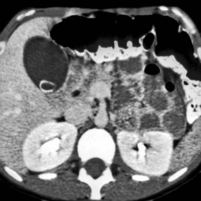
Hypervascular pancreatic mass with multiple peptic ulcers and thickened folds


 of the stomach.
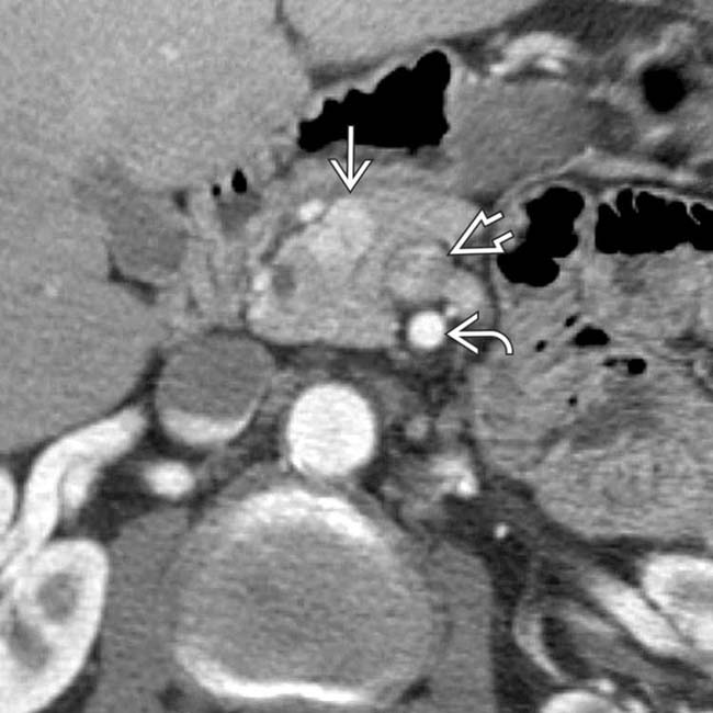
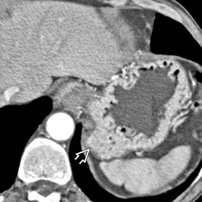
 in the pancreatic head. It is important to distinguish this from the superior mesenteric artery
  and superior mesenteric vein
  .
.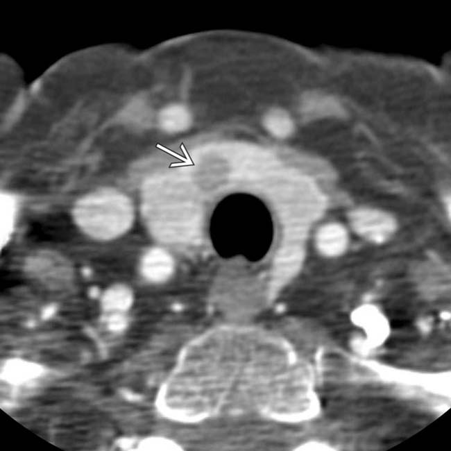
 , representing parathyroid adenomas or hyperplasia.
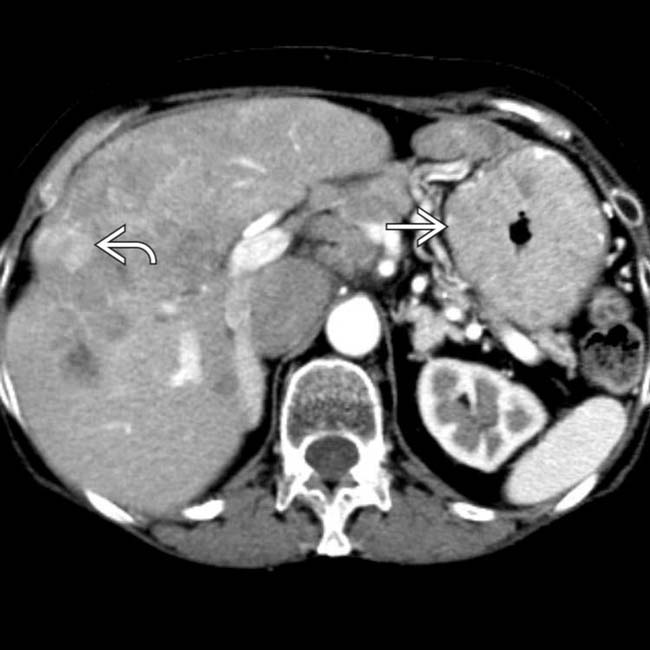
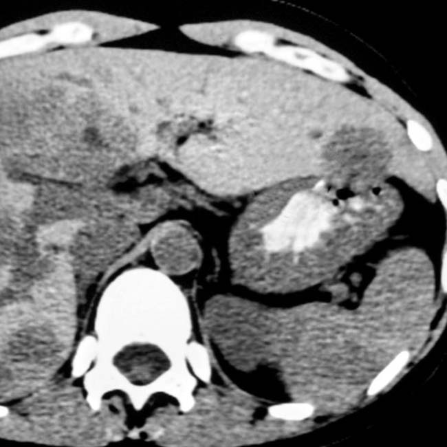
 . Multiple liver metastases are present
  . The serum gastrin levels were strikingly elevated, confirming ZES, though the gastrinoma was not identified on CT.
IMAGING
General Features


DIFFERENTIAL DIAGNOSIS
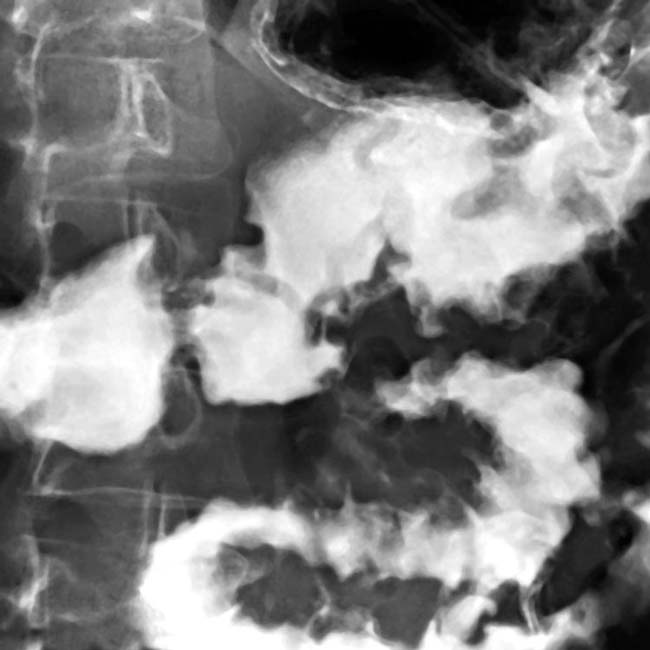
Gastritis


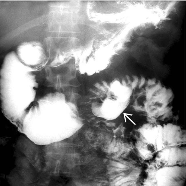
 resulting in a dilated duodenum due to spasm and edema. Gastric folds are thickened and the barium within the stomach is diluted and poorly adherent due to increased secretions. These are classic fluoroscopic features of ZES.
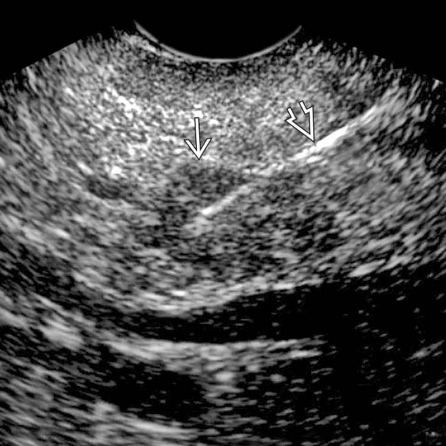
 that was biopsied during the same procedure. The biopsy needle is evident
  . This documented a gastrinoma, accounting for the patient’s signs and symptoms.
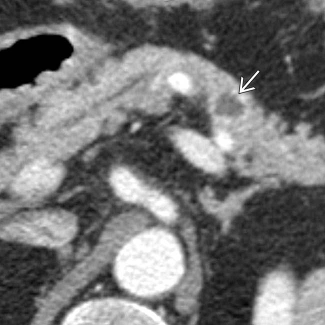
 with an enhancing, solid periphery and a cystic or necrotic center.
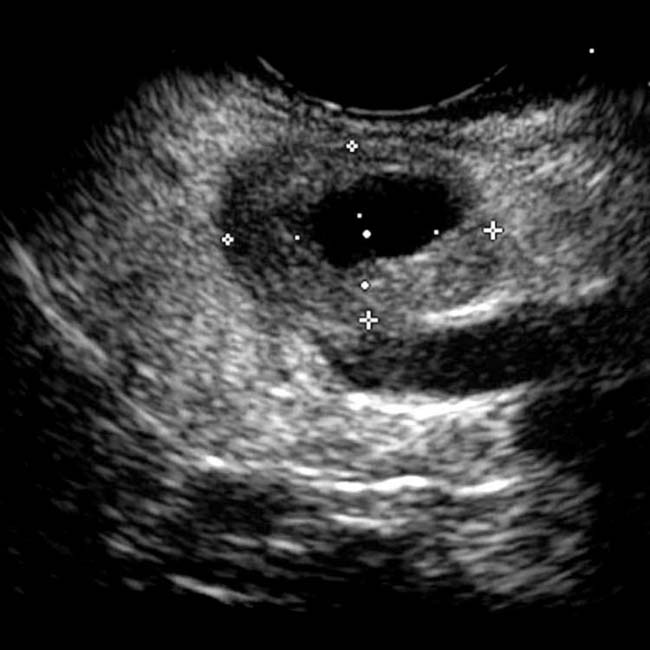
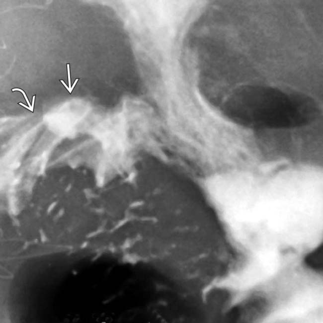
 at the site of the gastroenteric anastomosis. Note the radiating folds
  leading to the ulcer crater.
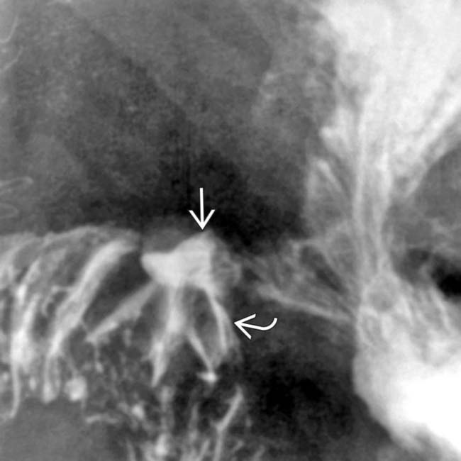
 and the radiating folds
  leading to the ulcer crater.
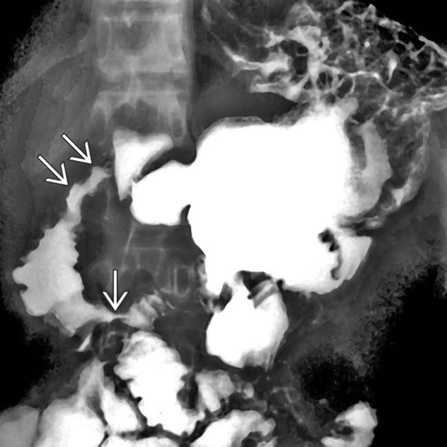
 .
.
 , probably from prior ulceration.
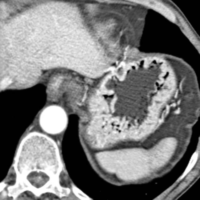
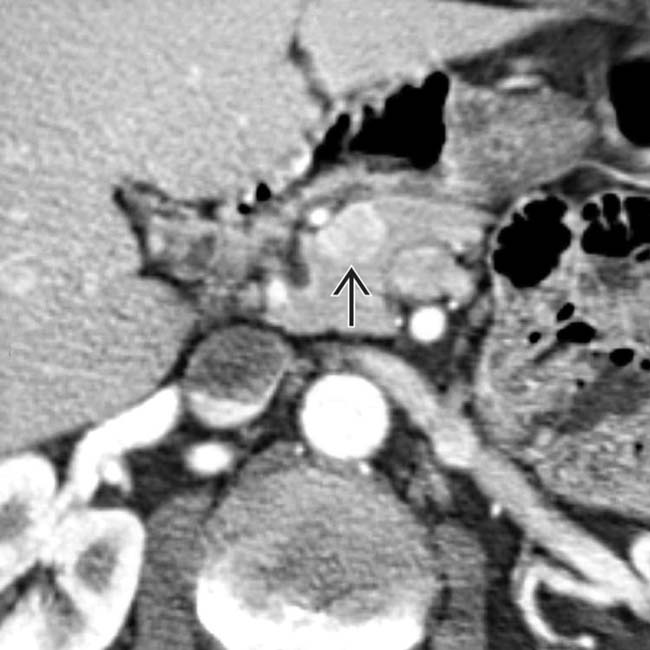
 in the pancreatic head which proved to be a gastrinoma at resection.
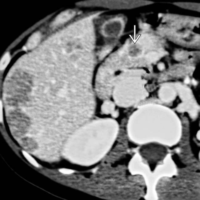
 in the pancreatic head and multiple subtle liver metastases.