X-Linked Juvenile Retinoschisis
Clinical Features:
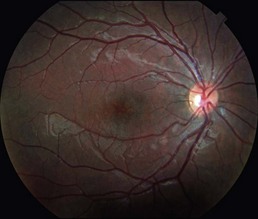
There is almost always schisis in the fovea, which is often accompanied by schisis in the peripheral retina (50% of affected eyes), usually inferotemporally. The foveal schisis leads to a characteristic clinical appearance similar to cystoid macular edema with a radial spoke-like pattern (Fig. 12.6.1).
OCT Features:
There is splitting within different retinal layers involving both the inner and outer retina. Within the macula, the inner nuclear layer is most commonly affected layer (Fig. 12.6.2), though the outer nuclear layer, ganglion cell layer, and nerve fiber layer can all be affected (Fig. 12.6.3). Unlike typical CME the splitting can occur well outside the foveal area.
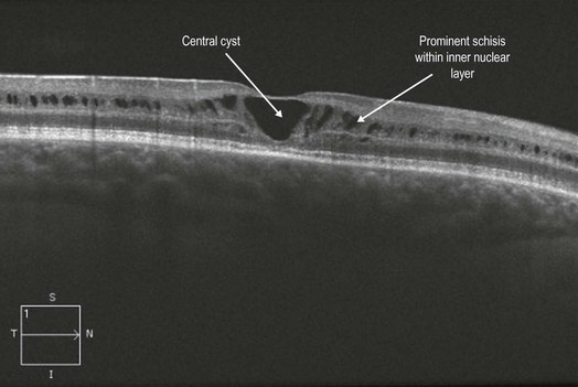
Figure 12.6.2 OCT shows prominent schisis mostly within the inner nuclear layer. There is a central cyst present.
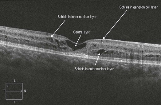
Figure 12.6.3 OCT (corresponding to Figure 12.6.1) shows schisis within the ganglion cell layer, inner nuclear layer, and outer nuclear layer. There is also a central cyst present.


