CHAPTER 9 Which block for which surgery?
Introduction
A recent editorial highlighted that ‘Regional anesthesia always works, provided we put the right dose of the right drug in the right place’.1 Locating the right place is key to providing a successful block. With recent developments in peripheral nerve blockade this has become easier. We now have high quality peripheral nerve stimulators in addition to portable ultrasound technology to aid us in nerve location.2–4 While accurate nerve location is key to successful peripheral nerve blockade, equally important is choosing an appropriate block for the planned surgery. This can only be obtained by discussing and understanding the planned surgery with the surgeon. With central neuraxial blockade we are aware that a low lumbar epidural would be of limited benefit for upper abdominal surgery, a thoracic epidural being of more benefit. Similarly, for wrist surgery, accepting ulnar nerve stimulation for an axillary block may be of limited benefit, if the surgical incision is on the dorsum of the wrist, where it would have been better to identify the radial nerve either with nerve stimulation or ultrasound, in addition to blocking other branches of the brachial plexus. Hence, it is not only important to block the correct plexus, but equally important to consider and ensure we block the necessary nerve branches for the surgical procedure. It is therefore essential that anesthetists have a comprehensive knowledge of applied anatomy for regional anesthesia.
Utilization of regional anesthesia has increased hugely over the last 20 years, with a recent study suggesting that it is now used in 37% of surgical cases amenable to a regional technique.5 This is likely to increase further in coming years. In recent years, we have seen a large number of peripheral nerve blocks described in the literature, with several descriptions for each approach for a given nerve block. Thankfully, the introduction of ultrasound has somewhat simplified this. While the ability to perform a wide variety of peripheral nerve blocks is desirable for successful regional anesthesia, the majority of regional anesthesiologists use only a limited number of blocks regularly. In addition, their block choice may also depend on the nerve location technique they use. For example, when performing brachial plexus anesthesia above the clavicle using nerve stimulation, many regional anesthesiologists will use an interscalene approach, while if they use ultrasound they may opt for an ultrasound guided supraclavicular approach. With experience, the anesthetist will be able to select the most appropriate technique (or combination of techniques) in addition to making the regional anesthesia technique work.
Applied anatomy
A thorough understanding of anatomy relevant to regional anesthesia is essential if it is to be performed with precision and accuracy and to avoid complications. While the exact location of nerves is important, the surrounding tissues and fascial planes are equally important, as the local anesthetic is placed in close proximity to the nerves. Traditionally, doctors have focused particularly on dermatomes, in addition to osteotomes and myotomes, when performing regional anesthetic techniques (Ch. 4). However, with peripheral nerve blocks it is more useful to think what nerve branches supply the surgical site and to ensure that we block these nerves.
Upper limb peripheral nerve blocks
The upper limb is supplied by nerves of the brachial plexus. The brachial plexus is formed by the anterior primary rami of the lower four cervical nerves (C5–C8) and the first thoracic nerve (T1).6,7 In some individuals there may also be contributions from the C4 and T2 nerve roots. The plexus supplies motor innervation to all the muscles of the upper limb with the exception of the trapezius and levator scapula muscles. It supplies cutaneous innervation to the upper limb with the exception of the area of the axilla (upper inner arm), which is supplied by the intercostobrachial nerve. Above the shoulder is supplied by the supraclavicular nerve (cervical plexus), and the dorsal scapular area is supplied by cutaneous branches of spinal nerves. The plexus has five terminal branches (median, ulnar, radial, musculocutaneous and axillary nerves) in addition to a number of smaller branches. The innervations of the major joints of the upper limb are highlighted in Table 9.1. A more detailed description of upper limb anatomy can be found in Chapter 14. It is possible to block the brachial plexus at various levels as it leaves the intervertebral foramina and traverses towards the periphery. The most proximal approach at the level of the nerve roots is the interscalene approach; the supraclavicular block is performed at the level of the trunks, while the infraclavicular block is performed at the level of the cords, with axillary approach at the level of the terminal branches. All of these blocks can be successfully performed with either nerve stimulation or with ultrasound guided techniques (Ch. 7).2–4
Table 9.1 Innervations of the major joints of the upper limb
| Acromioclavicular joint | Suprascapular, axillary and lateral pectoral nerves |
| Shoulder joint | Suprascapular, axillary and lateral pectoral nerves |
| Elbow joint | Musculocutaneous, radial, ulnar and median nerves |
| Wrist joint | Radial, ulnar and median nerves |
Notes: Median, ulnar, radial, musculocutaneous and axillary nerves are five terminal branches of the brachial plexus. The suprascapular nerve is a branch from the upper trunk while the lateral pectoral nerve is a branch from the lateral cord of the brachial plexus.
The interscalene approach is suitable for surgeries of the shoulder and upper arm, and can be performed with the arm in almost any position. It blocks the plexus close to the nerve roots at about the level of the cricoids cartilage (C6).6 This may help explain why there is less spread to the lower roots, resulting in poor block of the ulnar nerve (C8, T1). The posterior shoulder will not be anesthetized, requiring wound infiltration for the port site for shoulder arthroscopies. Equally, the anterior port for arthroscopy may also be missed because the skin is supplied by T2. A supplemental skin infiltration needs to be made over the distribution of T2 to ensure the anterior port or the lower end of a total shoulder incision is adequately anesthetized. The phrenic nerve is paralyzed with this approach as it lies on the anterior scalene muscle, so caution should be exercised in patients with severe respiratory co-morbidities. In addition, patients may develop hoarseness and ptosis. This is a superficial block and complications are rare. However, serious complications have been reported with this block, including spinal cord injury, vascular injection and pneumothorax.8
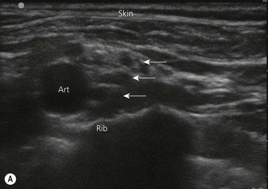
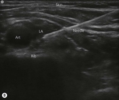
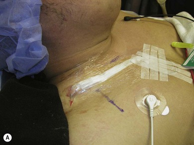
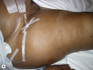
The supraclavicular approach is suitable for surgeries of the upper arm, elbow and hand, and is performed at the level of the trunks of the brachial plexus. It is frequently referred to as the spinal anesthetic of the upper limb as it can provide complete anesthesia of the limb below the shoulder in a consistent time-efficient manner. In the past, some anesthetists were cautious about performing this block because of the rare complication of pneumothorax, and the fact that the landmarks were not easy to ascertain, particularly in obese patients. However, the advent of ultrasound has resulted in a huge increase in the utilization of this block, where the plexus can be seen lying posterior and lateral to the subclavian artery on the first rib (Fig. 9.1). Local anesthetic can be preferentially injected around the lower trunk (C8, T1) of the plexus, particularly when used for hand surgery. Use of this block may still result in phrenic nerve paralysis (50%) and Horner’s syndrome.6 Placement of a continuous catheter at this site (supraclavicular fossa) for post operative analgesia is easy to secure and less likely to become dislodged (Fig. 9.2).
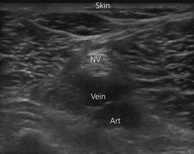
The axillary approach to the brachial plexus anesthetizes the terminal branches of the plexus, as they lie in close proximity to the axillary artery.6 It is ideal for surgeries below the elbow, particularly hand surgery. This block is safe and easy to perform and is therefore widely used. The anesthetist needs to be aware that there are multiple vessels (arteries and veins) in close proximity to the nerves and care should be taken to avoid intravascular injection. A single injection technique is associated with a significant failure, therefore a multiple injection technique should be used or the block performed under ultrasound guidance to ensure adequate spread of local anesthetic9 (Fig. 9.3). The musculocutaneous nerve lies between the biceps and coracobrachialis muscle and may be some distance away from the other branches of the plexus which lie close to the artery. It is critical to block the musculocutaneous nerve, as it supplies the flexor muscles of the arm and the skin over the lateral surface of the forearm.
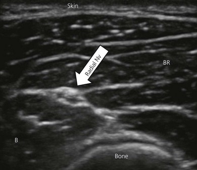
The utilization of elbow and forearm blocks is discussed in Chapters 21 and 22. These blocks are easy to perform, particularly when ultrasound guidance is used (Fig. 9.4). These techniques are useful both as the primary anesthetic, and also for postoperative analgesia. Consideration should be given as to whether the surgeon is using a tourniquet and in what location. Upper arm tourniquets can be used for short periods of time, but longer use will require anesthesia of the tourniquet location. In such cases, a distal forearm or elbow block performed with a long acting local anesthetic (to provide postoperative analgesia) can be used in combination with a more proximal block such as infraclavicular or axillary block, using a shorter-acting local anesthetic (lignocaine). The more distal block should be performed first to minimize the risk of unrecognized intraneural injection. These blocks are also frequently used as ‘rescue’ blocks, where following a more proximal block there is incomplete anesthesia in a branch of the plexus. This branch can then be blocked at the elbow or forearm. Again, caution should be exercised, as rescue blocks may be associated with a higher incidence of nerve injury.
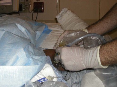
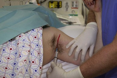
The intercostobrachial nerve (T2) supplies the area of the axilla (upper inner arm) which is important for certain upper arm procedures. In addition, this is where an upper arm tourniquet is typically located. While tourniquet pain is largely related to muscle ischemia, anesthetizing the skin in this area will improve patient comfort. This is easily achieved using subcutaneous infiltration of local anesthetic around the medial aspect of the upper arm (Fig. 9.5). The supraclavicular nerve block (cervical plexus) can be performed by injecting local anesthetic along the posterior border of the sternocleidomastoid muscle (Ch. 11).
Lower limb peripheral nerve blocks
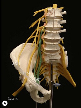
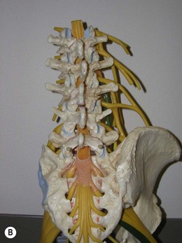
The lower limb has a dual nerve supply consisting of the lumbar plexus and the sacral plexus.7 The lumbar plexus is formed by the anterior rami of L1 to L4 with contributions from T12 in some individuals. The three main branches of the lumbar plexus related to lower limb peripheral nerve blocks are the femoral (L2,3,4), obturator (L2,3,4), and lateral femoral cutaneous nerves (L2,3); other branches include the iliohypogastric, ilioinguinal and the genitofemoral nerves. The sacral plexus is formed by the anterior rami of L4 to S4. The major nerve branches of the sacral plexus are the sciatic nerve, the posterior cutaneous nerve of the thigh and the pudendal nerve.7 The sciatic nerve splits into its 2 branches, the common peroneal and the tibial nerve, typically about 7–10 cm above the popliteal fossa. The sciatic nerve has numerous articular (hip and knee) and muscular branches. The innervations of the major joints of the lower limb are highlighted in Table 9.2. A more detailed description of lower limb anatomy can be found in Chapter 23. Blockade of peripheral nerves of the lower limb has been shown to provide comparable analgesia and functional recovery in the postoperative period to epidural analgesia, and superior analgesia and recovery to PCA opioids.10,11 To provide complete anesthesia of the lower limb using peripheral nerve blocks, we need to block both the lumbar and the sacral plexus. There is no single peripheral nerve block injection that can reliably block both of these together. Thus we need to consider which plexus provides the majority of the innervation and block this plexus first to allow time for the major block to setup. We also need to block either the other plexus or the significant branches. For above knee surgery, the major innervation is the lumbar plexus. In below knee surgery, the sciatic nerve (sacral plexus) is the dominant nerve supply (and should be anesthetized first), with only a small cutaneous distribution from the saphenous nerve (femoral) on the anterior medial surface of the leg to the medial malleolus. The saphenous nerve can then be blocked at the groin with a low volume femoral nerve block, in the thigh area, where it lies in the adductor canal behind the sartorius muscle or at the level of the tibial tuberosity.
Table 9.2 Innervations of the major joints of the lower limb
| Hip joint | Femoral, obturator and sciatic nerves |
| Knee joint | Femoral, obturator and sciatic (tibial, common peroneal) nerves |
| Ankle joint | Femoral (saphenous) and sciatic (tibial, superficial & deep peroneal) nerves |
Lumbar plexus
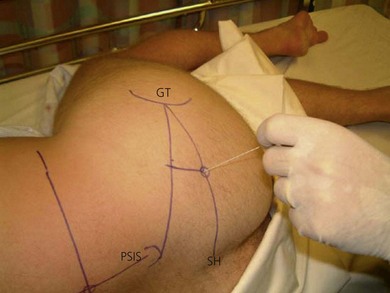
The most proximal approach to the lumbar plexus is the posterior approach (also called the psoas compartment block, Ch. 27). This block results in the most reliable anesthesia of the three important branches (femoral, obturator and lateral femoral cutaneous nerves) of the lumbar plexus. The branches of the lumbar plexus are spread out, and hence this block typically requires a large volume of local anesthetic (30 mL; Fig. 9.6). This block is ideal for providing both anesthesia and analgesia for hip surgery, surgery above the knee and knee surgery. It must be combined with blockade of the sacral plexus, or is frequently used in combination with spinal anesthesia to provide analgesia in the postoperative period for major joint surgery. This posterior approach is frequently used to place a continuous peripheral nerve catheter system, as it is a clean area and easy to fix the continuous catheter in the lumbar area. It is a deep block12 (6–10 cm, or greater in the obese) and more complex to perform, so it should not be used in people at risk of bleeding. As the plexus lies in a vascular area, it may be associated with a higher risk of inadvertent intravascular injection and local anesthetic toxicity.13 Because of the close proximity of the plexus to the spinal cord with this approach, and the volumes of local anesthetic used, it is not surprising that neuraxial spread of local anesthetic is well described. This can result in bilateral lower limb sensory and motor block, in addition to hypotension and cardiovascular compromise. Ultrasound facilitates the performance of many blocks, particularly the more superficial blocks. However, the lumbar plexus is difficult to accurately visualize with ultrasound. So while the posterior approach provides the most reliable blockade of the lumbar plexus, providing excellent anesthesia for hip and knee surgery, many anesthetists prefer the safer and easier anterior approach (femoral nerve block) to the lumbar plexus.14
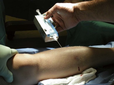
The femoral nerve block (Ch. 26) is indicated for patients undergoing hip surgery, surgery above the knee and knee surgery. It reliably results in blockade of the femoral nerve but less frequently results in blockade of the lateral femoral cutaneous nerve or obturator nerve.15 For this reason, it does not provide reliable anesthesia for knee replacement surgery even when combined with proximal sciatic nerve blockade. If the surgery is to be performed with peripheral nerve blocks alone, a psoas compartment block combined with a proximal sciatic nerve block would have the highest success rate. Alternately, the lateral femoral cutaneous and the obturator nerves can also be blocked. The femoral, lateral femoral cutaneous nerve, and obturator nerves can all be blocked with ultrasound guidance. A continuous peripheral nerve catheter is frequently placed to provide postoperative analgesia following lower limb joint replacement (Fig. 9.7). An alternative block to the femoral block is the fascia iliaca block (Ch. 28). This block is extremely easy and quick to perform and is ideal for analgesia for hip and knee surgery. Similar to the femoral nerve block, it provides reliable anesthesia to the femoral nerve, but variable anesthesia to the lateral femoral cutaneous and the obturator nerves. It is frequently used to provide analgesia both in the pre-operative hip fracture and postoperative patient.16–18
Sacral plexus
The main branch of the sacral plexus is the sciatic nerve, the most proximal approach being the parasacral approach, a posterior approach blocking the nerve as it exits the greater sciatic foramen. The classic approach to the sciatic nerve (Fig. 9.8) anesthetizes the nerve as it lies beneath the gluteal muscle, medial to the greater trochanter of the femur (Ch. 24). Sciatic nerve block is used for surgery on the knee, calf, ankle, and foot, when combined with block of the femoral or saphenous nerve. With these proximal approaches, the posterior cutaneous nerve of the thigh lies in close proximity to the sciatic nerve. These blocks are deep blocks and require adequate sedation, particularly when performed with nerve stimulation. Placement and securing continuous peripheral nerve catheters is ideal in these locations. As the patient is typically on their side for these procedures, they are frequently combined with a posterior approach to the lumbar plexus or spinal anesthetic. Ultrasound guidance can be used for these proximal approaches, but it is easier to visualize the sciatic nerve with the subgluteal and popliteal approaches. An anterior approach to the sciatic nerve (Ch. 25), when combined with a femoral and obturator block, also results in complete anesthesia of the knee and below. The anterior approach is a deeper block, which may be more difficult to perform and is associated with a higher incidence of complications.
A frequently used approach to the sciatic nerve is the popliteal block, performed proximal to the popliteal fossa before the sciatic nerve splits into its two branches, the common peroneal and the tibial nerve.19,20 This block can be performed with nerve stimulation or with ultrasound guidance, where it can be easily seen lying above the vascular structures (Fig. 9.9). It results in anesthesia below the knee, and can be combined with saphenous nerve block. It is ideal for ankle and foot surgery, although consideration has to be given to the location of the tourniquet. Again, this is an appropriate site to place a continuous catheter for postoperative analgesia after appropriate surgery.
A discussion on the utilization of paravertebral, intercostal and transversus abdominis plane (TAP) blocks is included in Chapters 32–34.
Making regional anesthesia work
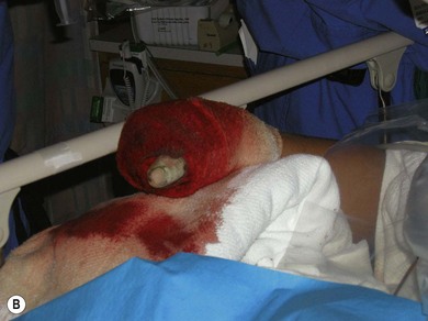
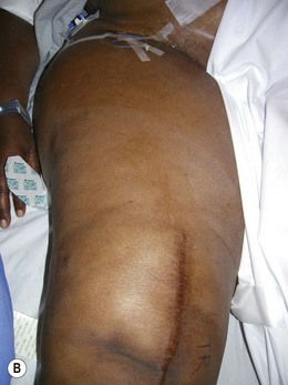
Choosing the appropriate peripheral nerve block for the surgery is the key to making regional anesthetic techniques work. This is particularly true when peripheral nerve block techniques are used for both surgical anesthesia and postoperative analgesia. However, success using peripheral nerve blocks starts long before the placement of the block needle. At the pre-anesthetic visit following full assessment of the patient (History, Examination and Investigations), the anesthetist informs the patient that the surgical procedure can be performed using a regional anesthetic technique, or a regional anesthetic may form part of the anesthetic plan (Box 9.1). Indications and contraindications to block placement are discussed in Chapter 4 of this book. They may also inform the patient of the potential benefits of a regional technique, including improved analgesia and decreased side-effects in the postoperative period. Early patient education is important to the acceptability and success of these techniques. Many surgeons now also inform their patients that the proposed surgery can be performed while using a regional anesthetic. Experienced surgeons may also caution the anesthetist when a peripheral nerve block technique may not be ideal (e.g. risk of compartment syndrome (Fig. 9.10) or when additional blocks may be necessary; for example, harvesting bone graft from the iliac crest.
Box 9.1
Making regional anesthesia work
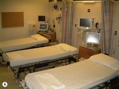
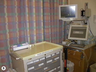
An important factor in making regional anesthesia work is maintaining operating theatre efficiency.21,22 This can largely be facilitated by being organized. The use of regional anesthesia block carts helps keep the necessary equipment together and improves efficiency. The use of induction rooms or ‘block areas’ allows peripheral nerve blockade to be performed ahead of entering the operating room, therefore saving time.21,22 (Fig. 9.11). This is especially important when using longer-acting local anesthetics (bupivacaine, levobupivacaine and ropivacaine), where it may take 30–45 minutes to achieve complete surgical anesthesia. Typically, higher concentrations of local anesthetics are used for anesthesia compared to postoperative analgesia. Bupivacaine 0.5% may be required for anesthesia, but it has been shown that the lower concentrations (i.e. bupivacaine 0.25%) are equally effective when used purely for postoperative analgesia, improving safety by decreasing the total dose of local anesthetic used. Mepivacaine and lignocaine have a faster onset but shorter duration of anesthesia and may be appropriate for shorter cases (Ch. 3). In addition, when regional anesthesia is performed prior to entering the operating room, the peripheral nerve block can be assessed and a rescue block performed, or alternate anesthetic planned if necessary. Regional anesthetic techniques also have advantages on completion of surgery where emergence and extubation are not required, thus improving operating room efficiency.21,22 Patients receiving these techniques may also have a shorter stay in the post-anesthetic care unit.
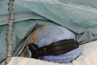
Performance of some peripheral nerve block techniques can be painful and may require sedation for block performance. This is particularly true for deeper peripheral nerve blocks such as infraclavicular blocks in muscular individuals, and the posterior approach to the lumbar plexus or some approaches to the sciatic nerve. In addition, the use of nerve stimulation is associated with increased discomfort compared with ultrasound-guided techniques. Appropriate use of sedation is associated with improved patient satisfaction. The use of low doses of opioid analgesics, such as alfentanil or fentanyl combined with midazolam, may facilitate the performance of the procedure. The aim is to provide analgesia and anxiolosis but maintain patient cooperation to try and minimize complications. The intra-operative care of patients who have received regional anesthetic techniques can be challenging and is important to the success of the block (Box 9.1). Depending on the patient, they may wish to be sedated during the surgery, or may prefer to listen to music rather than the noisy theatre environment. In addition they should be kept warm and comfortable (Fig. 9.12).
1 Denny NM, Harrop-Griffiths W. Location, location, location! Ultrasound imaging in regional anesthesia. British Journal of Anesthesia. 2005;94(1):1-3.
2 Pither C, Raj PP, Ford D. The use of peripheral nerve stimulators for regional anesthesia. A review of experimental characteristics, technique and clinical applications. Reg Anesth. 1985;10:49-58.
3 Soeding P, Eizenberg N. Review article: anatomical considerations for ultrasound guidance for regional anesthesia of the neck and upper limb. Can J Anaesth. 2009;56(7):518-533.
4 Chin KJ, Chan V. Ultrasound-guided peripheral nerve blockade. Curr Opin Anaesthesiol. 2008;21(5):624-631.
5 Hanna MN, Jeffries MA, Hamzehzadeh S, et al. Survey of the utilization of regional and general anesthesia in a tertiary teaching hospital. Reg Anesth Pain Med. 2009;34(3):224-228.
6 Neal JM, Gerancher JC, Hebl JR, et al. Upper extremity regional anesthesia: essentials of our current understanding, 2008. Reg Anesth Pain Med. 2009;34(2):134-170. Review
7 Moore KL. Clinically orientated anatomy. In Moore KL, Dalley AF, editors: Clinically orientated anatomy, 4th edn, Philadelphia: Lippincott, Williams & Wilkins, 1999.
8 Benumof JL. Permanent loss of cervical spinal cord function associated with interscalene block performed under general anesthesia. Anesthesiology. 2000;93(6):1541-1544.
9 O’Donnell BD, Ryan H, O’Sullivan O, Iohom G. Ultrasound-guided axillary brachial plexus block with 20 milliliters local anesthetic mixture versus general anesthesia for upper limb trauma surgery: an observer-blinded, prospective, randomized, controlled trial. Anesth Analg. 2009;109(1):279-283.
10 Singelyn FJ, Deyaert M, Joris D, et al. Effects of intravenous patient-controlled analgesia with morphine, continuous epidural analgesia, and continuous three-in-one block on postoperative pain and knee rehabilitation after unilateral total knee arthroplasty. Anesth Analg. 1998;87(1):88-92.
11 Capdevila X, Barthelet Y, Biboulet P, et al. Effects of perioperative analgesic technique on the surgical outcome and duration of rehabilitation after major knee surgery. Anesthesiology. 1999;91(1):8-15.
12 Capdevila X, Macaire P, Dadure C, et al. Continuous psoas compartment block for postoperative analgesia after total hip arthroplasty: new landmarks, technical guidelines, and clinical evaluation. Anesth Analg. 2002;94(6):1606-1613.
13 Breslin DS, Martin G, Macleod DB, et al. Central nervous system toxicity following the administration of levobupivacaine for lumbar plexus block: a report of two cases. Reg Anesth Pain Med. 2003;28(2):144-147.
14 Tran D, Clemente A, Finlayson RJ. A review of approaches and techniques for lower extremity nerve blocks. Can J Anaesth. 2007;54(11):922-934.
15 Morau D, Lopez S, Biboulet P, et al. Comparison of continuous 3-in-1 and fascia iliaca compartment blocks for postoperative analgesia: feasibility, catheter migration, distribution of sensory block, and analgesic efficacy. Reg Anesth Pain Med. 2003;28(4):309-314.
16 Foss NB, Kristensen BB, Bundgaard M, et al. Fascia iliaca compartment blockade for acute pain control in hip fracture patients: a randomized, placebo-controlled trial. Anesthesiology. 2007;106(4):773-778.
17 Cuignet O, Mbuyamba J, Pirson J. The long-term analgesic efficacy of a single-shot fascia iliaca compartment block in burn patients undergoing skin-grafting procedures. J Burn Care Rehabil. 2005;26(5):409-415.
18 Ganapathy S, Wasserman RA, Watson JT, et al. Modified continuous femoral three-in-one block for postoperative pain after total knee arthroplasty. Anesth Analg. 1999;89(5):1197-1202.
19 Hadzić A, Vloka JD. A comparison of the posterior versus lateral approaches to the block of the sciatic nerve in the popliteal fossa. Anesthesiology. 1998;88(6):1480-1486.
20 Perlas A, Brull R, Chan VW, et al. Ultrasound guidance improves the success of sciatic nerve block at the popliteal fossa. Reg Anesth Pain Med. 2008;33(3):259-265.
21 Williams BA, Kentor ML, Williams JP, et al. Process analysis in outpatient knee surgery: effects of regional and general anesthesia on anesthesia-controlled time. Anesthesiology. 2001;94(5):937-938.
22 Armstrong KP, Cherry RA. Brachial plexus anesthesia compared to general anesthesia when a block room is available. Can J Anaesth. 2004;51(1):41-44.