Chapter 76 What Is the Best Treatment of Displaced Talar Neck Fractures?
Talus fractures have been recognized in the scientific literature for centuries, with Fabricus’ 1608 description often cited as the first reference.1 Fractures of the talar neck comprise 3% of all foot fractures, but 50% of all talus fractures. They are usually high-energy injuries, and as such, often are associated with poor outcomes. Certain aspects of the talar anatomy further exacerbate this. For instance, the talus has no muscular attachments, with approximately 70% of its surface covered by articular cartilage. The talar neck itself has fewer trabeculae compared with the head or body, and these trabeculae have a different orientation.2 The precarious blood supply of the talus has been studied extensively, and it is vulnerable to disruption with fractures of the talar neck. This puts patients with displaced talar neck fracture at risk for the development of avascular necrosis (AVN).
CLASSIFICATION
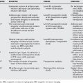
Hawkins3 introduced the popular classification system of talar neck fractures that remains commonly used today. This system is based on the degree of fracture fragment displacement and divides talar neck fractures into three groups. Type I fractures are nondisplaced. Type II fractures are displaced with subtalar subluxation or dislocation. Type III talar neck fractures are displaced with dislocation of the subtalar and tibiotalar joints. This classification system was modified by Canale and Kelly4 in 1978 to include type IV fractures that involve not only dislocation of the subtalar and tibiotalar joints but also dislocation of the talar head from the talonavicular joint.
CLOSED VERSUS OPEN MANAGEMENT
In the twentieth century, before Hawkins’s work, several clinical series on talus fractures were reported.5–8 In 1919, Anderson coined the term aviator’s astragalus after observing this injury in downed pilots.5 In 1952, Coltart6 reported a large Level IV clinical series examining this injury in British Air Force pilots during World War II. In this series, a poor outcome was associated with inadequate reduction of the subtalar joint, leading the author to advocate anatomic reduction with or without internal fixation.
Subsequently, Kenwright and Taylor7 examined 58 talus fractures in civilians. In 14 patients with talar neck fractures and subtalar subluxation, closed reduction and cast immobilization was attempted; however, in 10 patients (71%), satisfactory reduction could not be obtained or maintained, and open reduction was necessary. In some instances (the authors did not specify how many), the reduction was held with a Kirschner wire. Similar results were noted for talar neck fractures with tibiotalar dislocation. As such, the authors conclude that “a good result occurs only if accurate reduction is effected and maintained.”7
In 1970, Hawkins’3 landmark review and classification system of talar neck fractures was published. In this retrospective review (Level IV therapeutic study, Level II prognostic study), he drew attention to the incidence of AVN after talar neck fractures and emphasized the importance of open reduction. Nevertheless, in this series, Hawkins only peripherally mentions internal fixation in two patients but does not give details regarding fracture type or method of fixation. Of the 24 type II fractures in this series, 10 were treated with closed reduction and cast immobilization, whereas 14 were treated with open reduction.3 The overall incidence of AVN for type II fractures was 42%.3 For type III fractures, 2 of these injuries were treated with closed reduction and 20 with some form of open reduction for an overall AVN rate of 91%.3
Meanwhile, Canale and Kelly4 used closed reduction, as well as open reduction with and without internal fixation, in their historical series. Closed reduction was limited to those fractures in which adequate alignment (defined as less than 5 mm of displacement and less than 5 degrees of angular deformity) could be obtained. Of the 30 cases of type II fractures in this series, 19 were treated with closed reduction and casting, 1 presented late and was treated with only non–weight bearing, and 10 underwent open reduction with internal fixation. The overall AVN rate for type II fractures was 50%. Meanwhile, for type III fractures, 5 underwent closed reduction and 15 had open reduction (only 11 of which had internal fixation). The AVN rate in this group was 84%.4
Unfortunately, in both Hawkins’3 and Canale and Kelly’s4 series, the incidence and rate of AVN were not stratified by treatment type. Therefore, it is difficult to draw from these two studies any conclusions as to whether open treatment provides a lower AVN rate than closed reduction.
Other studies have reported varied rates of AVN. In a large Level IV review of 123 talar neck fractures, Lorentzen and colleagues9 found the overall incidence rate of AVN to be 21%. The majority of the fractures in this series were treated with cast immobilization. Among the 53 type II fractures, only 4 underwent open reduction. Thirty fractures were treated with closed reduction and casting, whereas 19 were treated with no reduction and casting only. Notably, although Lorentzen and colleagues report a 21% incidence rate of AVN, 44% of the fractures in their series were nondisplaced. The incidence rate of AVN was 35% when only the displaced talar neck fractures are considered.9
In those studies that examine the results of ORIF, the type of fixation often varies dramatically within the study. In a Level IV retrospective review, Grob and coworkers10 report the results of ORIF in 41 displaced talar neck fractures treated with malleolar screws, Kirschner wires, or cancellous screws. In this series, the incidence rate of AVN was only 16%.10 These encouraging data would seem to indicate that ORIF does reduce the incidence of AVN when compared with previous series in which patients were more commonly treated with closed techniques. However, in Vallier and colleagues’11 retrospective review (Level II prognostic study) of 102 displaced talar neck fractures in which all patients were treated with ORIF, the incidence of AVN was 39% for Hawkins group II fractures and 64% for Hawkins group III fractures.11
In addition to AVN, the development of malunion and post-traumatic osteoarthritis are two other clinical concerns associated with displaced talar neck fractures. Canale and Kelly4 report that subtalar degenerative changes occurred in approximately 50% of their patients, of which one third was said to have a malunion. Notably, in this series, 18 of 71 patients (25%) experienced development of a malunion. Because 11 of these 18 were initially treated with only closed reduction and casting, Canale and Kelly4 attributed subtalar arthritis to inadequate reduction or fixation. Comfort and colleagues’12 work supports the hypothesis that the incidence of malunion can be reduced with internal fixation. In this retrospective review of 28 patients with displaced talar neck fractures all treated with ORIF, there were no malunions and no nonunions. Radiographic evidence of ankle arthritis was noted in eight patients.12 Similarly, in the series reported by Vallier and coworkers,11 all 102 fractures were treated with ORIF. No patients experienced development of a malunion or nonunion. Although 54% of patients had radiographic evidence of arthritis, this was more common after comminuted and open fractures.11 Finally, Fleuriau Chateau and colleagues13 report a clinical series of displaced and highly comminuted talar neck fractures treated with open reduction and plate fixation. These authors note no varus malunions despite the fact that the fractures in this series were characterized by substantial comminution.
Although the aforementioned studies offer evidence that ORIF reduces the incidence of malunion, Sanders and colleagues’14 series does have a high malunion rate despite the use of internal fixation. In this series, 70 displaced talar neck fractures were treated with open reduction and screw fixation. Signs of radiographic arthritis were present in 40% of affected ankles and 78% of subtalar joints. The authors acknowledge malunion as “an important clinical problem” and note hindfoot malalignment in 30% of patients. When interpreting the results of this study, it is important to note that, in this series, surgery was performed or “directly supervised” by a consultant orthopedic trauma surgeon, and that eight fractures were treated by “other orthopedic surgeons at the trauma center.”14 In addition, malunion was seemingly not assessed by radiographs but rather hindfoot alignment alone.
Beyond these series, Grob and coauthors10 report osteoarthritis of the ankle joint in 32% of cases and of the subtalar joint in 37% of cases. All fractures in this series were treated with some form of ORIF. Unfortunately, data stratification with regard to post-traumatic arthritis and fracture type was not provided. No mention was made to malunion or nonunion. Pajenda and researchers15 reviewed 50 fractures of the talar neck and body (Level IV evidence), and reported osteoarthritis in 48% of the fractures. Severe ankle arthritis was noted in 11 of 30 displaced talar fractures (types II, III, and IV), but the form of fixation varied greatly including open reduction and internal with screws, external fixation, percutaneous screws, and closed reduction with percutaneous pinning using Kirschner wires.15
Collectively, these results justify a grade C recommendation that patients with displaced talar neck fractures should undergo ORIF of their displaced fractures.
TIMING OF SURGERY
At least three studies, two clinical series and one survey, have addressed this issue. In a recent clinical investigation, Vallier and colleagues11 performed a Level II retrospective review of 100 patients with 102 fractures of the talar neck. The average time to fixation was 3.4 days for patients who experienced development of AVN compared with 5.0 days for patients who did not. Rather than the timing of surgery, AVN was associated with talar neck comminution and the presence of an open injury. No correlation between surgical delay and the development of AVN could be identified.
These results are similar to those that Comfort and colleagues12 reported. In this retrospective review, the results of 28 patients who underwent ORIF of displaced talar neck fractures were reported and analyzed.12 Time to surgery was less than 12 hours for 18 patients, 1 to 7 days in 9, and 17 days in 1. Twelve of the 28 patients (43%) experienced development of AVN. Like Vallier and colleagues,11 the authors report that there was no trend between delay in operation and the degree of necrosis, and that the presence of necrosis correlated with the degree of displacement.
Recently, 89 expert orthopedic surgeons working at Level 1 trauma centers were surveyed regarding the current state of practice for the treatment of displaced talar neck fractures (Level V evidence).16 In this survey, 60% of respondents stated that, for displaced talar neck fractures, treatment after 8 hours is acceptable. Forty-six percent of respondents stated that treatment at or after 24 hours is acceptable. The authors of this study conclude that “most expert orthopedic trauma surgeons do not believe that an immediate operation is necessary to adequately treat a displaced talar neck fracture.”16
These studies, especially the work of Vallier and colleagues,11 offer fair (grade B) evidence that displaced talar neck fractures do not need to undergo ORIF on an urgent or emergent basis. Nevertheless, it should be noted that Vallier’s group did advocate emergent reduction of dislocations through either closed or percutaneous manipulation. Meanwhile, Comfort’s group12 recommends that ORIF “should be performed early,” although they did not expand on the definition of “early.”
METHOD OF FIXATION
The third question that must be answered in the treatment of displaced talar neck fractures is what form of surgical fixation should be used. Traditionally, surgical fixation of talar neck fractures has been achieved with Kirschner wires or screws placed in an anterior-to-posterior direction. Only a few clinical studies have examined other fixation constructs. Lemaire and Bustin17 analyzed their results using closed reduction followed by placement of a lag screw via the posterior tubercle. These authors suggest that the posterior approach allowed them to place the screw perpendicular to the fracture line. They also postulate that the posterior approach would have less effect on blood supply compared with an anterior approach. They report three excellent results and four good results, with AVN developing in two of the seven cases.17
Fayazi and coauthors18 describe a technique in which they closed reduced talar neck fractures and then performed percutaneous fixation using fluoroscopy and two partially threaded cannulated screws placed in opposite directions and parallel to each other. In this Level V technique article, the authors emphasize that if an anatomic reduction could not be obtained by closed methods, then open reduction was required. They state that their percutaneous approach resulted in shorter surgery, less pain, and minimal soft-tissue manipulation. Unfortunately, they report only on their technique and do not offer any results.18
Finally, Fleuriau Chateau and coworkers13 studied the effects of fixation of talar neck fractures using mini-fragment plates. In this retrospective review, 23 talar neck fractures were stabilized with a combination of mini-fragment plates augmented with lag screws if required. These fractures included type II, III, and IV injuries, and all were characterized by substantial comminution. The plates were placed on the more comminuted side of the fracture (either medial or lateral). With an average follow-up period of 20 months, the authors report no wound complications, a 100% union rate, and no evidence of varus malunion. Radiographically, there were two cases of tibiotalar arthritis and one case of subtalar arthritis. These patients were clinically asymptomatic. The authors argue that plate fixation minimizes varus malunion in highly comminuted fractures by avoiding excessive compression of the comminuted neck with lag screws.13
Although biomechanical studies do not provide direct clinical evidence, they are helpful in providing surgeons with basic principles that may provide guidance in the clinical setting. Several biomechanical studies have examined various methods of surgical fixation for talar neck fractures. Swanson and coworkers19 created a fracture model with cadaveric tali and compared four different fixation constructs: (1) Kirschner wires alone, (2) 4.0-mm cancellous screws placed from anterior to posterior, (3) 4.0-mm cancellous screws placed from posterior to anterior, and (4) a 6.5-mm cancellous screw placed from posterior to anterior combined with a Kirschner wire.19 Results showed that screw fixation was superior to Kirschner-wire fixation. Furthermore, screws placed in a posterior-to-anterior direction were found to provide stronger fixation than those placed in an anterior-to-posterior direction.19 More recently, Charlson and colleagues20 created a comminuted talar neck fracture model fixed with either two 4.0-mm cancellous screws placed in a posterior-to-anterior direction or a lateral mini-fragment plate augmented with a medial 2.7-mm lag screw.20 Their results showed superior fixation with the two-screw technique compared with plate fixation. Also, their results demonstrated failure at a load significantly below that shown by Swanson and coworkers.19 They attribute this to the comminuted component of the fracture model.20 Attiah and coauthors21 compared fixation among 3 3.5-mm cortical and 4.0-mm cancellous screws placed in an anterior-to-posterior direction, 2 4.0-mm cancellous screws placed in posterior-to-anterior direction, and a 2.7-mm blade plate placed on the medial aspect with a 4.0-mm cancellous screw placed laterally.21 Unlike Charlson and colleagues,20 these authors report no significant difference in the failure load between screw and plate fixation, but they did note that the screws failed by bending or pulling out, whereas the blade plate model failed by fracture of the talar head around the plate.21
Taken together, the combination of clinical evidence and biomechanical studies for fixation of talar neck fractures suggest the following: Screws placed in a posterior-to-anterior direction provide stronger fixation than those placed in the traditional anterior-to-posterior direction. Biomechanical studies have conflicting results whether screw fixation is equal to or superior to plate fixation. However, Fleuriau Chateau and colleagues13 offer limited Level IV clinical evidence that plate fixation may minimize varus malunion in highly comminuted fractures.
CONCLUSION
In conclusion, displaced talar neck fractures continue to pose a clinical challenge with an uncertain prognosis and potentially poor long-term morbidity. An evidenced-based review of the scientific literature regarding the optimal treatment of these injuries allows for several recommendations. First, such fractures should be treated with ORIF, rather than cast immobilization alone or open reduction without internal fixation. Second, surgery does not have to be performed on an emergent basis. Third, various fixation constructs are biomechanically advantageous, especially screws placed in a posterior-to-anterior direction and plate fixation for highly comminuted fractures. Table 76-1 provides a summary of recommendations for the treatment of displaced talar neck fractures.
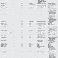
TABLE 76-1 Summary of Recommendations for the Treatment of Displaced Talar Neck Fractures
| RECOMMENDATIONS | LEVEL OF EVIDENCE/GRADE OF RECOMMENDATION |
|---|---|
1 Adelaar RS. Complex fractures of the talus. Instr Course Lect. 1997;46:323-338.
2 Ebraheim NA, Sabry FF, Nadim Y. Internal architecture of the talus: Implication for talar fracture. Foot Ankle Int. 1999;20:794-796.
3 Hawkins LG. Fractures of the neck of the talus. J Bone Joint Surg Am. 1970;52:991-1002.
4 Canale ST, Kelly FBJr. Fractures of the neck of the talus. Long-term evaluation of seventy-one cases. J Bone Joint Surg Am. 1978;60:143-156.
5 Anderson HGFM, Gotch OH, editors. The Medical and Surgical Aspects of Aviation. London: H Frowde, 1919.
6 Coltart WD. Aviator’s astragalus. J Bone Joint Surg Br. 1952;34-B:545-566.
7 Kenwright J, Taylor RG. Major injuries of the talus. J Bone Joint Surg Br. 1970;52:36-48.
8 Kleiger B. Fractures of the talus. J Bone Joint Surg. 1948;30A:735-744.
9 Lorentzen JE, Christensen SB, Krogsoe O, Sneppen O. Fractures of the neck of the talus. Acta Orthop Scand. 1977;48:115-120.
10 Grob D, Simpson LA, Weber BG, Bray T. Operative treatment of displaced talus fractures. Clin Orthop Relat Res.; 199; 1985; 88-96.
11 Vallier HA, Nork SE, Barei DP, et al. Talar neck fractures: Results and outcomes. J Bone Joint Surg Am. 2004;86-A:1616-1624.
12 Comfort TH, Behrens F, Gaither DW, et al. Long-term results of displaced talar neck fractures. Clin Orthop Relat Res.; 199; 1985; 81-87.
13 Fleuriau Chateau PB, Brokaw DS, Jelen BA, et al. Plate fixation of talar neck fractures: Preliminary review of a new technique in twenty-three patients. J Orthop Trauma. 2002;16:213-219.
14 Sanders DW, Busam M, Hattwick E, et al. Functional outcomes following displaced talar neck fractures. J Orthop Trauma. 2004;18:265-270.
15 Pajenda G, Vecsei V, Reddy B, Heinz T. Treatment of talar neck fractures: Clinical results of 50 patients. J Foot Ankle Surg. 2000;39:365-375.
16 Patel R, Van Bergeyk A, Pinney S. Are displaced talar neck fractures surgical emergencies? A survey of orthopaedic trauma experts. Foot Ankle Int. 2005;26:378-381.
17 Lemaire RG, Bustin W. Screw fixation of fractures of the neck of the talus using a posterior approach. J Trauma. 1980;20:669-673.
18 Fayazi AH, Reid JS, Juliano PJ. Percutaneous pinning of talar neck fractures. Am J Orthop. 2002;31:76-78.
19 Swanson TV, Bray TJ, Holmes GBJr. Fractures of the talar neck. A mechanical study of fixation. J Bone Joint Surg Am. 1992;74:544-551.
20 Charlson MD, Parks BG, Weber TG, Guyton GP. Comparison of plate and screw fixation and screw fixation alone in a comminuted talar neck fracture model. Foot Ankle Int. 2006;27:340-343.
21 Attiah M, Sanders DW, Valdivia G, et al. Comminuted talar neck fractures: A mechanical comparison of fixation techniques. J Orthop Trauma. 2007;21:47-51.