Chapter 39 What Is the Best Treatment for Simple Bone Cysts?
Simple bone cysts (SBCs) are benign bone tumors with a thin, cystlike lining and a fluid-filled cavity.1,2 They occur most commonly in the metaphyses of long bones in skeletally immature patients, although they have been reported in almost every bone of the skeleton.3 The proximal humerus and femur are the most common locations, with 50% to 80% humeral and 20% to 30% femoral in several large studies.4–6 Many cysts heal near skeletal maturity, thereby explaining the rarity in adults, but several large series have patients in their 40s and 50s.4,7, 8 Male-to-female ratios are varied, but two to three times greater in male than female individuals in most large series.4–69 SBCs are one of the most common bone tumors in children, representing 3% of all bone lesion biopsies.10–12 Many more lesions, however, have classic radiographic appearance and are never biopsied. The lesions, although characteristically metaphyseal and central, may rarely cross the physis.13–16 Most SBCs are centrally located and thought to originate from or near a physis. Various theories of cyst origin include physeal aberrations, venous obstruction, and synovial entrapment, or “local disturbance of bone growth and development.”2,7,17–20 SBCs were also called unicameral or solitary cysts. Although normally solitary, multiple concomitant cysts have been reported.21 Monolocular, or unicameral, is the common initial presentation, but fracture or treatment commonly results in septation and multiloculation. Rarely, SBC presents with more extensive ossification, confusing the diagnosis.22,23 Extension into the diaphysis is commonly reported and may represent a large active lesion that extends from the physis to mid-diaphysis, or a small latent diaphyseal cyst that has grown away from the physis.24,25 Cysts are radiolucent because of the fluid content, which is serous in nature but has some unique properties such as greater prostaglandin levels.18,26–30 This fluid under pressure could cause the endosteal erosion and bone expansion that is characteristic of these lesions.31 This expansion may also be due to bone weakness and the body’s attempt to preserve strength by increasing bone diameter.32,33 Cysts that occupy greater then 85% of the bone diameter are associated with increased risk for fracture.34 This is supported by the fact that healing cysts tend to “tubulate”, or narrow, and fragile cysts at risk for fracture are more expanded.35 Fracture in this weakened bone is common and may produce the classic “fallen-fragment” sign.36–38 This small fleck of bone fractures and settles in the cystic content. Fracture initiates a healing response and may cause opacification and septation with the cyst.39 Once the aggressive phase of fracture healing passes, however, the cyst, which appears to be healing, may again become more radiolucent and at risk for future fracture. Garceau and Gregory,40 in 1954, reported 15% cyst healing after fracture, but the true natural history of cyst healing has not been reported because of frequent and varied interventions and treatments. Lesions are reported to recur up to 4 years after initial healing.41 A natural history study was attempted by Neer and researchers16 but abandoned because of frequent fractures. Fractures may result in limb shortening and angular deformity. Physeal involvement may also cause the limb length inequality and angular deformity. Perceived risk for fracture prevents many children from participation in physical activities until cyst resolution. This can be disruptive to childhood for extended periods and limit activities.11,12
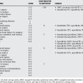
Bone cysts were first recognized by Virchow in 1891 and delineated more clearly in a case series by Bloodgood in 1910.42 Since these early times, numerous and various treatments have been proposed and combined. This chapter references more than 130 articles with greater than 20 treatment variations. The fact that so many variations and combinations of treatment for one bone lesion exists suggests the failure of any single current treatment for complete and permanent healing. Most articles are Level IV evidence and represent either single-center or multicenter case experiences with a single treatment. A few articles are comparison studies of two treatments and represent Level III evidence, but only one Level I evidence article is currently available. Another difficulty related to evaluation of cyst treatment is there is no universal system for defining a healed cyst. Neer proposed a classification that has been modified by many, but no study has outlined definitively what constitutes a healed cyst5,8,11,43–49 (Table 39-1). Most practitioners agree that even if a small cyst is present, as long as it does not pose a significant fracture risk and is not increasing in size, then observation is reasonable.4 Recent studies, however, have suggested that plain radiographs may be an inadequate measure of fracture risk and suggest computed tomographic (CT) scan as an alternative measure of fracture risk.32 Magnetic resonance imaging (MRI) study is also promising for assessing the load-carrying capacity of bones with osteolytic lesions.50 With poor outcome tools used for measurement of SBC healing, the comparison of treatment is arbitrary and difficult. Without having an accurate comparison of cysts before treatment, stratification for randomization is also arbitrary. Cysts are known to begin healing and then recur more than 2 to 4 years after healing.41,51 This may represent reactivation of a smaller remaining cystic area or recurrence of a completely healed or resected cyst.52 Capanna and coauthors8 described 12 of 90 patients treated with steroid injection whose cysts recurred after initial consolidation. Most studies have less than 2-year minimum follow-up and, therefore, show early promise for cyst healing, but later extensive follow-up demonstrates recurrence or persistence, or both, of these lesions.4,6, 41 Few articles evaluate the angular deformity and limb-length inequality that results from cysts.53–55 Some of this may be because of the presence of the cyst near the physis, and others may represent physeal damage secondary to treatment. Few studies evaluate whether patients have persistent pain or functional difficulties; however, one recent study does attempt to incorporate functional outcomes using the Activity Scale for Kids (ASK) and pain assessment using the OUCHER scale in a randomized prospective study of two treatments.49,56
TABLE 39-1 Summary of Criteria for Healing
| Graham (1951)43 | |
| Neer et al. (new) (1966)4 | Proposed classification: |
| Incomplete obliteration of the cyst after operation appears to be of little clinical significance, provided there is good bone strength | |
| Spence et al. (new) (1969)44 | |
| Baker (1970)45 | |
| Capanna et al. (modification of Neer Criteria) (1982)8 |
• Healed with residual: cyst consolidated with bone and the cortical margins thickened, but still small residual areas of osteolysis within cyst
|
| Chigara et al. (1983)17 | |
| Oppenheim and Galleno (1984)11 | |
| Lokiec et al. (1996)114 | Features of healing: |
| Weintroub (1989)104 | |
| Hashemi-Nejad and Cole (modification of Neer criteria; Reverse of Neer) (1997)47 | |
| Grade 1–2 = unsatisfactory healing | |
| Grade 3–4 = satisfactory healing | |
| Killian (modification of Neer criteria) (1998)48 | |
| Yandow et al. (1998)49 | Substantial healing |
| Partial healing | |
| Failure to respond | |
| Chang et al. (2002)6 |
Healed: cyst filled by formation of new bone with or without small static, radiolucent area(s) less than 1 cm in size
Healing with defect: static, radiolucent area(s) less than 50% of the diameter of bone with enough cortical thickness to prevent fracture
|
| Wright (modification of Neer and Cole criteria) (2008)56 |
Definition of healing: One of the single greatest difficulties in comparison of treatments and natural history is the definition of healing of simple bone cysts.
FACTORS THAT AFFECT CYST HEALING
Many factors related to the location of the cyst and its host have been proposed to affect the rate of healing.57,58 Most of the series that discuss cyst treatment qualify the results by separating these various factors, which are discussed later. This, however, does not separate the treatment groups before initiation of treatment and, therefore, does not provide equivalent groups for study purposes. More randomized prospective trials will be needed to clarify factors that may affect healing and then to compare headto-head the various treatments.
Lesion Size
Cysts can vary from less than 1 cm2 on two orthogonal views to large complex lesions extending from physis to mid-diaphysis, or more than 50% the length of the bone.49 Cyst index based on size and geography of the lesion was described by Kaelin and McEwen60 as a predictor for fracture risk, but a recent article suggests poor reliability for this technique as a predictor of fracture.59 Others have demonstrated that larger cyst areas correlate with poorer healing.8,56 Capanna and coauthors8 defined small cysts as less than 24 cm2. Spence and coworkers,44 in 1969, in a large multicenter review, showed 80% healing of small lesions, 49% of medium lesions, and 53% of large cysts. This makes excellent sense because larger lesions theoretically have been present for a longer time and may be recalcitrant to the body’s attempts at healing.
Patient Age and Sex
Cysts are uniformly reported in all series reviewed to occur more frequently in male than female individuals. The most common male-to-female ratio is 3:1.9,44 The only exception is 1 smaller series by Dorman121 in 2005 with a 1:1 ratio. The variation in number of male and female patients may affect the healing rates, as Spence suggested in his large multicenter series review of 177 cases. Cysts occur in all age groups. Originally, patients younger than 10 years were considered to have more active cysts.31 Capanna and coauthors,8 in 1982, found less recurrence of cysts after steroid treatment in patients 0 to 5 years of age and poorer healing in those older than 6. Spence and coworkers5 confirm in his large review the adverse effect of younger age on healing. Spence and coworkers also report that female patients had healed cysts in 77% of cases, whereas only 48% of male patients had healed cysts in his review (P < 0.001).44 Case series by Chigira46, Garceau40, Neer4, and Capanna8 include patients with cysts at older than 50 years. The overall lower frequency of this lesion in adulthood in many articles, however, helps us to understand that physeal closure and skeletal maturity does, in some unknown way, promote cyst healing.61 This is important because many of the reviewed articles vary in average age from 6.9 to 17.3 years.62,63 This variation of age may be the cause of variations in healing rates.
Proximity to Physis
Cysts were categorized as active and latent originally by Jaffe and Lichtenstein7 in 1942, based on proximity to the physis. Lesions less than 1 cm from the physis are considered active. Variation of healing response of active verses latent cysts exists in numerous reported series since that time.40,45, 64 Neer, however, in a large series of 175 cases, found no difference in healing of active verses latent cysts.4 Spence, in contrast, showed healing in 35% of active cysts and 67% of latent cysts.5 The data are inconsistent regarding whether location of cyst affects healing. Many articles even fail to evaluate this before treatment.65,66 No trials of similar treatment in randomized groups of active verses latent cysts have been reported to clearly compare location as a unique factor related to healing.
Cyst Fluid and Venous Outflow.
An early study of cyst fluid was conducted by Cohen in 1960.18 It was found to be either serous or bloody, but no correlation to healing was made. Needle perforation and measurement of manometric pressure was elucidated by Neer and coworkers4 in 1966. Active cysts had pressures of 30 cm of H2O and pulsated with Valsalva maneuver. Enneking,31 using this technique, also performed cystograms with injections of radiopaque contrast material attempting to correlate active lesions with rapid outflow of contrast into the venous circulation, and puddling and slow outflow with latent cysts. These are Level IV, observational, and Level V expert opinion articles. Draining or venting the cystic cavity is described in various articles with K-wires, cannulated screws, and intramedullary rods, and these articles are reported and level of evidence reviewed in this summary separately.
Bone Location
SBCs are reported in almost every bone; however, jaw lesions have a different histologic appearance and may be a separate entity. For the purposes of this chapter, these are not included. Greater than 90% of cysts occur in long bones. The calcaneus represents an unusual location, and treatment series for this bone are reported separately in this chapter. The proximal humerus is the most common location. In 195 cases reviewed by Mirra,10 44% were proximal humerus, and the second most common condition was proximal femur with 26% of cases. Hands are a rare location for SBCs. Neer’s series4 showed the femur as the most common location for cyst in adulthood, but fractures of adult cyst were also rarer. Healing rates for treatment of femoral cysts may be higher because of the weight-bearing status of the bone initiating a healing response because of added load. This weight-bearing bone, however, makes this location more at risk for fracture. Angular deformities are reported in numerous studies and may lead to the benefit of intramedullary devices for load sharing or external support during healing.65,67 Location also refers to the location of the cyst within the bone. Metaphysis is the most common initial presentation. Although called simple cysts, their geography and variability is extensive. Cross-sectional imaging (MRI and CT scan) is adding to a more detailed understanding of these lesions.68,69
Length of Follow-Up
Another of the significant difficulties in comparing healing rates of various treatments is the length of post-treatment follow-up. Follow-up varies from 6 months to 20 years in one series.70 Enneking31 states that fracture or intervention to the cyst initially stimulates an aggressive biologic fracture healing response. This promotes early bone formation and opacification of the cyst and “healing.” Once this process becomes more quiescent, the tumor’s biology may again become more active and cause “recurrence” or “visible reappearance” of the lesion.71 Many series report recurrence more than 2 years after treatment in lesions that initially “healed.”5,72 Docquier and Delloye73 had a recurrence at 89 months (7.4 years) after treatment. Therefore, uniform, longer length of follow-up may ruin early positive results. Lokiec and Wientroub12 report 100% healing with bone marrow injection in his preliminary report, but follow-up in some patients was only 12 months. Neer’s report carries such long-term value because of his insistence of a minimum 2-year follow-up period.
Capanna and coauthors describe an interesting phenomenon not studied by others since his articles in 1982.8 Recurrences occurred at varying rates based on healing patterns:
TRANSFORMATION OF SIMPLE BONE CYSTS
Six cases of malignant conversion of SBC to sarcoma have been reported, but 5 are unproved because of lack of biopsy confirmation of the SBC before transformation, and therefore a lesser level of certainty.74 There is 1 report of malignant transformation to chondrosarcoma from a biopsy-proved SBC.75 This biopsy confirmation of SBC, and later biopsy confirmation of sarcoma, is valuable for the understanding that malignant conversion is a true risk, but not for estimating its incidence, which must be low. SBC can transform into another benign lesion, aneurysmal bone cyst (ABC).76,77 ABC may represent a spectrum of the same disease as SBC, as Neer and coauthors discussed in 1973.78–81 Separation of the two may be difficult even with cross-sectional imaging. Biopsy may be indicated for definitive diagnosis.13,82, 83 Recognition of this possibility is important because ABC does not respond to many forms of treatment proposed for SBC.84 Injections of steroids into ABC were tried by Scaglietti and investigators85 and abandoned because of poor healing in the original series of patients in 1982.
HISTORICAL SUMMARY OF TREATMENT RECOMMENDATIONS AND LEVEL OF EVIDENCE COMPARISON
Although variation of characteristics of the SBC exists, variations of treatment recommended in the literature are also widely variable and difficult to compare.86 There is only 1 Level I evidence article on the treatment of SBC; the remaining reports, no matter how large, are either case series Level IV or, at best, retrospective comparative Level III.6,9, 11, 66 Lokiec’s series114 in 1996 is prospective but not comparative and may be considered Level II evidence regarding prognosis, but only Level IV evidence regarding selection of therapy because no comparison was made.
Early Case Series (Level IV)
The earliest articles were summaries of case descriptions by Bloodgood in 1910,42 Phemister20 and Gordon in 1926, James and colleagues41 in 1942, Graham43 in 1951, and Garceau and Gregory40 in 1954. They focus on the pathology of the lesion, and Garceau states a 15% healing rate after fracture with observation alone. Blount61 reports in Fractures in Children that most cysts heal during childhood. A natural history study following fracture of cysts was further attempted by Neer4 in 1966 but abandoned during the study because of greater than 70% refracture in both humeral and femoral locations. Active verses latent cysts were compared with little variation of refracture rate. These case series are Level IV evidence, but they are important for historical significance.
Intralesional Curettage and Grafting (Level IV)
The largest group of articles in the literature on the treatment of SBC reports results for intralesional curettage and grafting. Graft materials were initially autograft, but subsequent series have recommended allograft and a host of other nonbiologic materials, which are discussed separately.87,88 In early studies after curettage, the lining was removed; then the residual rim was treated with phenol or zinc chloride, followed by bone grafting. Curettage provided cyst lining to pathology, and detailed descriptions of findings by Geschickter and Copeland,90 Jaffe,89 and Jaffe and Lichtenstein7 delineated the histologic findings of simple cysts. Some are cases series Level IV and others comparative and Level III. Neer and coworkers4 showed no difference in those treated with or without adjuvant chemicals; therefore, this step was abandoned, but later series did advocate cryosurgery to ablate the lining.91 Neer’s series was the largest reported at that time and carefully outlined many variables contributing to treatment results including location, age, and proximity to the physis. He also proposed one of the first grading scales for cyst healing, which serves as the basis for most other authors. Variations of this are still used today, and a variation was utilized in the current randomized, prospective trial by Wright.4,8, 47, 56 The problems with intralesional curettage and grafting are the need for a larger open procedure, the necessity of harvesting bone graft and its risk, the risk for physeal damage with curetting near the physis in active cysts, and universally, the reports of recurrence despite initial healing.9,11,40,43,45,92–94 The complications of autograft harvesting and the shortage of bone available to fill the cyst led some authors to recommend allograft and bone substitutes. Peltier and Jones,95 in 1978, reported a new technique of curettage followed by filling the lesion with sterile plaster of Paris(Ethicon). Ninety-two percent of these patients healed, but 8% recurred and 12% (3/26) either had post-operative drainage or infection. The technique did not spread widely into the literature. Spence and coworkers,44 in 1969, reported a Level IV multicenter large series of 177 patients treated with freeze-dried allograft. Recurrence or failure of healing, however, was 45%, and this technique was not popularized, likely because of the high recurrence rate. Because of the recurrence seen with intralesional curettage and grafting with or without bone, radical or subtotal resection of the cysts were next historically recommended.
Radical or Subtotal Cyst Resection (Level IV)
Agerholm and Goodfellow96 described the technique of subperiosteal diaphysectomy in 1965; the first 20 case series was published in 1973 by Fahey and O’Brien,70 12 cases by Gartland and Cole62 in 1975, and another series of 21 patients was published by McKay and Nason98 in 1977.97 This technique was proposed because of the recurrence rate of 21% to 50% with intralesional curettage and grafting70 (Table I summary from Fahey and O’Brien’s70 article). Despite this aggressive resection without grafting, McKay and Nason98 had a 9% rate of recurrence. It would seem that aggressively removing the entire cyst subperiosteally could still result in later recurrences. It is important to understand that McKay’s recurrences occurred greater than 1 year after initial treatment, and this aggressive surgery was also complicated by postoperative fractures and limb shortening.98 These aggressive procedures, with their complications and recurrences, were the spawning ground of percutaneous injection techniques that would follow.
Percutaneous Injection with Steroid (Methylprednisolone Acetate)
A landmark article of innovative treatment appeared from Pisa, Italy, by Dr. Scaglietti and investigators99 in 1979. It provided Level IV evidence of a new treatment utilizing percutaneous injection of methylprednisolone acetate (MPA) into 72 cysts. The initial article had up to six injections of MPA varying from 40 to 200 mg. Follow-up period was a minimum of 18 months; 60% of patients healed completely, and 36% healed “more or less complete,” with only a 4% failure to heal rate. Campanacci and Capanna9 from Rizzoli Institute in Bologna, Italy, reported results in 90 patients treated similarly and followed a minimum of 1 year (Level IV evidence) with 46 of 90 patients (51%) healed, 26 of 90 (29%) healed with small residual cysts, 12 of 90 (13%) with recurrence, and 6 of 90 (7%) had no response. They emphasize that smaller cyst size (<24 cm2) and monoloculation positively affected the healing outcome (P = 0.002 and P < 0.001, respectively). Scaglietti and investigators reported final follow-up in 1982 and strongly cautioned that “patients must be observed until termination of their skeletal growth before evaluating the final results of the injection treatment. Otherwise, further extension of the cyst is possible.”85,100 Subsequent Level III articles comparing steroid with intralesional curettage and grafting showed only 74% healing with steroid and 54% healing with curettage and grafting.11 Bovill et al,86 also with Level III studies, compared steroid injection with open curettage and grafting, and found similar levels of efficacy for both treatments, but less complications in the injections group. Percutaneous injection offered the other advantage of a smaller intervention for residual or recurrent cysts, but the disadvantage of chemical injection near the physis and reports of local and systemic effects of the steroids and osseous necrosis of the femoral head after injection.8,9,11,101–103
Bone Marrow Aspiration and Percutaneous Injection.
Bone marrow transplant for malignancy and bone-marrow aspiration and injection to promote fracture healing laid the groundwork for bone-marrow aspiration and percutaneous injection into SBCs to promote healing.104–110 In 1985, Burwell111 discussed the expanded understanding of the biology of bone marrow as an important osteoinductive factor in bone graft incorporation. Many subsequent articles furthered our understanding of the osteoinductive power of bone marrow.106,109, 112, 113 The negative host effects of open bone graft harvesting added to the appeal of a less invasive technique of obtaining the osteogenic cells by aspiration.92,94 A preliminary (Level of Evidence IV) prospective case series report by Lokiec and coworkers114 of 10 patients with 100% healing of SBCs treated by bone marrow injection was published in 1996. This injection was combined with indirect venting of the cyst using a custom curved needle into the medullary canal.114 A similar injection of bone marrow into cysts, but obtained by small 3-mL aliquot aspiration to potentiate osteoblast concentration and without venting into the medullary canal, showed similar positive results to Lokiec and Weintroub’s initial reports, but 2 of 12 patients (17%) did not respond to marrow injection.49,81, 105 Because of these reports, marrow injection became a popular technique with minimal invasion to promote cyst healing.115–117 Chang and coauthors,6 in a Level III review of 79 patients treated with bone marrow (14 patients) verses steroid (65 patients) injection, showed no superiority of 1 technique over the other (P > 0.05). Docquier and Delloye73,118 reported a Level IV series of 17 cases with 76% healing rate, 12% recurrence rate, and 12% failure of response rate. None of the bone marrow studies had uniform greater than 2-year follow-up until Wright’s56 study in 2007. Currently, this is the only Level I randomized, prospective, multicenter study that compares bone marrow with steroid injection. Seventy-seven of 90 patients completed 2-year follow-up. Radiologists, using Cole’s modification of Neer’s criteria for healing, demonstrated only 42% healing with steroids and 23% healing with bone marrow (P = 0.01). No cases were vented into the medullary canal before injection. The radiologists grading the healing were blind to treatment, and this is the first report of outside grading of the surgical outcome for cyst treatment. The great disparity to prior reports may be related to past evaluation bias, the longer length of follow-up in this study, or the failure to vent cysts into the medullary canal in the recent study.
Mechanical Cyst Disruption with and without Fixation.
Although the literature is full of exogenous materials that can promote cyst healing, many authors have outlined that simple mechanical disruption of the cyst can promote fracture-like biologic response and healing of cysts.34,119, 120 Chigira and colleagues,17 in a 1983 Level IV study, utilized Kirschner-wire disruption of the cyst to decrease the pressure within the cyst, but their 1986 review reported recurrence if the K-wire was removed.46 Komiya and coauthors’27 1991 Level IV study recommends “trepanation” of cysts percutaneously with a K-wire to promote healing. This article offers an evaluation of cyst fluid content and theory of its negative effect on bone formation. Although the first report of drainage of the cyst into the medullary canal was Ober’s122 in 1944, many subsequent articles offer drainage either intramedullary or cortically.65,121, 123 A Level III comparative study by Tsuchiya66 in 2002 recommends continuous cyst decompression after curettage by either a cannulated screw or a hydroxyapatite cannulated pin, with 100% healing in the hydroxyapatite pin group. Follow-up, however, was 8 to 60 months in this group. Similar ideas of mechanical disruption with intramedullary rodding and the added advantage of fracture fixation and prophylaxis of possible future fracture were introduced by Catier and researchers124 in 1981 and further by Santori and investigators125 in 1988. These ideas were even further popularized by Roposch and colleagues65 in 2000 and de Sanctis and Andreacchio126 in 2006. Healing occurred in all 32 patients of this Level IV study, but 2 recurrences resulted from nail removal. Roposch and colleagues65 emphasize in their review the tenacity and prevalence of cyst recurrence after treatment. Givon and coauthors,127 in a technique article, utilizes a titanium elastic nail to indirectly open the cyst into the medullary canal but removes the nail and fills the defect with a bone substitute, Osteoset. Again, combinations of treatment obscure what actually is promoting cyst healing, and short follow-up negates the later recurrences seen throughout cyst treatment history.
Alternative Graft Materials for Treatment of Simple Bone Cysts.
Gazdag and colleagues88 wrote an excellent summary of alternatives to autogenous bone grafting. Many of the substitutes from hydroxyapatite cubes to demineralized bone matrix, Ethibloc and bioactive glass (Norion), have been utilized to encourage healing in bone cysts.48,128–130 Demineralized bone matrix and bone marrow were combined with trephination in a percutaneous injection technique by Rougraff and Kling87 reported in 2002. Twenty-three patients in this Level IV evidence study were observed for 30 to 81 months with 100% healing. In 2005, Kanellopoulos and coworkers131 further amplified the technique with the addition of intramedullary perforation of the cyst and similar DBM and bone marrow injection. All patients healed, but 2 required repeat procedures to achieve this outcome. Follow-up period was 12 to 42 months, and Neer’s criteria were used for healing. The historical return to calcium sulfate (Osteoset) but with a newer addition of venting into the medullary canal and indirect curettage, is reported in Dormans and researchers’121 Level IV series. Twelve of 28 patients were managed for at least 24 months. Eleven of these achieved complete healing and 1 partial healing. In contrast with Peltier and Jones’s95 study in 1978 utilizing plaster of Paris, only 1 of Dormans’s patients had a stitch abscess. A risk of alternative graft material is the possibility of rapid venous outflow from some active cysts.31,132 Reports have demonstrated central Doppler flow disturbances from marrow and steroids injected into cyst with large venous outflow.49,83, 132 Smaller particulate materials used as graft substitutes could also exit these venous channels and pose a similar risk.
Calcaneal Bone Cysts
The calcaneus is the sixth most common location for SBCs, which are relatively rare. In large series reviewing SBCs, they represent a small percentage of overall cyst locations.22 Behavior of calcaneal cysts related to fracture risk and treatment is somewhat unique. Pogoda and researchers133 studied 50 cysts in 47 patients and found cysts reaching a critical size, defined as a 100% intracalcaneal cross section in the coronal plane and at least 30% in the sagittal plane, are at risk for fracture. Calcaneal cysts tend to occur in older patients and, because of the weight-bearing nature of the bone, when larger, cause pain with daily activities.133 Radiologic assessment with plain films, CT scans, and/or MRI offer a characteristic pattern that aids with diagnosis. Radiographically, atypical cysts should undergo biopsy to prevent misdiagnosis and confusion with intraosseous lipomas, pseudocysts, or cysts secondary to other osseous or chondral lesions.134 Intralesional curettage and bone grafting, either with allograft or autograft, has been the mainstay of treatment.134–136 Curettage and grafting, in several studies, has shown superior healing to steroid injection.137,138 Saraph’s117,134 articles include an excellent table providing a comprehensive overview of the literature to date with methods of treatment for calcaneus cysts. All of the articles are Level IV evidence and are retrospective summaries of techniques used with outcomes of healing reported. This summary Level IV evidence article outlines their excellent result with continuous decompression of the cyst using titanium cannulated screws and no curettage or grafting. Eight of nine healed completely with a minimum of 2-year follow-up. Patients healed with a residual cyst. This technique supports the theory that cyst fluid decompression facilitates and/or promotes healing in all cyst locations.
SUMMARY
Clearly, numerous treatments and combinations of treatment have been utilized for treatment of SBCs. The unique features of the host, such as age, sex, and activity, have been reported to affect healing. Tumor variables such as lesion size, proximity to the physis, and venous outflow are also reported to affect response to treatment and healing. Surgical treatment variables of curettage, septation disruption, medullary or cortical venting, and fixation have been shown to affect healing. Graft materials, both biologic and nonbiologic, osteoinductive and conductive, are important factors. A standardized scale for healing and clear risk for fracture must be determined. Length of follow-up, ideally to skeletal maturity, is important in outcome success. Unless these variables are controlled in a randomized, prospective study, the levels of evidence will remain as multiple case series and be difficult, if not impossible, to compare. Level IV evidence supports injection or venting techniques using a variety of specific procedures as outlined earlier. The only Level I evidence reports a greater healing rate with steroid injection than with bone marrow injection, but fewer than 50% of the cysts in either arm of the randomized trial healed. More randomized, prospective, multicenter studies are needed to guide treatment recommendations for patients and families. Table 39-2 provides a summary of recommendations for calcaneal SBC treatment.
TABLE 39-2 Summary of Recommendations for Calcaneal Simple Bone Cyst Treatment
| STATEMENT | LEVEL OF EVIDENCE/GRADE OF RECOMMENDATION | REFERENCES |
|---|---|---|
SBC, simple bone cyst.
1 Campanacci M. Pseudotumors of bone. Bone and Soft Tissue Tumors. New York, Wien: Springer-Verlag, 1990;709-724.
2 Cohen J. Etiology of simple bone cyst. J Bone Joint Surg Am. 1970;52:1493-1497.
3 Johnson L, Kindred R. The anatomy of bone cysts. J Bone Joint Surg Am. 1958;40:1440.
4 Neer CN, Francis K, Marcove R. Treatment of unicameral bone cyst. A follow-up study of one hundred seventy-five cases. J Bone Joint Surg Am. 1966;48:731-745.
5 Spence KJ, Bright R, Fitzgerald S. Solitary unicameral bone cyst: Treatment with freeze-dried crushed cortical-bone allograft. A review of one hundred and forty-four cases. J Bone Joint Surg Am. 1976;58:636-641.
6 Chang C, Stanton R, Glutting J. Unicameral bone cysts treated by injection of bone marrow or methylprednisolone. J Bone Joint Surg Am. 2002;84:407-412.
7 Jaffe H, Lichtenstein L. Solitary unicameral bone cyst with emphasis on the roentogen picture, the pathologic appearance and the pathogenesis. Arch Surg. 1942;44:288-293.
8 Capanna R, Dal Monte A, Gitelis S. The natural history of unicameral bone cyst after steroid injection. Clin Orthop Relat Res. 1982;166:204-211.
9 Campanacci M, Capanna R, Picci P. Unicameral and aneurysmal bone cysts. Clin Orthop Relat Res.; 204; 1986; 25-36.
10 Mirra J. Bone Tumors Diagnosis and Treatment. Philadelphia: J.B. Lippincott, 1980.
11 Oppenheim W, Galleno H. Operative treatment versus steroid injection in the management of unicameral bone cysts. J Pediatr Orthop. 1984;4:1-7.
12 Lokiec F, Wientroub S. Simple bone cyst: Etiology, classification, pathology and treatment modalities. J Pediatr Orthop B. 1998;7:262-273.
13 Malawer MM, Markle B. Unicameral bone cyst with epiphyseal involvement: Clinicoanatomic analysis. J Pediatr Orthop. 1982;2:71-79.
14 Ovadia D, Ezra E, Segev E. Epiphyseal involvement of simple bone cysts. J Pediatr Orthop. 2003;23:222-229.
15 Gupta A, Crawford A. Solitary bone cyst with epiphyseal involvement: Confirmation with magnetic resonance imaging. J Bone Joint Surg Am. 1996;78:911-915.
16 Nelson J, Foster R. Solitary bone cyst with epiphyseal involvement. Clin Orthop Relat Res. 1976;118:147-150.
17 Chigira M, Maehara S, Arita S. The aetiology and treatment of simple bone cysts. J Bone Joint Surg Am. 1983;65:633-637.
18 Cohen J. Simple bone cysts. Studies of cyst fluid in six cases with a theory of pathogenesis. Am J Orthop. 1960;42-A:609-616.
19 Allredge R. Localized fibrocystic disease of bone. Results of treatment in one hundred and fifty-two cases. J Bone Joint Surg Am. 1942;24:795-804.
20 Phemister. The etiology of solitary bone cyst. JAMA. 1926;87:1429-1433.
21 Abdel-Wanis ME, Tsuchiya H, Minato H, et al. Bilateral symmetrical cysts in the upper tibiae in a skeletally mature patient: Might they be simple bone cysts? J Orthop Sci. 2001;6:595-600.
22 Mirra J, Bernard G, Bullough P. Cementum-like bone production in solitary bone cysts (so-called “cementoma” of long bones). Report of three cases. Electron microscopic observations supporting a synovial origin to the simple bone cyst. Clin Orthop Relat Res.; 135; 1978; 295-307.
23 Amling M, Werner M, Pösl M, et al. Calcifying solitary bone cyst: Morphological aspects and differential diagnosis of sclerotic bone tumours. Virchows Arch. 1995;426:235-242.
24 Cohen J. Unicameral bone cysts. A current synthesis of reported cases. Orthop Clin North Am. 1977;8:715-736.
25 Makley J, Joyce M. Unicameral bone cyst (simple bone cyst). Orthop Clin North Am. 1989;20:407-415.
26 Komiya S, Inoue A. Development of a solitary bone cyst: A report of a case suggesting its pathogenesis. Arch Orthop Trauma Surg. 2000;120:455-457.
27 Komiya S, Minamitani K, Sasaguri Y. Simple bone cyst. Treatment by trepanation and studies on bone resorptive factors in cyst fluid with a theory of its pathogenesis. Clin Orthop Relat Res. 1993:204-211.
28 Komiya S, Tsuzuki K, Mangham DC, et al. Oxygen scavengers in simple bone cysts. Clin Orthop Relat Res.; 308; 1994; 199-206.
29 Gerasimov A, Toporova S, Furtseva L. The role of lysosomes in the pathogenesis of unicameral bone cysts. Clin Orthop Relat Res. 1991;266:53-63.
30 Shindell R, Connolly JF, Lippiello L. Prostaglandin levels in a unicameral bone cyst treated by corticosteroid injection. J Pediatr Orthop. 1987;7:210-212.
31 Enneking W. Lesions of Uncertain Origin Originating in Bone. Musculoskeletal Tumor Surgery.; vol. 2; 1983; Churchill Livingston: New York.
32 Snyder BD, Hauser-Kara DA, Hipp JA, et al. Predicting fracture through benign skeletal lesions with quantitative computed tomography. J Bone Joint Surg Am. 2006;88:55-70.
33 Hong J, Cabe GD, Tedrow JR, et al. Failure of trabecular bone with simulated lytic defects can be predicted non-invasively by structural analysis. J Orthop Res. 2004;22:479-486.
34 Ahn JI, Park JS. Pathological fractures secondary to unicameral bone cysts. Int Orthop. 1994;18:20-22.
35 Hipp JA, Springfield DS, Hayes WC. Predicting pathologic fracture risk in the management of metastatic bone defects. Clin Orthop Relat Res.; 312; 1995; 120-135.
36 McGlynn F, Mickelson M, El-Khoury G. The fallen fragment sign in unicameral bone cyst. Clin Orthop Relat Res. 1981:157-159.
37 Reynolds J. “The fallen fragment sign” in the diagnosis of unicameral bone cyst. J Radiol. 1969;92:949-953.
38 Struhl S, Edelson C, Pritzker H, et al. Solitary (unicameral) bone cyst. The fallen fragment sign revisited. Skeletal Radiol. 1989;18:261-265.
39 Suei Y, Tanimoto K, Wada T. Simple bone cyst. Evaluation of contents with conventional radiography and computed tomography. Oral Surg Oral Med Oral Pathol. 1994;77:296-301.
40 Garceau G, Gregory C. Solitary unicameral bone cyst. J Bone Joint Surg Am. 1954;36:267-280.
41 James A, Coley B, Higinbotham N. Solitary (unicameral) bone cyst. Arch Surg. 1942;57:137-147.
42 Bloodgood J. Benign bone cysts, ostititis fibrosa, giant-cell sarcoma and bone aneurism of the long pipe bones. Ann Surg. 1910;52:145-185.
43 Graham J. Solitary unicameral bone cyst. A follow-up study of thirty-one cases with proven pathological diagnosis. Bull Hosp Joint Dis. 1952;13(1):106-130.
44 Spence KJ, Sell K, Brown R. Solitary bone cyst: Treatment with freeze-dried cancellous bone allograft. J Bone Joint Surg Am. 1969;51:87-96.
45 Baker D. Benign unicameral bone cyst. A study of forty-five cases with long-term follow up. Clin Orthop Relat Res. 1970;71:140-151.
46 Chigira M, Shimizu T, Arita S, et al. Radiological evidence of healing of a simple bone cyst after hole drilling. Arch Orthop Trauma Surg. 1986;105:150-153.
47 Hashemi-Nejad A, Cole W. Incomplete healing of simple bone cysts after steroid injections. J Bone Joint Surg Br. 1997;79:727-730.
48 Killian J, Wilkinson L, White S. Treatment of unicameral bone cyst with demineralized bone matrix. J Pediatr Orthop. 1998;18:621-624.
49 Yandow SM, Lundeen GA, Scott SM, Coffin C. Autogenic bone marrow injections as a treatment for simple bone cyst. J Pediatr Orthop. 1998;18:616-620.
50 Hong J, et al. Magnetic resonance imaging measurements of bone density and cross-sectional geometry. Calcif Tissue Int. 2000;66:74-78.
51 Kleinberg S. The solitary bone cyst. A report of a case of twenty years’ duration. J Bone Joint Surg Am. 1944;26:337-344.
52 Bowen RE, Morrissy RT. Recurrence of a unicameral bone cyst in the proximal part of the fibula after en bloc resection. A case report. J Bone Joint Surg Am. 2004;86:154-158.
53 Stanton R, Abdel-Motá al M. Growth arrest resulting from unicameral bone cyst. J Pediatr Orthop. 1998;18:198-201.
54 Violas P, Salmeron F, Chapuis M, et al. Simple bone cysts of the proximal humerus complicated with growth arrest. Acta Orthop Belg. 2004;70:166-170.
55 Norman-Taylor FH, Hashemi-Nejad A, Gillingham BL, et al. Risk of refracture through unicameral bone cysts of the proximal femur. J Pediatr Orthop. 2002;22:249-254.
56 Wright JG, Yandow S, Donaldson S, et al. A randomized trial comparing intralesional bone marrow and steroid injections for simple bone cysts. J Bone Joint Surg Am. 2008;90(4):722-730.
57 Wilkins RM. Unicameral bone cysts. J Am Acad Orthop Surg. 2000;8:217-224.
58 Biermann JS. Common benign lesions of bone in children and adolescents. J Pediatr Orthop. 2002;22:268-273.
59 Vasconcellos D, Yandow SM, Grace AM, et al. Cyst index: A nonpredictor of simple bone cyst fracture. J Pediatr Orthop. 2007;27:307-310.
60 Kaelin AJ, MacEwen GD. Unicameral bone cysts. Natural history and the risk of fracture. Int Orthop. 1989;13:275-282.
61 Blount W. Fractures in Children. Philadelphia: Lippincott Williams & Wilkins, 1955;251-252.
62 Gartland J, Cole F. Modern concepts in the treatment of unicameral bone cysts of the proximal humerus. Orthop Clin North Am. 1975;6:487-498.
63 Inoue O, Ibaraki K, Shimabukuro H. Packing with high-porosity hydroxyapatite cubes alone for treatment of simple bone cyst. Clin Orthop Relat Res. 1993:287-292.
64 Farber J, Stanton R. Treatment options in unicameral bone cysts. Orthopedics. 1990;13:25-32.
65 Roposch A, Saraph V, Linhart W. Flexible intramedullary nailing for the treatment of unicameral bone cysts in long bones. J Bone Joint Surg Am. 2000;82:1447-1453.
66 Tsuchiya H, Abdel-Wanis M, Uehara K. Cannulation of simple bone cysts. J Bone Joint Surg Br. 2002;84:245-248.
67 Taroni A, Faccini R, Ventre T. Pathologic fracture on bone cyst of the femoral neck treated by external fixator. Chirurgia Degli Organi di Movimento. 1997;82:91-94.
68 Margau R, Babyn P, Cole W, et al. MR imaging of simple bone cysts in children: Not so simple. Pediatr Radiol. 2000;30:551-557.
69 Woertler K. Benign bone tumors and tumor-like lesions: Value of cross-sectional imaging. Eur Radiol. 2003;13:1820-1835.
70 Fahey J, O’Brien E. Subtotal resection and grafting in selected cases of solitary unicameral bone cyst. J Bone Joint Surg Am. 1973;55:59-68.
71 Enneking W, Yandow SM: (personal communication) Editor. 1990.
72 James A, Coley B, Higinbotham N. Solitary (unicameral) bone cyst. Arch Surg. 1948:137-147.
73 Docquier P, Delloye C. Treatment of simple bone cysts with aspiration and a single bone marrow injection. J Pediatr Orthop. 2003;23:766-773.
74 Johnson L, Vetter H, Pautschar WG. Sarcomas arising in bone cysts. Virchows Arch Path Anat. 1962;335:428-451.
75 Grabias S, Mankin H. Chondrosarcoma arising in histologically proved unicameral bone cyst. J Bone Joint Surg Am. 1974;56:1501-1509.
76 Vergel De Dios A, et al. Aneurysmal bone cyst: A clinicapathologic study of 238 cases. Cancer. 1992;69:2921-2931.
77 Dahlin D, Unni K. Bone Tumors: General Aspects and Data on 8,542 Cases. Springfield, IL: Charles C. Thomas, 1986.
78 Neer C, Francis K, Johnston A. Current concepts on the treatment of solitary unicameral bone cyst. Clin Orthop Relat Res. 1973;97:40-51.
79 Hillerup S, Hjorting-Hansen E. Aneurysmal bone cyst—simple bone cyst, two aspects of the same pathologic entity? Int J Oral Surg. 1978;7:16-22.
80 Abdel-Wanis M, Tsuchiya H. Simple bone cyst is not a single entity: Point of view based on a literature review. Med Hypotheses. 2002;58:87-91.
81 Yandow SM, Coffin C, Perkins S, et al: Aneurysmal (ABC) & Solitary (SBC) Bone Cyst. Separate Entities or a Pathologic Continuum? POSNA (Poster Presentation). Salt Lake City, UT, May 4, 2002.
82 Woertler K, Brinkschmidt C. Imaging features of subperiosteal aneurysmal bone cyst. Acta Radiol. 2002;43:336-339.
83 Yandow SM, Crim J, Donaldson S, et al: The Not So Simple Bone Cyst: Appearance on MRI. POSNA (Poster Presentation). Ottawa, Ontario, Canada, May 13–15, 2005.
84 Cole W. Treatment of aneurysmal bone cysts in childhood. J Pediatr Orthop. 1986;6:326-329.
85 Scaglietti O, Marchetti P, Bartolozzi P. Final results obtained in the treatment of bone cysts with methylprednisolone acetate (depo-medrol) and a discussion of results achieved in other bone lesions. Clin Orthop Relat Res.; 165; 1982; 33-42.
86 Bovill DF, Skinner HB. Unicameral bone cysts. A comparison of treatment options. Orthop Rev. 1989;18:420-427.
87 Rougraff B, Kling T. Treatment of active unicameral bone cysts with percutaneous injection of demineralized bone matrix and autogenous bone marrow. J Bone Joint Surg Am. 2002;84:921-929.
88 Gazdag A, Lane J, Glaser D. Alternatives to autogenous bone graft: Efficacy and indications. J Am Acad Orthop Surg. 1995;3:1-8.
89 Jaffe H. Tumors and tumorous conditions of the bones and joints. Philadelphia: Lea & Febiger, 1958;629.
90 Geschickter C, Copeland M. Tumors of Bone, 3rd ed. Philadelphia: JB Lippincott, 1949.
91 Schreuder HB, Conrad EU3rd, Bruckner JD, et al. Treatment of simple bone cysts in children with curettage and cryosurgery. J Pediatr Orthop. 1997;17:814-820.
92 Goulet J, Senunas LE, DeSilva GL, Greenfield ML. Autogenous iliac crest bone graft. Clin Orthop Relat Res. 1997;339:76-81.
93 Lane J, Sandhu H. Current approaches to experimental bone grafting. Orthop Clin North Am. 1987;18:213-225.
94 Younger E, Chapman M. Morbidity at bone graft donor sites. J Orthop Trauma. 1989;3:192-195.
95 Peltier L, Jones R. Treatment of unicameral bone cysts by curettage and packing with plaster-of-Paris pellets. J Bone Joint Surg Am. 1978;60:820-822.
96 Agerholm J, Goodfellow J. Simple cysts of the humerus treated by radical excision. J Bone Joint Surg Br. 1965;47:714-717.
97 MacKenzie D. Treatment of solitary bone cysts by diaphysectomy and bone grafting. S Afr Med J. 1980;58:154-158.
98 McKay D, Nason S. Treatment of unicameral bone cysts by subtotal resection without grafts. J Bone Joint Surg Am. 1977;59:515-518.
99 Scaglietti O, Marchetti P, Bartolozzi P. The effects of methylprednisolone acetate in the treatemtn of bone cysts. Results of three years follow-up. J Bone Joint Surg Br. 1979;61:200-204.
100 Yu C, D’Astous J, Finnegan M. Simple bone cysts. The effects of methylprednisolone on synovial cells in culture. Clin Orthop Relat Res. 1991:34-41.
101 Colville M, Aronson DD, Prcevski P, Crissman JD. The systemic and local effects of an intramedullary injection of methylprednisolone acetate in growing rabbits. J Pediatr Orthop. 1986;7:412-414.
102 Taneda H, Azuma H. Avascular necrosis of the femoral epiphysis complicating a minimally displaced fracture of solitary bone cyst of the neck of the femur in a child. Clin Orthop Relat Res. 1994;304:172-175.
103 Nakamura T, Takagi K, Kitagawa T, Harada M. Microdensity of solitary bone cyst after steroid injection. J Pediatr Orthop. 1988;8:566-568.
104 Weintroub S, Goodwin D, Khermosh O. The clinical use of autologous marrow to improve osteogenic potential of bone grafts in pediatric orthopedics. J Pediatr Orthop. 1989;9:186-190.
105 Batinić D, Marusić M, Pavletić Z, et al. Relationship between differing volumes of bone marrow aspirates and their cellular composition. Bone Marrow Transplant. 1990;6:103-107.
106 Majors A, Boehm C, Nitto H. Characterization of human bone marrow stromal cells with respect to osteoblastic differentiation. J Orthop Res. 1997;15:546-557.
107 Connolly J, Guse R, Lippiello L. Development of an osteogenic bone-marrow preparation. J Bone Joint Surg Am. 1989;71:684-691.
108 Connolly J, Guse R, Tiedeman J. Autologous marrow injection as a substitute for operative grafting of tibial nonunions. Clin Orthop Relat Res. 1991;266:259-270.
109 Beresford JN. Osteogenic stem cells and the stromal system of bone and marrow. Clin Orthop Relat Res. 1989;240:270-280.
110 Healey J, Zimmerman P, McDonnell J. Percutaneous bone marrow grafting of delayed union and nonunion in cancer patients. Clin Orthop Relat Res. 1990;256:280-285.
111 Burwell R. The function of bone marrow in the incorporation of a bone graft. Clin Orthop Relat Res. 1985:125-141.
112 Muschler G, Boehm C, Easley K. Aspiration to obtain osteoblast progenitor cells from human bone marrow: The influence of aspiration volume. J Bone Joint Surg Am. 1997;79:1699-1709.
113 Maniatopoulos C, Sodek J, Melcher A. Bone formation in vitro by stromal cells obtained from bone marrow of young adult rats. Cell Tissue Res. 1988;254:317-330.
114 Lokiec F, Ezra E, Khermosh O. Simple bone cysts treated by percutaneous autologous marrow grafting. A preliminary report. J Bone Joint Surg Br. 1996;78:934-937.
115 Arazi M, Senaran H, Memik R, Kapicioglu S. Minimally invasive treatment of simple bone cysts with percutaneous autogenous bone marrow injection. Orthopedics. 2005;28:108-112.
116 Köse N, Göktürk E, Turgut A, et al. Percutaneous autologous bone marrow grafting for simple bone cysts. Bull Hosp Jt Dis. 1999;58:105-110.
117 Saraph V. Treatment of simple bone cyst using bone marrow injection. J Pediatr Orthop. 2004;24:449.
118 Docquier P, Delloye C. Autologous bone marrow injection in the management of simple bone cysts in children. Acta Orthop Belg. 2004;70:204-213.
119 Badgley C. Unicameral cyst of the long bones. Treatment by crushing cystic walls and onlay grafts. In Proceedings of the AOA. J Bone Joint Surg Am. 1957;39:1429-1430.
120 Kuboyama K, et al. Therapy of solitary unicameral bone cyst with percutaneous trepanation. Rinsho Seikei Geka (Japanese). 1981;16:288.
121 Dormans J, et al. Percutaneous intramedullary decompression, curettage, and grafting with medical-grade calcium sulfate pellets for unicameral bone cysts in children: A new minimally invasive technique. J Pediatr Orthop. 2005;25:804-811.
122 Ober FR. Discussion of the solitary bone cyst by Samuel Kleinberg. J Bone Joint Surg Am. 1944;26:343.
123 Linhart W, Roposch A, Reitinger T. Flexible but stable intramedullary nailing for unicameral bone cysts of the humerus in juveniles. Orthop Traumatol. 2002;10:60-69.
124 Catier P, Bracq H, Canciani JP, et al. The treatment of upper femoral unicameral bone cysts in children by Ender’s nailing technique. Rev Chir Orthop Reparatrice Appar Mot. 1981;67:147-149.
125 Santori F, Ghera S, Castelli V. Treatment of solitary bone cysts with intramedullary nailing. Orthopedics. 1988;11:873-878.
126 de Sanctis N, Andreacchio A. Elastic stable intramedullary nailing is the best treatment of unicameral bone cysts of the long bones in children? Prospective long-term follow-up study. J Pediatr Orthop. 2006;26:520-525.
127 Givon U, Sher-Lurie N, Schindler A. Titanium elastic nail—A useful instrument for the treatment of simple bone cyst. J Pediatr Orthop. 2004;24:317-318.
128 Sponer P, Urban K. Treatment of juvenile bone cysts by curettage and filling of the cavity with BAS-0 bioactive glass-ceramic material. Acta Chir Orthop Traumatol Cech. 2004;71:214-219.
129 Adamsbaum C, Kalifa G, Seringe R, Dubousset J. Direct Ethibloc injection in benign bone cysts: Preliminary report on four patients. Skeletal Radiol. 1993;22:317-320.
130 Wilkins RM, Kelly CM, Giusti DE. Bio-assayed demineralized bone matrix and calcium sulfate: Use in bone-grafting procedures. Ann Chir Gynaecol. 1999;88:180-185.
131 Kanellopoulos A, Yiannakopoulos C, Soucacos P. Percutaneous reaming of simple bone cysts in children followed by injection of demineralized bone matrix and autologous bone marrow. J Pediatr Orthop. 2005;25:671-675.
132 Yandow SM, Galloway K, Fillman R, et al: Precordial Doppler evaluation of simple bone cyst injections. POSNA (Poster Presentation). St. Louis, MO, April 28-May 4, 2004. J Pediatr Orthop (accepted).
133 Pogoda P, Priemel M, Linhart W. Clinical relevance of calcaneal bone cysts: A study of 50 cysts in 47 patients. Clin Orthop Relat Res. 2004:202-210.
134 Saraph V, Zwick E, Maizen C. Treatment of unicameral calcaneal bone cysts in children: Review of literature and results using a cannulated screw for continuous decompression of the cyst. J Pediatr Orthop. 2004;24:568-573.
135 Moreau G, Letts M. Unicameral bone cyst of the calcaneus in children. J Pediatr Orthop. 1994;14:101-104.
136 Smith R, Smith C. Solitary unicameral bone cyst of the calcaneus. A review of twenty cases. J Bone Joint Surg Am. 1974;56:49-56.
137 Fantini A, Ruggieri P, Biagini R. Cisti ossee di calcagno (studio di 14 osservazioni). Chir Org Mov. 1985;70:315-320.
138 Glaser D, Dormans J, Stanton R. Surgical management of calcaneal unicameral bone cysts. Clin Orthop Relat Res. 1999:231-237.


