Chapter 41 What Is the Best Treatment for Hip Displacement in Nonambulatory Patients with Cerebral Palsy?
Hip displacement in cerebral palsy (CP) is a common problem, particularly in nonambulant patients.1 Depending on age and severity of involvement, the incidence rate of hip displacement varies between 10% and 70%.2 Lack of stimulation to the femoral head and acetabulum by weight bearing, muscle spasticity, and asymmetrical posture may explain the greater incidence of displacement in the nonambulant children. The displacement is gradual, and secondary changes occur on both sides of the joint. The femoral head becomes oval-shaped, whereas acetabular dysplasia develops gradually with the formation of a grove-shaped deformity proximally. The direction of the displacement is superoposterior in the majority of patients. Posterior acetabular defects are more often seen in patients who progress to subluxation, whereas global defects are more common in patients with fully dislocated hips.3
It has been recommended that the migration percentage should be used to quantify the severity of the displacement,4 but the acetabular index has also been used.5 However, problems with the reliability of both measurements have been reported.1,6 Three-dimensional imaging, particularly as part of preoperative planning may be appropriate in investigating the direction of instability and acetabular deficiency.3
SCREENING FOR HIP DISPLACEMENT IN CEREBRAL PALSY
A retrospective review of the notes and radiographs of 462 patients with CP showed an overall dislocation rate of 10%. Measurement of the acetabular index by a method that allowed for rotation of the pelvis was the single-most important predictor of dislocation. A normal index at the age of 3 years would predict normal development of the hips, provided that clinical examination remained normal and no scoliosis developed5 (Level of Evidence 2).
A similar finding was presented in a prospective, population-based study of hip displacement in CP.4 Migration percentage was thought, however, to be the best guide to hip surveillance. The recommendation was that all children with bilateral CP should undergo a standardized position radiograph of their hips at the age of 30 months, to predict the risk for dislocation (Level of Evidence 2).
A population-based study in southern Sweden claimed that, with adequate screening and early intervention, the incidence of dislocation declined significantly, when compared with historical controls (Level IV evidence).7 The screening was based on a register of children with CP and, therefore, relied on its efficacy.
Radiologic surveillance of children with hips at risk was recommended by a retrospective (Level III evidence) study, which demonstrated a high incidence of dislocation in Gross Motor Function Classification System (GMFCS) level IV and V patients.1 Similarly, a greater risk for dislocation was identified in quadriplegic children who were not walking by the age of 5 years, when compared with diplegic patients.8
In conclusion, two of the studies4,5 suggest that a radiograph of the hips should be taken around the age of 2 to 3 years to assess the risk for hip dislocation. The presence of an abnormal acetabular index of more than 30 degrees or an abnormal migration percentage of more than 15% would indicate a significant risk for dislocation. A normal radiograph at this age would be reassuring, provided no changes occur on clinical examination of the hips over time. There is consensus in these articles that nonambulant children (GMFCS levels IV and V) are at greater risk and should be screened thoroughly (grade C).
Natural History and Indications for Treatment
Hip displacement in CP is a slow process, which allows secondary structural changes in the femoral head and the acetabulum.9 It has been suggested that these secondary changes occur early and, in some cases, before the evolution of hip instability.10
Pain, poor sitting balance, as well as prevention of windswept posture and the resulting pain have been suggested as indications for treatment of the displaced hip in CP.11 Loss of mobility in ambulant children, as well as decubitus ulceration and secondary deformity of the spine in the nonambulant ones, have also been suggested as indications.12 However, a comparison between quadriplegic children with scoliosis and hip displacement and children with the same diagnosis and spinal deformity but no hip displacement showed no effect of the hip displacement on the progression of the spinal curvature (Level III evidence). This was despite the greater incidence of pelvic obliquity in the group with hip displacement.13
Preventing dislocation, or reducing dislocated hips, requires that the treated hip represent an improvement on the natural history of the untreated hip. In 1990, Pritchett14 reported a cross-sectional study of 100 mature patients with total body involvement CP. Fifty had had hip surgery, and 50 had had no treatment. No differences were found in the level or frequency of pain, the sitting ability, pelvic obliquity, scoliosis, or nursing care between the groups. Nursing care difficulties and the ability to sit did not depend on the status of the hip. The conclusion drawn was that the surgical treatment of already dislocated hips of patients with severe CP is not helpful (Level of Evidence III).
A similar cross-sectional study of 77 patients older than 21 years with total body involvement CP attempted to correlate the radiographic status of the hip (dislocated, subluxed, arthritic) with clinical symptoms of hip pain, seating difficulty, and contractures.15 Clinical symptoms were obtained by caregiver interview and physical examination. Hip status was determined by radiographs with evaluators blinded to clinical status. The Ashworth scale of knee muscle spasticity was strongly correlated with hip pain, and with radiographic dislocation and osteoarthritis. The overall incidence rate of hip pain was low with only 18% definitely painful. The radiographic status of the hip (dislocated, subluxed, or osteoarthritic) was a poor predictor of which hips were painful. The authors conclude that surgical treatment of the hip be based on the presence of pain or contractures, and not on the radiographic status of the hip. This study can be considered Level II evidence regarding prognosis because it did not directly address the results of therapy.
PREVENTION OF HIP DISPLACEMENT AND THE ROLE OF SOFT-TISSUE SURGERY
It has been suggested that soft-tissue surgery around the hips can result in long-term radiographic stability in previously displaced hips.16–19 Preventative surgery in a cohort of 22 children followed up prospectively for a minimum of 5 years showed that 39 hips remained enlocated, whereas 5 were subluxed. None of the hips that was radiographically normal before surgery deteriorated in the follow-up period20 (Level of Evidence 4). The effectiveness of adductor tenotomy alone compared with more extensive releases and obturator neurectomy remains a controversial issue.21,22 However, agreement exists that a preoperative migration percentage of less than 40% would predict successful outcome22–24 (Level of Evidence 2). It has also been suggested that early surgery, before the age of 6 years, and postoperative bracing contribute to good results24,25 (Level of Evidence 4). Finally, nonsurgical postural control has also been claimed to limit hip displacement.26 These studies are limited by the small numbers of patients assessed, the lack of control subjects, and their retrospective design (Level IV evidence).
In a retrospective study of 65 children treated within a well-defined surgical protocol and followed up for a minimum of 8 years, it was found that prevention of dislocation was achieved in 67% of patients. Migration percentage at 1 year after surgery was a good predictor of outcome. Diplegic pattern and the ability to walk were also positive prognostic factors.27 A similar study of 78 patients followed-up for a mean of 10 years showed a favorable outcome in two thirds of the patients. Preoperative migration percentage of less than 50% was associated with a good result.28 However, a failure rate of 58% with continuing or recurrent subluxation was found in another retrospective study with 8 years of mean follow-up29 (Level of Evidence 4).
Iliopsoas transfer has also been suggested for the treatment of hip instability. A study of 39 patients over a mean of 8 years demonstrated that 45 of the 47 displaced hips remained enlocated. However, sitting ability had deteriorated in 50% of the patients.30 Disappointing results of the same operation have also been reported31 (Level of Evidence 4).
In a population-based study from Sweden, it was shown that the introduction of preventative measures to treat spasticity or dystonia reduced the incidence of hip dislocation, compared with historical control subjects. Preventative measures included selective dorsal rhizotomy, continuous intrathecal Baclofen infusion, botulinum toxin injections, and nonsurgical treatment of contractures.7,32 A prospective, open-label case series has also demonstrated that controlling spasticity with continuous intrathecal Baclofen infusion led to improvement or no deterioration of more than 90% of displaced hips within the first 12 months from implantation of the baclofen pump. The observed changes were not associated with age or severity of involvement33 (Level of Evidence 4). A retrospective study of patients with spastic diplegia before and after selective dorsal rhizotomy showed that 93% of all hips were stable after surgery. However, the follow-up time was short, and no control subjects were used.34
An American Academy of Cerebral Palsy and Developmental Medicine evidence report concludes that the published evidence on the effects of adductor surgery in CP should be “regarded as preliminary at best.”35 The lack of long-term studies and comparison with control subjects was highlighted. The need to study the reliability and validity of the radiographic methods was also discussed (grade I).
The existing literature suggests that soft-tissue surgery may prevent hip displacement in young children with bilateral CP. Low preoperative migration index and young age may be associated with good results. Approximately two thirds of patients maintain hip stability at 8 to 10 years from surgery. The evidence supporting these interventions is generally of low quality (case series) and primarily reports radiographic rather than patient-related results. In light of the natural history studies showing that radiographic results are not predictive of either pain or function,14,15 the clinician is left uncertain whether “preventive” treatment is warranted by the current information available.
BONE PROCEDURES FOR HIP DISPLACEMENT: CHOICE AND RESULTS
Femoral varus/derotation osteotomy was shown to lead to improvement of hip displacement in 20 patients (20 hips) with CP followed up for a minimum of 7 years. However, some degree of subluxation persisted in 4 hips and 1 remained dislocated.36 A retrospective study of 65 patients treated with 79 varus femoral osteotomies showed that 72% of those were still stable at 5.2 years of follow-up. Age at surgery and degree of preoperative displacement determined the postoperative results.15 Another retrospective study of 99 patients (144 hips) showed good results in 43% of patients at 7.7 years of follow-up. Pain was still present in 8% of patients, and 8% of hips redislocated.37 Prolonged rehabilitation should be expected after femoral osteotomy, and patients take a mean of 7 months to achieve preoperative functional level.38
A retrospective review of 42 patients (58 hips) treated with open reduction, femoral osteotomy, and a variety of acetabular osteotomies showed a redislocation rate of 11% at 5.9 years of follow-up when a Pemberton or Salter osteotomy was used. This compared favorably with patients treated with a Chiari osteotomy.9 However, another study of 11 patients (12 hips) treated by femoral and Chiari osteotomies and soft-tissue releases, and followed up for 14.1 years showed significant improvement in radiographic parameters. Two hips showed early degenerative changes.39 A further study of 44 patients (5 hips) treated by femoral varus rotational and Pemberton pelvic osteotomies showed redisplacement in 1 hip at 4 years of follow-up. All hips remained asymptomatic.40 Another study using a similar surgical protocol showed improvement of the migration index at 10 years of follow-up.41 A study of 21 patients (23 hips) treated with a variety of pelvic osteotomies showed that 4 hips had redisplaced at 6.1 years of follow-up.42
A combination of femoral osteotomy with a triple pelvic osteotomy to improve acetabular coverage has also been suggested. The results were evaluated in 11 ambulant patients with CP. All hips remained enlocated at follow-up, and the migration index improved significantly.43 Satisfactory results were also reported after shelf acetabuloplasty. Twenty hips in 17 patients with CP were followed up for 41.5 months. Only 1 failure, requiring reoperation, was reported.44
Dega’s transiliac osteotomy performed in isolation or combined with open reduction led to satisfactory early results in 25 patients (30 hips). However, at 12 years of follow-up, 7 hips had displaced. The authors conclude that the procedure should be performed in combination with varus shortening derotation femoral osteotomy.45 Several modifications of the Dega procedure have been suggested in combination with femoral varus shortening derotation osteotomy to treat hip displacement in CP. The common principle of these procedures is based on leaving the medial cortex of the ilium intact and centering the osteotomy on the center of the triradiate cartilage. The claimed advantage of the procedure includes the reduction in volume of the acetabulum, the provision of posterolateral cover by articular cartilage, and the preservation of the medial wall of the ilium. Consistent satisfactory results in more than 90% of patients at 5 to 10 years of follow-up were reported in retrospective series that examined clinical and radiographic outcomes.46–48
Unilateral surgery to treat hip displacement in nonambulant patients with CP was shown to be associated with progressive deformity and displacement of the contralateral nonoperated hip.49 A further study of 35 patients with CP treated for unilateral hip displacement and followed up for 4.2 years demonstrated dislocation in 10 contralateral hips and subluxation in a further 16 hips. The authors recommend bilateral surgery for the majority of patients.15 Bilateral pelvic and femoral osteotomies were shown to carry similar risk for postoperative complications compared with unilateral pelvic and bilateral femoral operations. Postoperative anemia and respiratory problems were the most common early postoperative complications observed.50
Nonambulant patients with CP and particularly those with reflux, absent gag reflex, and respiratory problems are more at risk for perioperative complications, particularly respiratory problems, and risk for perioperative death. It has been suggested that 25% of patients with CP undergoing hip osteotomies suffer at least 1 complication.51 Complications of bony hip reconstruction include heterotopic ossification (HO). In a study of 219 patients with CP who underwent a combination of femoral and pelvic osteotomies for hip displacement, HO was observed in 16%. However, only 2% were symptomatic. Spastic quadriplegia was highly associated with HO, compared with diplegia or hemiplegia.52 Another study showed greater incidence rates of skin sores, instrumentation failure, reoperation, and infection in patients who were casted after surgery compared with those who were not.53 Epiphyseal changes suggestive of osteonecrosis were reported in 10% of patients who underwent femoral osteotomy and 46% of those who underwent a combination of pelvic and femoral osteotomy. The clinical significance of those radiographic changes were unclear.54
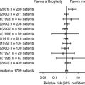
The untreated established hip displacement in CP may progress to the development of secondary degenerative changes and pain. This may, in turn, result in further functional compromise and affect quality of life. However, severe and definite hip pain is a relatively rare outcome among adults with CP and correlates poorly with radiographic status of the hip.15 Furthermore, cross-sectional studies find little difference in pain or quality of life among totally involved adults regardless of whether the hips have been treated with surgery.14 Hip replacement surgery, excision/interposition arthroplasty with or without valgus femoral osteotomy, and hip arthrodesis have been suggested.55–57 However, the existing reports in the literature include small numbers of patients, no control subjects, and limited follow-up. Therefore, it is not possible to draw any useful conclusions on the optimal management of these patients.
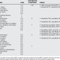
Deciding on the best treatment for the dislocated or at-risk hip in severe CP is difficult given the current state of the literature. Most of the available material regarding treatment consists of uncontrolled case series, and radiographic outcomes predominate over clinical ones. The only prognostic studies of high quality14,15 suggest that, among adult patients, the radiographic status of the hip is a poor predictor of clinical outcome, the incidence of severe pain is low, and the clinical status of patients is no better among those with operated hips than those without. The complication rates are high in all the reported series. Some consensus exists in the Level IV literature regarding the particular techniques to soft-tissue releases (for prevention) and open reductions with femoral and pelvic osteotomies (for treatment). However, from the current literature, we cannot confidently conclude whether patients with severe CP should have hip operations. Table 41-1 provides a summary of recommendations.
| STATEMENT | LEVEL OF EVIDENCE/GRADE OF RECOMMENDATION | REFERENCES |
|---|---|---|
| Surveillance | ||
| Nonambulant children with CP should be screened for hip displacement with a pelvic radiograph at the age of approximately 3 years. A normal radiograph at this age would rule out hip pathology, provided no changes occur on clinical examination of the hips over time. | B | 4 |
| Natural History | ||
| Hip displacement in CP is a slow process, which allows secondary changes to the femoral head and acetabulum to develop. | B | 9, 11, 12 |
| Hip dislocation does not increase the risk for progression of scoliosis. | B | 13 |
| Hip pain, seating function, and contractures are unrelated to the radiographic status of the hip (dislocated, subluxed, or osteoarthritic) | B | 14, 15 |
| Soft-Tissue Surgery | ||
| Soft-tissue surgery prevents hip displacement at 8 to 10 years in approximately two thirds of young children with bilateral CP. | C | 27 |
| Low preoperative migration index and young age are associated with good results. | B | 22–25 |
| Bony Surgery | ||
| Combined femoral and pelvic osteotomies lead to better results in the mid term, compared with femoral osteotomy or soft-tissue surgery in isolation. | C | 9, 42–44 |
| The Dega osteotomy and its modifications address posterolateral instability and carry advantages over conventional pelvic osteotomies performed for developmental hip displacement. | C | 45–48 |
| In the majority of children, the contralateral hip should be treated to prevent progressive deformity and displacement. | C | 15 |
| A high risk for complications is associated with this type of surgery. | C | 50, 51 |
CP, cerebral palsy.
1 Morton RE, Scott B, McClelland V. Dislocation of the hips in children with bilateral spastic cerebral palsy, 1985-2000. Dev Med Child Neurol. 2006;48:555-558.
2 Murray AW, Robb JE. The hip in cerebral palsy. Curr Orthop. 2006;20:286-293.
3 Chung CY, Park MS, Choi IH, et al. Morphometric analysis of acetabular dysplasia in cerebral palsy. J Bone Joint Surg Br. 2006;88B:243-247.
4 Scrutton D, Baird G. Surveillance measures of the hips of children with bilateral cerebral palsy. Arch Dis Child. 1997;56:381-384.
5 Cooke P, Cole W, Carey R. Dislocation of the hip in cerebral palsy. J Bone Joint Surg Br. 1989;71B:441-446.
6 Spencer JD, Sait MS. The ‘true’ acetabular index in children with cerebral palsy. Ann R Coll Surg Engl. 2004;86:371-374.
7 Hägglund G, Andersson S, Düppe H, et al. Prevention of severe contractures might replace multilevel surgery in cerebral palsy: Results of a population-based health care programme and new techniques to reduce spasticity. J Pediatr Orthop B. 2005;14:269-273.
8 Terjesen T. Development of the hip joints in unoperated children with cerebral palsy: 3a A radiographic study of 76 patients. Acta Orthop. 2006;77:125-131.
9 Brunner R, Baumann JU. Clinical benefit of reconstruction of dislocated or subluxated hip joints in patients with spastic cerebral palsy. J Pediatr Orthop. 1994;14:290-294.
10 Heinrich SD, MacEwen GD, Zembo MM. Hip dysplasia, subluxation, and dislocation in cerebral palsy: An arthrographic analysis. J Pediatr Orthop. 1991;11:488-493.
11 Bos CF, Rozing PM, Verbout AJ. Surgery for hip dislocation in cerebral palsy. Acta Orthop Scand. 1987;58:638-640.
12 Cigala F, Marmo C, Lotito FM, et al. Hip surgery in cerebropalsy. Chir Organi Mov. 2003;88:23-32.
13 Senaran H, Shah SA, Glutting JJ, et al. The associated effects of untreated unilateral hip dislocation in cerebral palsy scoliosis. J Pediatr Orthop. 2006;26:769-772.
14 Pritchett JW. Treated and untreated unstable hips in severe cerebral palsy. Dev Med Child Neurol. 1990;32:3-6.
15 Noonan KJ, Jones J, Pierson J, et al. Hip function in adults with severe cerebral palsy. J Bone Joint Surg Am. 2004;86-A:2607-2613.
16 Bowen JR, MacEwen GD, Mathews PA. Treatment of extension contracture of the hip in cerebral palsy. Dev Med Child Neurol. 1981;23:23-29.
17 Schultz RS, Chamberlain SE, Stevens PM. Radiographic comparison of adductor procedures in cerebral palsied hips. J Pediatr Orthop. 1984;4:741-744.
18 Spruit M, Fabry G. Psoas and adductor release in children with cerebral palsy. Acta Orthop Belg. 1997;63:91-93.
19 Wheeler ME, Weinstein SL. Adductor tenotomyobturator neurectomy. J Pediatr Orthop. 1984;4:48-51.
20 Moreau M, Cook PC, Ashton B. Adductor and psoas release for subluxation of the hip in children with spastic cerebral palsy. J Pediatr Orthop. 1995;15:672-676.
21 Cobeljic G, Vukasinovic Z, Djoric I. Surgical prevention of paralytic dislocation of the hip in cerebral palsy. Int Orthop. 1994;18:313-316.
22 Cornell MS, Hatrick NC, Boyd R, et al. The hip in children with cerebral palsy. Predicting the outcome of soft tissue surgery. Clin Orthop. 1997;340:165-171.
23 Cottalorda J, Gautheron V, Metton G, et al. Predicting the outcome of adductor tenotomy. Int Orthop. 1998;22:374-379.
24 Onimus M, Allamel G, Manzone P, Laurain JM. Prevention of hip dislocation in cerebral palsy by early psoas and adductors tenotomies. J Pediatr Orthop. 1991;11:432-435.
25 Houkom JA, Roach JW, Wenger DR, et al. Treatment of acquired hip subluxation in cerebral palsy. J Pediatr Orthop. 1986;6:285-290.
26 Pountney T, Green EM. Hip dislocation in cerebral palsy. BMJ. 2006;332:772-775.
27 Presedo A, Oh CW, Dabney KW, Miller F. Soft-tissue releases to treat spastic hip subluxation in children with cerebral palsy. J Bone Joint Surg Am. 2005;87A:832-841.
28 Terjesen T, Lie GD, Hyldmo AA, Knaus A. Adductor tenotomy in spastic cerebral palsy. A long-term follow-up study of 78 patients. Acta Orthop. 2005;76:128-137.
29 Turker RJ, Lee R. Adductor tenotomies in children with quadriplegic cerebral palsy: Longer term follow-up. J Pediatr Orthop. 2000;20:370-374.
30 Erken EH, Bischof FM. Iliopsoas transfer in cerebral palsy: The long term outcome. J Pediatr Orthop. 1994;14:295-298.
31 Uematsu A, Bailey HL, Winter WGJr, Brower TD. Results of posterior iliopsoas transfer for hip instability caused by cerebral palsy. Clin Orthop Relat Res.; 126; 1977; 183-189.
32 Hägglund G, Andersson S, Düppe H, et al. Prevention of dislocation of the hip in children with cerebral palsy. The first ten years of a population-based prevention programme. J Bone Joint Surg Br. 2005;87:95-101.
33 Krach LE, Kriel RL, Gilmartin RC, et al. Hip status in cerebral palsy after one year of continuous intrathecal baclofen infusion. Pediatr Neurol. 2004;30:163-168.
34 Park TS. Selective dorsal rhizotomy: An excellent option for spastic cerebral palsy. Clin Neurosurg. 2000;47:422-439.
35 Stott NS. Effects of surgical adductor releases for hip subluxation in cerebral palsy: An AACPDM evidence report. Dev Med Child Neurol. 2004;46:628-645.
36 Hoffer MM, Stein GA, Koffman M, Prietto M. Femoral varus-derotation osteotomy in spastic cerebral palsy. J Bone Joint Surg Am. 1985;67:1229-1235.
37 Settecerri JJ, Karol LA. Effectiveness of femoral varus osteotomy in patients with cerebral palsy. J Pediatr Orthop. 2000;20:776-780.
38 Stasikelis PJ, Davids JR, Johnson BH, Jacobs JM. Rehabilitation after femoral osteotomy in cerebral palsy. J Pediatr Orthop B. 2003;12:311-314.
39 Debnath UK, Guha AR, Karlakki S, et al. Combined femoral and Chiari osteotomies for reconstruction of the painful subluxation or dislocation of the hip in cerebral palsy: A long-term outcome study. J Bone Joint Surg Br. 2006;88B:1373-1378.
40 Gordon JE, Capelli AM, Strecker WB, et al. Pemberton pelvic osteotomy and varus rotational osteotomy in the treatment of acetabular dysplasia in patients who have static encephalopathy. J Bone Joint Surg. 1996;78:1863-1871.
41 Shea KG, Coleman SS, Carroll K, et al. Pemberton pericapsular osteotomy to treat a dysplastic hip in cerebral palsy. J Bone Joint Surg Am. 1997;79:1342-1351.
42 Pope DF, Bueff HU, De Luca A. Pelvic osteotomies for subluxation of the hip in cerebral palsy. J Pediatr Orthop. 1994;14:724-730.
43 Jerosch J, Senst S, Hoffstetter I. Combined realignment procedure (femoral and acetabular) of the hip joint in ambulatory patients with cerebral palsy and secondary hip dislocation. Acta Orthop Belg. 1995;61:92-99.
44 Zuckerman JD, Staheli LT, McLaughlin JF. Acetabular augmentation for progressive hip subluxation in cerebral palsy. J Pediatr Orthop. 1984;4:436-442.
45 Jozwiak M, Marciniak W, Piontek T, Pietrzak S. Dega’s transiliac osteotomy in the treatment of spastic hip subluxation and dislocation in cerebral palsy. J Pediatr Orthop B. 2000;9:257-264.
46 Roposch A, Wedge JH. A modified periacetabular osteotomy to treat spastic hip dysplasia. J Bone Joint Surg Br. 2003;90B(supp I):53-54.
47 Miller F, Girardi H, Lipton G, et al. Reconstruction of the spastic hip with peri-ilial pelvic and femoral osteotomy followed by immediate mobilization. J Pediatr Orthop. 1997;17:592-602.
48 Mubarak SJ, Valencia FG, Wenger DR. One stage correction of the spastic dislocated hip. J Bone Joint Surg Am. 1992;74A:1347-1357.
49 Carr CR, Boyd BM. Correctional osteotomy for metatarsus primus varus and hallux valgus. J Bone Joint Surg Am. 1968;50:1353-1367.
50 Inan M, Harma A, Ertem K, et al. Successful treatment of high congenital dislocated hips in older children by open reduction, pelvic and femoral osteotomy with external fixator stabilization (average 8.2 years of age). J Pediatr Orthop B. 2005;14:405-409.
51 Stasikelis PJ, Lee DD, Sullivan CM. Complications of osteotomies in severe cerebral palsy. J Pediatr Orthop. 1999;19:207-210.
52 Inan M, Chan G, Dabney K, Miller F. Heterotopic ossification following hip osteotomies in cerebral palsy: Incidence and risk factors. J Pediatr Orthop. 2006;26:551-556.
53 Lubicky JP, Bernotas S, Herman JE. Complications related to postoperative casting after surgical treatment of subluxed/dislocated hips in patients with cerebral palsy. Orthopedics. 2003;26:407-411.
54 Stasikelis PJ, Ridgeway SR, Pugh LI, Allen BLJr. Epiphyseal changes after proximal femoral osteotomy. J Pediatr Orthop B. 2001;10:25-29.
55 Blake SM, Kitson J, Howell JR, et al. Constrained total hip arthroplasty in a paediatric patient with cerebral palsy and painful dislocation of the hip: A case report. J Bone Joint Surg Br. 2006;88B:655-657.
56 de Moraes Barros Fucs PM, Svartman C, de Assumpcao RM, Kertzman PF. Treatment of the painful chronically dislocated and subluxated hip in cerebral palsy with hip arthrodesis. J Pediatr Orthop. 2003;23:529-534.
57 McHale KA. Bilateral spontaneous arthrodesis of the hip after combined shelf acetabular augmentation and femoral varus osteotomies. J Pediatr Orthop. 1991;11:108-111.