Chapter 73 What Is the Best Treatment for Hallux Valgus?*
Theories on the pathology and appropriate treatment of hallux valgus have been extensively described in the orthopedic literature. The wealth of information on the surgical management of hallux valgus has been molded into frequently taught treatment algorithms and principles. Although these algorithms and principles aim to provide consistency in treating symptomatic hallux valgus, the wide variety of approaches to treating hallux valgus suggests that they are far from commonly accepted. The purpose of this current concept review is to provide a balanced representation of current thinking on the pathomechanics, assessment, and treatment of hallux valgus. Orthopedic management of hallux valgus remains challenging. Despite the appeal of establishing universally accepted treatment protocols and algorithms, a critical review of the literature suggests that the surgeon treating hallux valgus deformity should individualize management to the particular patient.
HISTORY OF THE CONDITION AND OVERVIEW OF CAUSATIVE FACTORS
Bunion, a term evolving from the Latin word bunio, meaning “turnip,” poorly defines the condition hallux valgus. To our knowledge, the first published reference to hallux valgus is by Carl Hueter in 1870.1 Hallux valgus is commonly thought to develop because of unaccommodative shoe wear. Some support this conclusion,2,3 but consistently sufficient evidence does not exist to confirm unaccommodative shoe wear as a causative factor in the development of hallux valgus. Conversely, the observation that many individuals do not experience development of hallux valgus despite wearing nonphysiologic shoe wear for many years implies that some individuals may have an incompletely defined predisposition to hallux valgus. Other studies report that hallux valgus develops in some unshod individuals, implying a congenital predisposition.4–10 Hallux valgus in juveniles, adolescents, or male individuals whose feet have not been subjected to shoes with narrow toe boxes supports a congenital predisposition. An association between hallux valgus and female sex is also suggested.11,12 It has also been proposed that there is a familial predisposition to development of hallux valgus.6,13–15 The exact cause leading to the development of a hallux valgus deformity remains unclear and may be multifactorial. However, as the bunion deformity tends to develop over time, it seems reasonable to conclude that repetitive forces applied to the first metatarsal phalangeal joint leads to hallux valgus.
CAUSATIVE FACTORS AND PATHOMECHANICS (PROPOSED THEORIES)
Anatomic Considerations
Repetitively forcing the hallux into a valgus position, particularly with weight bearing and ambulation, is believed to eventually result in a valgus deformity at the first MTP joint. The summation of ground reactive forces and dynamic muscular forces eventually leads to attenuation of the medial joint capsule, contractures of the lateral joint capsule and adductor tendons, with a resultant medial deviation of the first metatarsal head (“bunion deformity”).
Pes Planus and Hallux Valgus
Pes planus may lead to hallux valgus because of increased forefoot abduction that creates a nonphysiologic load on the plantarmedial aspect of the great toe during heel rise. The association between pes planus and hallux valgus is controversial. Although some authors suggest that patients with pes planus have a greater tendency to experience development of hallux valgus than patients with maintained arches,16–23 others fail to support this association.14,24–26 The combination of conflicting reports and consistently Level III-V evidence provide insufficient evidence (grade I) to prove or disprove an association between pes planus and hallux valgus.
Hypermobility of the First Tarsometatarsal Joint
Mobility of the first TMT joint is observed in the sagittal and transverse plane.27 The actual prevalence of medial column hypermobility continues to be controversial. It is theorized that hypermobility could lead to the development of hallux valgus in two ways. First, greater than physiologic dorsal subluxation of the first metatarsal could result in pes planus alignment, increased forefoot abduction, and a nonphysiologic load on the plantarmedial aspect of the great toe during heel rise. Second, greater than physiologic medial subluxation of the first metatarsal could result in increasing the 1-2 intermetatarsal angle (IMA), promoting metatarsus primus varus. Some foot and ankle surgeons maintain that hypermobility of the first TMT joint or lack of stability of the foot’s medial column contributes to the development of hallux valgus28–32 and resultant pain,33 a theory popularized by Morton.9,34–37 Lapidus38–40 supports this theory and suggests surgical correction with a first TMT joint arthrodesis. Although a convincing argument in theory, no evidence exists to support such a correlation, and in fact, other investigators have demonstrated that hypermobility of the first TMT joint is not directly associated with hallux valgus.37,41–45 Insufficient evidence (Level III-V) exists to support or disprove the contribution of first TMT joint hypermobility to the development of hallux valgus (grade I).
Distal Metatarsal Articular Angle
Hallux valgus may exist with a congruent/symmetric relation between the first proximal phalanx and the first metatarsal head, suggesting a congenital predisposition in select patients with an increased distal metatarsal articular angle (DMAA).46–48 Richardson and colleagues49 note that the DMAA ranged from 6.3 to 18 degrees; as the angle increases, so does the propensity for hallux valgus, albeit congruent/symmetric. Coughlin14 adds that the DMAA tends to be greater in patients with juvenile hallux valgus younger than 10 years when compared with those older than 10. Although Richardson and colleagues49 suggest that the DMAA can be reliably determined radiographically, others have reported poor interobserver reliability.50–52
Medial Capsular Integrity
Uchiyama and investigators53 demonstrate, in a cadaveric model, that feet with hallux valgus have a different organization of collagen fibrils than that observed in normal feet. These findings may be in response to abnormal stress repetitively applied to this part of the joint capsule. Alternatively, abnormal mechanical properties of the medial capsule such as those seen in patients with conditions such as rheumatoid arthritis may increase the propensity to develop hallux valgus.
CLINICAL MANIFESTATION/TYPICAL PRESENTATION
Physical Examination
The severity of hallux valgus deformity and pes planus are assessed with the patient weight bearing. To illustrate appropriateness of shoe wear, the physician may wish to contrast an outline of the patient’s foot with nonphysiologic shoe wear. Medial eminence tenderness, first MTP joint range of motion, and first TMT hypermobility may be evaluated with the patient seated.43 Limited first MTP joint range of motion with or without crepitance should alert the physician to potential first MTP joint degenerative changes.
Because physiologically normal values of first TMT joint mobility have not been defined, first ray hypermobility remains a controversial finding and a diagnostic challenge, despite some authors providing methods to objectively quantitate first TMT joint motion.54 Even though a validated Klaue device exists to measure first ray mobility,55,56 it is not particularly practical in the clinical setting. Moreover, first TMT joint hypermobility may not only occur in the sagittal plane but also in the transverse plane.27 Clinical evaluation may not be adequately specific to isolate the first TMT joint, and therefore may assess only medial column mobility. Physical examination should also include the evaluation of the second MTP joint for presence of synovitis, metatarsal head overload, and/or second toe deformity, all of which are often associated with hallux valgus.
Imaging Studies
Radiographic Measurements Pertinent to Hallux Valgus.
Several parameters measured from AP radiographs aid in the basic characterization of a hallux valgus deformity. The hallux valgus angle (HVA), defined as the angle formed by the intersection of longitudinal axes of the diaphyses of the first metatarsal and the proximal phalanx, quantifies the malalignment of the first MTP joint. Several authors have suggested that the upper limit of normal for this measurement is 15 degrees.6,57–59 The IMA represents the angle formed between the diaphyses of the first and second metatarsals. This measurement quantifies the extent of metatarsus primus varus. The upper limit of normal for the IMA is 9 degrees.6,57–59 The interphalangeal angle, which measures the angle between the metaphysis and diaphysis of the proximal phalanx, determines the amount of hallux valgus interphalangeus (HVI). The physiologic upper limit of normal for this parameter is 10 degrees.51,58, 59 The DMAA assesses the angular relation between the articular surface of the head and diaphysis of the first metatarsal. The upper limit of normal DMAA is 10 degrees.37,49, 51 The literature suggests that, although preoperative intraobserver and interobserver reliability for the HVA and IMA is excellent (<5 degrees, 95% confidence interval),51,59–61 assessment of the DMAA remains a diagnostic challenge, with poor intraobserver and interobserver reliability.47,49–52
Radiographic Measurements Purported to Suggest Hypermobility.
Second metatarsal diaphyseal hypertrophy, a medially oriented first TMT joint, and first TMT joint obliquity have been suggested to indirectly determine hypermobility of the first ray. Diaphyseal hypertrophy of the second metatarsal, particularly the medial cortex, has been suggested as a sign of hypermobility of the first ray.28,29, 31, 62, 63 No investigation has demonstrated a correlation between radiographic changes in the second metatarsal and hypermobility.37,44, 64 However, one study demonstrated a marginal correlation between the IMA and dorsal mobility of the TMT joint in patients with hallux valgus.65 A medially oriented obliquity of the first TMT joint has proposed as an associated sign of a hypermobility, but Brage and colleagues66 demonstrated that changing the inclination of the x-ray beam relative to the floor created wide variation in the measurement of the first TMT joint obliquity. Based on these findings, Brage and colleagues66 concluded that first TMT joint obliquity was not a reliable indication for first TMT arthrodesis in the management of hallux valgus. An investigation observed significant dorsal translation and dorsiflexion of this joint in a series of patients with moderate-to-severe hallux valgus compared with normal control subjects.67 The appearance of plantar gapping at the TMT joint has been attributed to radiographic projection and discounted as an indication of hypermobility.33,71 Currently, no investigation has correlated these presumed radiographic abnormalities of the TMT joint with clinical hypermobility.
Correlating Physical and Radiographic Findings.
Thordarson and coworkers68 evaluated 285 women, with an average age of 49 years, scheduled for corrective surgery for hallux valgus. Validated AAOS foot-specific outcomes data collection questionnaires were used. Preoperative radiographic data (HVA and IMA) were stratified into degree of deformity. The data were stratified into age groups consistent with those reported for the 36-Item Short Form Health Survey (SF-36), and the results were compared with the SF-36 for the general population. The global foot and ankle score and the shoe comfort score were compared with the general population, and the severity of the preoperative deformity was correlated with the baseline scores. Bodily pain scores were uniformly worse for hallux valgus patients compared with the general population, with significantly lower global foot and ankle and shoe comfort scores, a finding that suggests that the bodily pain score from the SF-36 represents a sensitive measure of the difficulties experienced by patients undergoing corrective hallux valgus surgery. The preoperative radiographically determined severity of deformity failed to correlate with any scores measured.
CLASSIFICATION
NONOPERATIVE TREATMENT
Symptoms of pain are best treated with shoe wear and activity modification. Shoes with a wider toe box and a comfortable upper are often helpful. Padding over the medial eminence or adjustments to the shoe to create more space medially can be helpful. However, nonoperative management cannot reverse hallux valgus deformity, and successful surgery may lead to an improved functional outcome. A randomized, controlled trial in 209 consecutive patients with symptomatic hallux valgus treated in four Finnish general community hospitals demonstrated that, although orthoses provided short-term symptomatic relief, surgical management of hallux valgus led to superior functional outcome and patient satisfaction as compared with orthotic management at a minimum follow-up of 12 months.69 Surgical correction also led to better functional outcome and patient satisfaction than observation (“watchful waiting”), suggesting that the natural history of symptomatic hallux valgus deformity, at 12 months, is not one of improvement (Level I evidence). Although this prospective, randomized study demonstrates benefits of surgical correction of hallux valgus when compared with nonoperative treatment, insufficient evidence exists to support that corrective bunion surgery should be favored over nonoperative management (grade I).
MILD-TO-MODERATE DEFORMITY
Distal Procedures
Incongruent Hallux Valgus.
Simple Bunionectomy.
Few recent orthopedic articles report on simple bunionectomy (medial eminence resection with medial capsular plication). In a retrospective review of simple bunionectomy, Kitaoka and coauthors70 note high recurrence and high patient dissatisfaction rates. Given the limited Level IV evidence for simple bunionectomy, no specific recommendation (grade I) can be made for medial eminence resection in hallux valgus correction.
Modified McBride Procedure (Distal Soft-Tissue Procedure).
The McBride distal soft-tissue procedure, although common as an adjunct procedure in many hallux valgus corrective surgeries, has also been described as an isolated procedure for hallux valgus correction. In 1923, Silver22 reported the combination of medial eminence resection, lateral capsular release, adductor hallucis tendon release, and medial capsular plication for the treatment of hallux valgus deformity. The modified McBride procedure includes medial capsulotomy (and subsequent plication), division of the ligament between the lateral capsule and fibular sesamoid, adductor hallucis release, lateral capsular fenestration, and a controlled varus stress to the first MTP joint.71–73 A concern about hallux varus after the original McBride procedure74 prompted the preservation of the fibular sesamoid in the modification.
Few recent orthopedic articles report on isolated modified McBride procedures for the correction of hallux valgus deformity. In a retrospective review, Mann and Pfeffinger72 note acceptable patient satisfaction rates and improvement in hallux alignment (Level IV evidence). However, a selection bias to patients with mild and flexible deformities was suggested.75 Johnson and coworkers71 retrospectively compared the modified McBride procedure and distal chevron osteotomy, with the two groups matched for age, severity of deformity, and length of follow-up (Level III evidence). Although postoperative satisfaction rates were not significantly different, the distal chevron group exhibited significantly better correction of alignment. Given the limited Level III and IV evidence for the modified McBride procedure, no specific recommendation (grade I) can be made for the McBride distal soft-tissue procedure when used in isolation for hallux valgus correction.
Distal Chevron Osteotomy.
The distal chevron osteotomy is a V-shaped osteotomy of the first metatarsal, described by Corless,76 Johnson and coworkers,77 and Austin and Leventen.78 The capital fragment is shifted laterally to narrow the forefoot. An anatomic study suggested that the capital fragment can be safely shifted laterally 6.0 mm in men and 5.0 mm in women, and still maintain greater than 50% bony apposition of the fragments.79 The procedure has been performed with or without fixation of the shifted capital fragment.80–85 The symmetric orientation of the distal chevron osteotomy78 has undergone several modifications to accommodate fixation.77,80 The combination of a medial closing wedge osteotomy of the first proximal phalanx (Akin) and a distal chevron osteotomy has been described when hallux valgus with metatarsus primus varus is associated with HVI.86,87 The distal chevron osteotomy also has been combined with a lateral capsular or adductor tendon release, or both.83,85,88–90
For mild-to-moderate hallux valgus correction, the effectiveness of the distal chevron osteotomy in providing favorable outcomes and patient satisfaction, regardless of fixation method, addition of lateral soft-tissue release, length of follow-up, or patient age, is supported by numerous retrospective reviews (Level IV evidence).80–85,90–97 The average preoperative IMA was less than 15 degrees in all studies. DeOrio and Ware81 report satisfactory outcomes and patient satisfaction with a low complication rate with bioabsorbable fixation (Level IV evidence). Crosby and Bozarth93 and Gill and colleagues82 note no significant differences in favorable outcomes or patient satisfaction and minimal complications in case series comparisons of screw, Kirschner wire, and no fixation and Kirschner wires versus bioabsorbable fixation, respectively (Level IV evidence).
Although the addition of a lateral release to a distal chevron osteotomy may improve global correction of hallux alignment, patient satisfaction is similar in patients who have distal chevron osteotomies with or without lateral release. Resch and investigators’89 Level I evidence investigation compares distal chevron osteotomy with and without adductor tenotomy. Although the clinical appearance and radiographic alignment were significantly improved in the group with adductor release, patient satisfaction was not. Mann and Donatto,80 in a small case series (Level IV evidence), note satisfactory outcome for distal chevron osteotomy without lateral release: similar to results of Level IV evidence studies of distal chevron osteotomies with lateral release.92,95, 97
Two recently published Level IV case series of distal chevron osteotomies with lateral release83,85 note that the results were maintained with longer follow-up: Trnka and coworkers’83 follow-up period was 2 to 5 years, and Schneider and colleagues’85 follow-up period was 5.6 to 12.7 years. Furthermore, both studies suggest that results were equal for patients older and younger than the arbitrarily chosen age of 50 years.
Congruent Hallux Valgus.
Although subject to poor interobserver reliability,47,50–52 the DMAA47,49 can be decreased by combining the distal chevron osteotomy with Akin osteotomy80 or by making a biplanar distal chevron osteotomy in mild hallux valgus deformity46,98 (Level IV evidence). Whereas the goal of a combination of distal chevron and Akin osteotomies is to improve clinical alignment through extra-articular correction, the biplanar distal chevron osteotomy aims to simultaneously correct hallux valgus and decrease the DMAA. In the biplanar distal chevron procedure, two different osteotomy configurations can be used. With the conventional, symmetric pattern of two osteotomy limbs of equal length, a second oblique wedge resection for each cut allows a reduction in the DMAA in combination with the lateral shift of the metatarsal head46 (Level IV evidence). With a short, relatively vertical dorsal limb and long, horizontal plantar limb, a second wedge resection dorsally permits redirection of the metatarsal head simultaneous with the lateral shift98 (Level IV evidence).
Risk for Osteonecrosis of the First Metatarsal Head with Distal Chevron Osteotomy.
Adding a lateral capsular release to a chevron osteotomy may improve deformity correction,84,88, 95, 96, 99, 100 but this type of release may increase the risk for osteonecrosis of the first metatarsal head90 (Level IV evidence). A distal chevron osteotomy disrupts the intraosseous blood supply to the metatarsal head, and the medial capsular release eliminates a substantial portion of the blood supply to the metatarsal head.88 Retrospective reviews83–85,94–96 (Level IV evidence) suggest that a lateral capsular release or adductor tenotomy can be safely combined with a distal chevron osteotomy. Moreover, in a prospective, randomized study (Level I evidence), Resch and investigators99 used scintigraphy to demonstrate that an adductor hallucis tenotomy performed with a distal chevron osteotomy did not lead to an increased circulatory disturbance to the first metatarsal head compared with distal chevron osteotomies performed without a lateral soft-tissue procedure. Kuhn and colleagues88 prospectively (Level III evidence) utilized an intraoperative laser Doppler probe to demonstrate that the combination of chevron osteotomy, medial capsular release, and lateral release plus adductor tenotomy resulted in a cumulative decrease in blood flow to the metatarsal head of 71%, with the greatest insult being attributed to the medial capsular release (45%). None of the 20 metatarsal heads analyzed in Kuhn and colleagues’88 study experienced development of osteonecrosis. Some investigations have noted initial radiographic findings suggestive of avascular change in the first metatarsal head, but with further follow-up these changes resolved in most patients.80,96, 99 Even after chevron osteotomy without lateral release, subtle findings suggestive of osteonecrosis may be identified, but these rarely have long-term sequelae80,99 (Level I and IV evidence). Jones and coauthors100 propose an overpenetration with the saw blade through the lateral first metatarsal cortex and the lateral capsule as the technical error potentially leading to osteonecrosis.
Levels of Evidence for Distal Chevron Osteotomy.
Given the numerous positive Level IV evidence investigations and one Level I evidence study in the orthopedic literature, a grade B treatment recommendation can be made to support the use of a distal chevron osteotomy for correction of mild-to-moderate hallux valgus deformity. One Level I evidence study and multiple Level IV evidence investigations that provide grade B evidence that hallux alignment and functional outcome may be better after a distal chevron osteotomy with a lateral soft-tissue procedure than without suggest that patient satisfaction is no different in these two groups. Moreover, consistently positive Level IV evidence and one Level I evidence investigation allow a grade B recommendation that a lateral capsular or adductor hallucis tendon release can be done with a distal chevron osteotomy without increased risk for first metatarsal head osteonecrosis. Two relatively recent Level IV evidence studies exist for the distal chevron and Akin osteotomies and the biplanar distal chevron osteotomy to correct mild-to-moderate hallux valgus associated with an increased DMAA.46,86, 87, 98 Although functional outcomes and patient satisfaction for these case series are favorable, only grade C evidence supports their use in the management of mild-to-moderate hallux valgus with an increased DMAA.
Keller Resection Arthroplasty.
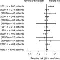
Several Level IV retrospective case series have been published on the Keller resection arthroplasty.21,101–107 The Keller resection arthroplasty is the resection of the first proximal phalanx base to correct hallux valgus deformity. In a prospective comparison, Turnbull and Grange101 demonstrate better HVA and IMA correction, hallux MTP joint motion, and metatarsal head relation with the sesamoid complex for the distal chevron osteotomy compared with the Keller procedure (Level II evidence). Although some authors have reported satisfactory results with the Keller resection arthroplasty, these authors note that acceptable results were achieved because of ancillary procedures. Specifically, Donley and researchers108 note acceptable postoperative alignment when the resection arthroplasty is combined with a fibular sesamoidectomy (Level IV). Likewise, several authors105,106,109–111 attribute improved outcome applying a cerclage fibreux (described by LeLievre and LeLievre112) distal soft-tissue procedure or tendon transfer in conjunction with the resection (Level IV). Zembsch and investigators,102 in a retrospective, uncontrolled comparative case series, demonstrate worse results with the Keller procedure than with proximal metatarsal closing wedge osteotomy, with significantly better correction of hallux valgus deformity in the proximal osteotomy group. Both groups had high rates of transfer metatarsalgia (Level IV evidence). Anecdotally, most authors suggest that the Keller procedure be used only in older patients with limited functional expectations who may be at risk if subjected to corrective surgery. Given that no more than Level IV evidence exists in the orthopedic literature, only grade C evidence exists for recommending the Keller procedure in the management of hallux valgus deformity.
MODERATE-TO-SEVERE DEFORMITY
Incongruent Hallux Valgus Deformity
Proximal Procedures and First Metatarsophalangeal Joint Arthrodesis.
Whereas crescentic113–117 and closing wedge osteotomies are done through a dorsal approach, proximal chevron, opening wedge Ludloff, and scarf osteotomies are done from the medial aspect of the proximal first metatarsal.118–121 Several authors suggest that dorsiflexion malunion is less likely to occur with proximal osteotomies done from the medial aspect of the first metatarsal than from the dorsal aspect.115,118, 121 Most proximal first metatarsal osteotomies require full transection of the first metatarsal; the opening and closing wedge procedures maintain lateral and medial cortical hinges, respectively. For all proximal osteotomies combined with a distal soft-tissue procedure, potential complications include recurrence, hallux varus, first MTP joint stiffness, malunion, nonunion, and infection.
Proximal Crescentic Osteotomy.
The proximal crescentic osteotomy for correction of hallux valgus associated with metatarsus primus varus has been popularized by Mann and colleagues.122 Unique to the proximal crescentic osteotomy is the use of a crescentic saw blade. A commonly cited potential complication of the proximal crescentic osteotomy is dorsiflexion malunion.115,116, 122, 123 Jones and coauthors124 describe a technique to aid surgeons in properly orienting the crescentic saw blade in the coronal plane to minimize the risk for initial dorsiflexion malpositioning.
Several case series (Level IV evidence) reported satisfactory radiographic correction, high rates of satisfaction, and significant improvement in functional outcomes with the proximal crescentic osteotomy at intermediate- to long-term follow-up periods.113,114, 116, 117, 122, 123, 125, 126 A prospective, randomized comparison115 (Level II evidence) of the proximal crescentic and proximal chevron osteotomies suggested favorable radiographic correction and clinical outcomes for both procedures, with American Orthopaedic Foot and Ankle Society (AOFAS) outcome scores and radiographic correction of HVA and IMA improving significantly at an average follow-up of 24 (proximal crescentic) and 20 months (proximal chevron). Dorsiflexion malunion was observed in 17% of the proximal crescentic cohort. Mann and colleagues,122 in one of the original articles describing this procedure, note dorsiflexion malunions in 28% of cases. The consistently favorable results from several case series and one Level II evidence study impart a grade B recommendation for the use of the proximal crescentic osteotomy in the surgical management of hallux valgus.
Proximal Chevron Osteotomy.
The proximal chevron osteotomy, first reported by Sammarco and researchers,118 relies not simply on lateral translation of the distal fragment, as with the distal chevron procedure, but concomitantly incorporates an opening wedge principle.115,118–120 The large contact area is relatively stable, and recommended fixation is with a combination of a screw and a Kirschner wire, two screws, or a plate.115,118–120,127,128 The procedure has been described using a single-119,120 or two-incision115,118 technique.
Three case series118,120 (Level IV evidence) and one prospective, randomized comparative study115 (Level II evidence) reported favorable radiographic correction, high rates of satisfaction, and significant improvements in the functional outcomes with the proximal chevron osteotomy at intermediate follow-up. In the Level II investigation comparing the proximal chevron and crescentic osteotomies, healing time was shorter and the tendency for first metatarsal shortening was less for the proximal chevron cohort. Dorsiflexion malunion was observed in 0% and 17% for the proximal chevron and crescentic cohorts, respectively. Taking the limited (but favorable) Level IV evidence and the positive Level II evidence into consideration, a grade B recommendation for the use of the proximal chevron osteotomy in the operative management of hallux valgus can be made.
Opening Wedge Proximal First Metatarsal Osteotomy.
The opening wedge proximal first metatarsal osteotomy was described by Trethowen in 1923129 but was largely abandoned because of concerns of stability and nonunion. With improved fixation techniques, including fixed-angle plating, the opening wedge has regained acceptance in some centers. It probably is inaccurate to state that an opening wedge proximal first metatarsal osteotomy lengthens the metatarsal, but it may maintain length, a feature that may be beneficial when the first metatarsal is relatively short compared with the second metatarsal. Healing rarely is problematic despite the gap that is created; minimal periosteal stripping and an intact lateral cortex allow relatively rapid incorporation of local autograft, allograft, or bone graft substitutes.
Proximal Oblique (“Ludloff”) Osteotomy.
The proximal oblique first metatarsal osteotomy was introduced in 1918 by Ludloff,130 but it failed to gain acceptance because the original description did not include fixation. More recently, a modified technique included fixation with two screws.121 The first screw is placed before the osteotomy is completed, allowing the surgeon to maintain full control of the osteotomy throughout the procedure.121 Beischer and coauthors131 report the optimal geometric parameters of the modified Ludloff osteotomy in a three-dimensional computer analysis. Specifically, they determined that first metatarsal shortening and rotational malalignment can be controlled if the osteotomy is started dorsally at the first TMT joint and extended distally to the plantar first metatarsal, just proximal to the sesamoid complex. They also explain that first metatarsal elevation is avoided by tilting the osteotomy 10 degrees plantarward, thereby directing the distal fragment plantarward during correction.
A few orthopedic clinical series (Level IV evidence) have analyzed the modified Ludloff osteotomy combined with a distal soft-tissue procedure.121,132, 133 At intermediate follow-up, in prospective case series (Level IV evidence), Hofstaetter and colleagues,132 Petroutsas and Trnka,133 and Chiodo and coworkers121 report significant improvement in the AOFAS Hallux-IP joint score, favorable patient satisfaction, and significant correction of radiographic hallux alignment. Based on the limited clinical series of Level IV articles, a grade B recommendation exists for the modified Ludloff osteotomy and distal soft-tissue procedure in the operative management of moderate-to-severe hallux valgus deformity.
Closing Wedge Proximal First Metatarsal Osteotomy.
A proximal closing wedge osteotomy perpendicular to the first metatarsal longitudinal axis is not universally accepted by orthopedic foot and ankle surgeons because of concerns regarding complications, including shortening and dorsiflexion malunion.102,134, 135 Perhaps by utilizing an oblique wedge orientation (as has been espoused at various meetings), these risks might be diminished. To date, however, no peer-reviewed published data exist in support of this in the orthopedic literature. The proximal closing wedge first metatarsal osteotomy is done through a dorsal approach, with the base of the resected segment directed laterally. Despite a medial hinge being maintained, most published series acknowledge a risk for dorsiflexion malunion. Given the natural propensity for the wedge osteotomy to shorten the ray, theoretically, this osteotomy may be uniquely suitable for patients with a relatively long first metatarsal.
Several retrospective case series (Level IV evidence) of proximal first metatarsal closing wedge osteotomies and distal soft-tissue procedures with intermediate- to long-term follow-up periods have been reported in the orthopedic literature.102,134–136 Trnka and researchers135 report long-term retrospective results (follow-up range, 10–22 years) of basal metatarsal closing wedge osteotomies. Despite good-to-excellent results in 85% of patients who returned for follow-up, a considerable number of complications occurred, including dorsiflexion malunion, first metatarsal shortening (mean, 5 mm), transfer metatarsalgia, and hallux varus. In an uncontrolled comparative study (Level IV evidence), the same authors report that a subset of the same patients compared favorably with a group of similar patients undergoing Keller resection arthroplasties.102 Although the incidence of transfer metatarsalgia was equal in the two groups, the basal closing wedge osteotomy had significantly better AOFAS Hallux-IP joint scores and radiographic outcomes. Again, the frequency of dorsiflexion malunion and hallux varus was high. In an intermediate follow-up case series, Resch and investigators134 cite a long average time to healing of the osteotomy and a 20% incidence rate of dorsiflexion malunion associated with transfer metatarsalgia.
Granberry and Hickey136 report a retrospective, uncontrolled comparative study (Level III evidence) of proximal first metatarsal closing wedge and Akin osteotomies with or without a distal soft-tissue procedure. Although the same combination of osteotomies was done in the two groups, only one group had release of the lateral joint capsule from the sesamoid and transfer of the adductor tendon into the first metatarsal neck. The significantly better radiographic correction of hallux valgus in those with a distal soft-tissue procedure was accompanied by significantly less first MTP joint motion. Several dorsiflexion malunions were observed in the entire group of patients. With the Level III and IV evidence, a grade B recommendation can be made for the closing wedge proximal first metatarsal osteotomy in the correction of hallux valgus, with the observation that dorsiflexion malunion and considerable shortening of the first metatarsal are frequent.
Scarf Osteotomy.
The scarf osteotomy is not a proximal first metatarsal osteotomy per se but is commonly used outside of the United States for moderate-to-severe hallux valgus deformity.137–141 The configuration of the osteotomy with a distal dorsal limb (virtually identical to that of a traditional distal chevron osteotomy), a long transverse cut, and a proximal limb (similar to the distal extension of the Ludloff osteotomy) confers stability and permits fixation with two screws. It is designed primarily as an osteotomy that laterally translates the distal fragment, but with slight modification of the bone cuts, rotation also can be imparted to the distal fragment to further correct the IMA increased DMAA. A potential complication unique to the scarf osteotomy is “troughing” (i.e., an impaction of the two osteotomy fragments, resulting in loss of metatarsal height).
Several prospective and retrospective case series (Level IV evidence) have reported favorable radiographic correction, high rates of patient satisfaction, and significant improvements in the functional outcomes and pedobaric foot pressure analyses with the scarf osteotomy.137–145 Aminian and coauthors,142 in their retrospective review, note no transfer metatarsalgia pattern in foot pressure analysis. Prospectively, Lorei and coworkers141 observed a redistribution from the lateral forefoot to the first ray after scarf osteotomies in their case series using pedobarographic analysis (Level IV evidence). Jones and coauthors,139 prospectively, and Crevoisier and investigators,137 retrospectively, reported favorable outcomes and radiographic correction using a combination of scarf and Akin osteotomies (Level IV evidence).
In contrast, Coetzee146 reports poor AOFAS outcome scores and an alarming complication rate in a prospective case series of scarf osteotomies (Level IV evidence). Complications included “troughing” (35%), rotational malunion (30%), metatarsal fracture (10%), and early recurrence of deformity (25%). Although many authors have acknowledged the complexity of this osteotomy, several have provided technique tips and experience to accelerate the learning curve in mastering the procedure.138,143,145,147–149
First Tarsometatarsal Joint Arthrodesis (Modified Lapidus Procedure).
Lapidus38 originally described an arthrodesis between the bases of the first and second metatarsals and the first intercuneiform joint to correct metatarsus primus varus in patients with hallux valgus. Currently, the modified Lapidus procedure incorporates an isolated arthrodesis of the first TMT joint with a lateral and plantar based closing wedge osteotomy of the medial cuneiform.31,138,150–152 This procedure has been indicated for the correction of metatarsus primus varus in patients with moderate-to-severe hallux valgus and hypermobility of the first ray. First ray hypermobility has been controversial and often is questioned.37,42, 45, 152
Several retrospective case series (Level IV evidence) have collectively reported excellent radiographic correction, high rates of satisfaction, and significant improvements in functional outcomes with the modified Lapidus procedure.28–30,32,153,154 Faber and coauthors,152 in a prospective, randomized study comparing the Hohmann procedure (distal first metatarsal osteotomy) with the modified Lapidus procedure in 101 feet (Level I evidence), found no significant differences in clinical outcomes, radiographic correction, or patient satisfaction. Feet with preoperatively identified hypermobility had equally favorable outcomes with either procedure when compared with feet without hypermobility. A prospective study (Level II evidence) evaluated the efficacy of the modified Lapidus procedure in the treatment of recurrent hallux valgus. Significant decreases in the pain score, IMAs, and HVAs, and increases in the AOFAS clinical ratings score were associated with an 81% satisfaction rate at 2 years after surgery.155
Early reports identified nonunion rates of 10% to 12% with the modified Lapidus procedure.29,31, 156 However, a more recent large clinical series reported a 4% nonunion rate and a 2% revision rate in feet treated with the Lapidus procedure.32 The authors reported that five of the eight patients with nonunions had previous bunion surgery, and two patients smoked. Another study reported no nonunions with the use of the modified Lapidus procedure for the primary correction of hallux valgus.157 The uniformly successful results from numerous case series, supported by one Level I evidence study, justify a grade B recommendation for the use of the modified Lapidus procedure in the treatment of primary hallux valgus. Although the results of Coetzee and coworkers155,158 suggest that the modified Lapidus procedure also is an effective salvage procedure for recurrent hallux valgus, this evidence from a single Level II study is insufficient (grade I) to make a recommendation.
Congruent Hallux Valgus Deformity
Double/Triple Osteotomies for Hallux Valgus Correction.
A large IMA in combination with an increased DMAA cannot be corrected with a proximal osteotomy alone; reducing the IMA will effectively increase the DMAA. In hallux valgus with a large IMA and increased DMAA, a double osteotomy, with a proximal first metatarsal osteotomy or medial opening wedge osteotomy of the first cuneiform (Cotton procedure) to correct the increased IMA and a distal medial closing wedge osteotomy of the metatarsal head (Reverdin osteotomy) to reduce the DMAA, may be considered. With severe deformity, an opening wedge medial cuneiform osteotomy (Cotton procedure) can be added to proximal MT osteotomy and Reverdin to further correct the IMA, creating a triple osteotomy. When associated with HVI, an Akin osteotomy also should be added because none of the aforementioned osteotomies directly corrects angulation in the hallux proximal phalanx.159 One Level IV evidence investigation suggested favorable outcomes in a limited number of patients undergoing multiple osteotomies for a more comprehensive hallux valgus correction.159 Several case series (Level IV evidence) have reported favorable outcomes with Akin osteotomies added to distal chevron osteotomies86,87 and more proximal osteotomies.137,139 The limited published Level IV evidence available for double and triple osteotomies is insufficient (grade I) to make a recommendation for their use in the management of hallux valgus deformity.
First Metatarsophalangeal Joint Arthrodesis.
Arthrodesis of the first MTP joint generally is reserved for patients with severe hallux valgus deformity, or patients in whom hallux valgus is associated with arthrosis of the joint, failed prior surgical correction, or a neuromuscular disorder; it also is used as part of the reconstruction of a rheumatoid forefoot. Coughlin and coauthors160 (Level IV evidence) report significant decreases in pain after fusion for moderate-tosevere hallux valgus. There were no dissatisfied patients, and most patients were able to wear conventional or comfort shoe wear at an average of 8 years after surgery. However, radiographic progression of arthritis was evident at the interphalangeal joint in 7 of the 21 feet in their series.160 Grimes and Coughlin151 report satisfactory outcomes at an average follow-up period of 8 years in a retrospective case series of first MTP joint arthrodeses performed for failed hallux valgus procedures. When fusion was compared with resection of the first MTP joint for reconstruction of the rheumatoid forefoot (Level II evidence), no differences were detected other than a significant increase in the duration of the procedure for the group undergoing arthrodesis.161 Salvage of a failed Keller procedure with fusion was associated with a greater rate of satisfaction and AOFAS clinical rating score, and avoided the onset of postoperative valgus or cock-up deformity when compared with isolated soft-tissue release (Level III evidence).162 A case series (Level IV evidence) evaluating the results of arthrodesis for hallux valgus in children with cerebral palsy reported high rates of fusion and satisfaction with the procedure. Together, these investigations favor a grade B recommendation for the use of arthrodesis in the management of a wide spectrum of hallux valgus deformities.
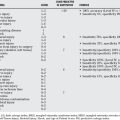
Our evidence-based recommendations for the treatment of hallux valgus are as follows (Table 73-1):
| STATEMENT | LEVEL OF EVIDENCE/GRADE OF RECOMMENDATIONS | REFERENCE |
|---|---|---|
1 Hueter C. [Klinik der Gelenkkrankungen mit Einschluss der Orthopaedie]. Leipzig: Vogel, 1870.
2 Kato TWS. The etiology of hallux valgus in Japan. Clin Orthop Relat Res. 1981;158:78.
3 Lam SL, Hodgson AR. A comparison of foot forms among the non-shoe and shoe-wearing Chinese population. J Bone Joint Surg. 1958;40:1058.
4 MacLennan R. Prevalence of hallux valgus in a neolithic New Guinea population. Lancet. 1966;1:1398.
5 Creer W. The feet of the industrial worker: Clinical aspect; relation to footwear. Lancet. 1938;2:1482.
6 Hardy RH, Clapham JC. Observations on hallux valgus; based on a controlled series. J Bone Joint Surg Br. 1951;33-B:376-391.
7 Barnicot NA, Hardy RH. The position of the hallux in West Africans. J Anat. 1955;89:355.
8 James CS. Footprints and feet of natives of Soloman Islands. Lancet.; 2; 1939; 1390.
9 Engle ET, Morton DJ. Notes on foot disorders among natives of the Belgian Congo. J Bone Joint Surg. 1931;13:311.
10 Wells LE. The foot of the south African native. Am J Phys Anthropol. 1931;15:185.
11 Wilkins E. Feet with particular reference to school children. Med Officer. 1941;66:5. 13,21,9,
12 Hewitt DS, Stewart AM, Webb JW. The prevalence of foot defects among wartime recruits. Br Med J. 1953;2:745.
13 Glynn MK, Dunlop JB, Fitzpatrick D. The Mitchell distal metatarsal osteotomy for hallux valgus. J Bone Joint Surg Br. 1980;62-B:188-191.
14 Coughlin M. Juvenile hallux valgus: Etiology and treatment. Foot Ankle. 1995;16:682-697.
15 Johnson O. Further studies of the inheritance of hand and foot anomalies. Clin Orthop Relat Res. 1956;8:146-160.
16 Hohmann G. Der hallux valgus und die uebrigen Zehenverkruemmungen. Ergeb Chir Orthop. 1925;18:308-348.
17 Anderson RL. Hallux valgus: report of end results. South Med Surg. 1929;91:74-78.
18 Craigmile D. Incidence, origin, and prevention of certain foot defects. Br Med J. 1953;2:749.
19 Galland WI, Jordan H. Hallux valgus. Surg Gunecol Obstet. 1938;66:95.
20 Joplin RJ. Sling procedure for correction of splayfoot, metatarsus primus varus, and hallux valgus. J Bone Joint Surg. 1950;32:779.
21 Rogers WAJ, Joplin RJ. Hallux valgus, weak foot, and the Keller operations: An end-result study. Surg Clin North Am. 1947;27:1295-1302.
22 Silver D. The operative treatment of hallux valgus. J Bone Joint Surg. 1923;5:225.
23 Stein HC. Hallux valgus. Surg Gunecol Obstet. 1938;66:889-898.
24 Mann RA, Coughlin MJ. Hallux valgus: Etiology, anatomy, treatment and surgical considerations. Clin Orthop Relat Res. 1981;157:31.
25 Kilmartin TE, Wallace WA. The significance of pes planus in juvenile hallux valgus. Foot Ankle. 1992;13:53-56.
26 Canale PB, Aronsson DD, Lamont RL, Manoli A2nd. The Mitchell procedure for the treatment of adolescent hallux valgus. A long-term study. J Bone Joint Surg Am. 1993;75:1610-1618.
27 Faber FW, Kleinrensink GJ, Verhoog MW, et al. Mobility of the first tarsometatarsal joint in relation to hallux valgus deformity: Anatomical and biomechanical aspects. Foot Ankle Int. 1999;20:651-656.
28 Bednarz PA, Manoli A2nd. Modified lapidus procedure for the treatment of hypermobile hallux valgus. Foot Ankle Int. 2000;21:816-821.
29 Sangeorzan BJ, Hansen STJr. Modified Lapidus procedure for hallux valgus. Foot Ankle. 1989;9:262-266.
30 Kopp FJ, Patel MM, Levine DS, Deland JT. The modified Lapidus procedure for hallux valgus: A clinical and radiographic analysis. Foot Ankle Int. 2005;26:913-917.
31 Myerson M. Metatarsocuneiform arthrodesis for treatment of hallux valgus and metatarsus primus varus. Orthopedics. 1990;13:1025-1031.
32 Thompson IM, Bohay DR, Anderson JG. Fusion rate of first tarsometatarsal arthrodesis in the modified Lapidus procedure and flatfoot reconstruction. Foot Ankle Int. 2005;26:698-703.
33 Ito H, Shimizu A, Miyamoto T, et al. Clinical significance of increased mobility in the sagittal plane in patients with hallux valgus. Foot Ankle Int. 1999;20:29-32.
34 Morton D. Hypermobility of the first metatarsal bone: The interlinking factor between metatarsalgia and longitudinal arch strains. J Bone Joint Surg Am. 1928;10:187-196.
35 Morton D. Significant characteristics of the Neanderthal foot. Natural History. 1926;26:310-314.
36 Morton D. The human foot: Its evolution, physiology and functional disorders. New York: Columbia University Press, 1935.
37 Grebing BR, Coughlin MJ. Evaluation of Morton’s theory of second metatarsal hypertrophy. J Bone Joint Surg Am. 2004;86-A:1375-1386.
38 Lapidus PW. Operative correction of the metatarsus varus primus in hallux valgus. Surg Gynecol Obstet. 1934;58:183-191.
39 Lapidus PW. A quarter of a century experience with the operative correction of the metatarsus varus primus in hallux valgus. Bull Hosp Joint Dis. 1956;17:404-421.
40 Lapidus PW. The author’s bunion operation from 1931 to 1959. Clin Orthop Relat Res. 1960;16:119-135.
41 Coughlin MJ, Jones CP, Viladot R, et al. Hallux valgus and first ray mobility: A cadaveric study. Foot Ankle Int. 2004;25:537-544.
42 Coughlin MJ, Shurnas PS. Hallux valgus in men. Part II: First ray mobility after bunionectomy and factors associated with hallux valgus deformity. Foot Ankle Int. 2003;24:73-78.
43 Grebing BR, Coughlin MJ. The effect of ankle position on the exam for first ray mobility. Foot Ankle Int. 2004;25:467-475.
44 Prieskorn DW, Mann RA, Fritz G. Radiographic assessment of the second metatarsal: Measure of first ray hypermobility. Foot Ankle Int. 1996;17:331-333.
45 Glasoe WM, Coughlin MJ. A critical analysis of Dudley Morton’s concept of disordered foot function. J Foot Ankle Surg. 2006;45:147-155.
46 Chou LB, Mann RA, Casillas MM. Biplanar chevron osteotomy. Foot Ankle Int. 1998;19:579-584.
47 Coughlin MJ. Hallux valgus in men: Effect of the distal metatarsal articular angle on hallux valgus correction. Foot Ankle Int. 1997;18:463-470.
48 Coughlin MJ. Roger A. Mann Award. Juvenile hallux valgus: Etiology and treatment. Foot Ankle Int. 1995;16:682-697.
49 Richardson EG, Graves SC, McClure JT, Boone RT. First metatarsal head-shaft angle: A method of determination. Foot Ankle. 1993;14:181-185.
50 Chi TD, Davitt J, Younger A, et al. Intra- and inter-observer reliability of the distal metatarsal articular angle in adult hallux valgus. Foot Ankle Int. 2002;23:722-726.
51 Coughlin MJ, Freund E. Roger A. Mann Award. The reliability of angular measurements in hallux valgus deformities. Foot Ankle Int. 2001;22:369-379.
52 Vittetoe DA, Saltzman CL, Krieg JC, Brown TD. Validity and reliability of the first distal metatarsal articular angle. Foot Ankle Int. 1994;15:541-547.
53 Uchiyama E, Kitaoka HB, Luo ZP, et al. Pathomechanics of hallux valgus: Biomechanical and immunohistochemical study. Foot Ankle Int. 2005;26:732-738.
54 Voellmicke KV, Deland JT. Manual examination technique to assess dorsal instability of the first ray. Foot Ankle Int. 2002;23:1040-1041.
55 Jones CP, Coughlin MJ, Pierce-Villadot R, et al. The validity and reliability of the Klaue device. Foot Ankle Int. 2005;26:951-956.
56 Glasoe WM, Grebing BR, Beck S, et al. A comparison of device measures of dorsal first ray mobility. Foot Ankle Int. 2005;26:957-961.
57 Steel MW3rd, Johnson KA, DeWitz MA, Ilstrup DM. Radiographic measurements of the normal adult foot. Foot Ankle. 1980;1:151-158.
58 Mann RA. Bunion surgery: Decision making. Orthopedics. 1990;13:951-957.
59 Saltzman CL, Brandser EA, Berbaum KS, et al. Reliability of standard foot radiographic measurements. Foot Ankle Int. 1994;15:661-665.
60 Schneider W, Csepan R, Knahr K. Reproducibility of the radiographic metatarsophalangeal angle in hallux surgery. J Bone Joint Surg Am. 2003;85-A:494-499.
61 Smith RM, Reynolds JC, Stewart MJ. Hallux valgus assessment: Report of research committee of American Orthopaedic Foot and Ankle Society. Foot Ankle. 1984;5:92-103.
62 Hansen STJr. Functional reconstruction of the foot and ankle. Philadelphia: Lippincott Williams & Wilkins, 2000.
63 Myerson MS, Badekas A. Hypermobility of the first ray. Foot Ankle Clin. 2000;5:469-484.
64 Faber FW, Kleinrensink GJ, Mulder PG, Verhaar JA. Mobility of the first tarsometatarsal joint in hallux valgus patients: A radiographic analysis. Foot Ankle Int. 2001;22:965-969.
65 Glasoe WM, Allen MK, Saltzman CL. First ray dorsal mobility in relation to hallux valgus deformity and first intermetatarsal angle. Foot Ankle Int. 2001;22:98-101.
66 Brage ME, Holmes JR, Sangeorzan BJ. The influence of x-ray orientation on the first metatarsocuneiform joint angle. Foot Ankle Int. 1994;15:495-497.
67 King DM, Toolan BC. Associated deformities and hypermobility in hallux valgus: An investigation with weightbearing radiographs. Foot Ankle Int. 2004;25:251-255.
68 Thordarson DB, Ebramzadeh E, Rudicel SA, Baxter A. Age-adjusted baseline data for women with hallux valgus undergoing corrective surgery. J Bone Joint Surg Am. 2005;87:66-75.
69 Torkki M, Malmivaara A, Seitsalo S, et al. Surgery vs orthosis vs watchful waiting for hallux valgus: A randomized controlled trial. Jama. 2001;285:2474-2480.
70 Kitaoka HB, Franco MG, Weaver AL, Ilstrup DM. Simple bunionectomy with medial capsulorrhaphy. Foot Ankle. 1991;12:86-91.
71 Johnson JE, Clanton TO, Baxter DE, Gottlieb MS. Comparison of Chevron osteotomy and modified McBride bunionectomy for correction of mild to moderate hallux valgus deformity. Foot Ankle. 1991;12:61-68.
72 Mann RA, Pfeffinger L. Hallux valgus repair. DuVries modified McBride procedure. Clin Orthop Relat Res.; 272; 1991; 213-218.
73 Pfeffinger LL. The modified McBride procedure. Orthopedics. 1990;13:979-984.
74 McBride ED. The conservative operation for bunions. J Bone Joint Surg. 1928;10:735.
75 Mann R, Coughlin MJ. Adult hallux valgus. In: Coughlin M, Mann RA, editors. Surgery of the Foot and Ankle. Philadelphia: Mosby; 1999:150-269.
76 Corless JR. A modification of the Mitchell procedure. J Bone Joint Surg. 1976;55:138.
77 Johnson KA, Cofield RH, Morrey BF. Chevron osteotomy for hallux valgus. Clin Orthop Relat Res.; 142; 1979; 44-47.
78 Austin DW, Leventen EO. A new osteotomy for hallux valgus: A horizontally directed “V” displacement osteotomy of the metatarsal head for hallux valgus and primus varus. Clin Orthop Relat Res.; 157; 1981; 25-30.
79 Badwey TM, Dutkowsky JP, Graves SC, Richardson EG. An anatomical basis for the degree of displacement of the distal chevron osteotomy in the treatment of hallux valgus. Foot Ankle Int. 1997;18:213-215.
80 Mann RA, Donatto KC. The chevron osteotomy: A clinical and radiographic analysis. Foot Ankle Int. 1997;18:255-261.
81 Deorio JK, Ware AW. Single absorbable polydioxanone pin fixation for distal chevron bunion osteotomies. Foot Ankle Int. 2001;22:832-835.
82 Gill LH, Martin DF, Coumas JM, Kiebzak GM. Fixation with bioabsorbable pins in chevron bunionectomy. J Bone Joint Surg Am. 1997;79:1510-1518.
83 Trnka HJ, Zembsch A, Easley ME, et al. The chevron osteotomy for correction of hallux valgus. Comparison of findings after two and five years of follow-up. J Bone Joint Surg Am. 2000;82-A:1373-1378.
84 Trnka HJ, Zembsch A, Wiesauer H, et al. Modified Austin procedure for correction of hallux valgus. Foot Ankle Int. 1997;18:119-127.
85 Schneider W, Aigner N, Pinggera O, Knahr K. Chevron osteotomy in hallux valgus. Ten-year results of 112 cases. J Bone Joint Surg Br. 2004;86:1016-1020.
86 Mitchell LA, Baxter DE. A Chevron-Akin double osteotomy for correction of hallux valgus. Foot Ankle. 1991;12:7-14.
87 Tollison ME, Baxter DE. Combination chevron plus Akin osteotomy for hallux valgus: Should age be a limiting factor? Foot Ankle Int. 1997;18:477-481.
88 Kuhn MA, Lippert FG3rd, Phipps MJ, Williams C. Blood flow to the metatarsal head after chevron bunionectomy. Foot Ankle Int. 2005;26:526-529.
89 Resch S, Stenstrom A, Reynisson K, Jonsson K. Chevron osteotomy for hallux valgus not improved by additional adductor tenotomy. A prospective, randomized study of 84 patients. Acta Orthop Scand. 1994;65:541-544.
90 Meier PJ, Kenzora JE. The risks and benefits of distal first metatarsal osteotomies. Foot Ankle. 1985;6:7-17.
91 Caminear DS, Pavlovich RJr, Pietrzak WS. Fixation of the chevron osteotomy with an absorbable copolymer pin for treatment of hallux valgus deformity. J Foot Ankle Surg. 2005;44:203-210.
92 Chen YJ, Hsu RW, Shih HN, et al. Distal chevron osteotomy with intra-articular lateral soft-tissue release for treatment of moderate to severe hallux valgus deformity. J Formos Med Assoc. 1996;95:776-781.
93 Crosby LA, Bozarth GR. Fixation comparison for chevron osteotomies. Foot Ankle Int. 1998;19:41-43.
94 Peterson DA, Zilberfarb JL, Greene MA, Colgrove RC. Avascular necrosis of the first metatarsal head: Incidence in distal osteotomy combined with lateral soft tissue release. Foot Ankle Int. 1994;15:59-63.
95 Pochatko DJ, Schlehr FJ, Murphey MD, Hamilton JJ. Distal chevron osteotomy with lateral release for treatment of hallux valgus deformity. Foot Ankle Int. 1994;15:457-461.
96 Thomas RL, Espinosa FJ, Richardson EG. Radiographic changes in the first metatarsal head after distal chevron osteotomy combined with lateral release through a plantar approach. Foot Ankle Int. 1994;15:285-292.
97 Trnka HJ, Hofmann S, Salzer M, Ritschl P. Clinical and radiological results after Austin bunionectomy for treatment of hallux valgus. Arch Orthop Trauma Surg. 1996;115(3-4):171-175.
98 Nery C, Barroco R, Ressio C. Biplanar chevron osteotomy. Foot Ankle Int. 2002;23:792-798.
99 Resch S, Stenstrom A, Gustafson T. Circulatory disturbance of the first metatarsal head after Chevron osteotomy as shown by bone scintigraphy. Foot Ankle. 1992;13:137-142.
100 Jones KJ, Feiwell LA, Freedman EL, Cracchiolo A3rd. The effect of chevron osteotomy with lateral capsular release on the blood supply to the first metatarsal head. J Bone Joint Surg Am. 1995;77:197-204.
101 Turnbull T, Grange W. A comparison of Keller’s arthroplasty and distal metatarsal osteotomy in the treatment of adult hallux valgus. J Bone Joint Surg Br. 1986;68:132-137.
102 Zembsch A, Trnka HJ, Ritschl P. Correction of hallux valgus. Metatarsal osteotomy versus excision arthroplasty. Clin Orthop Relat Res.; 376; 2000; 183-194.
103 Jordan HH, Brodsky AE. Keller operation for hallux valgus and hallux rigidus. Arch Surg. 1951;62:586-596.
104 Richardson EG. Keller resection arthroplasty. Orthopedics. 1990;13:1049-1053.
105 Sarda G, Bertini G, Celenza M, et al. [Review of 64 cases of hallux valgus surgically treated with the Keller-Leviere technique]. Minerva Med. 1990;81(7-8 suppl):121-122.
106 Schneider W, Knahr K. Keller procedure and chevron osteotomy in hallux valgus: Five-year results of different surgical philosophies in comparable collectives. Foot Ankle Int. 2002;23:321-329.
107 Viladot R, Rochera R, Alvarez F, Pasarin A. [Resection arthroplasty in the treatment of hallux valgus]. Orthopade. 1996;25:324-331.
108 Donley BG, Vaughn RA, Stephenson KA, Richardson EG. Keller resection arthroplasty for treatment of hallux valgus deformity: Increased correction with fibular sesamoidectomy. Foot Ankle Int. 2002;23:699-703.
109 Bardelli M, Gusso MI, Allegra M. [Hallux valgus treated by the Keller-Lelievre-Viladot technic: indications and results]. Arch Putti Chir Organi Mov. 1983;33:201-211.
110 Grandi A, Neri M. [Critical review of cases operated for hallux valgus using the Keller-Lelievre-Viladot method]. Chir Organi Mov. 1986;71:115-117.
111 Capasso G, Testa V, Maffulli N, Barletta L. Molded arthroplasty and transfer of the extensor hallucis brevis tendon. A modification of the Keller-Lelievre operation. Clin Orthop Relat Res.; 308; 1994; 43-49.
112 LeLievre J, LeLievre J-F. [Technique chirurgicale de l’avant pied]. In: LeLievre J, editor. Pathologie due Pied. Paris: Masson et Cie; 1967:809-812.
113 Mann RA. Distal soft tissue procedure and proximal metatarsal osteotomy for correction of hallux valgus deformity. Orthopedics. 1990;13:1013-1018.
114 Dreeben S, Mann RA. Advanced hallux valgus deformity: Long-term results utilizing the distal soft tissue procedure and proximal metatarsal osteotomy. Foot Ankle Int. 1996;17:142-144.
115 Easley ME, Kiebzak GM, Davis WH, Anderson RB. Prospective, randomized comparison of proximal crescentic and proximal chevron osteotomies for correction of hallux valgus deformity. Foot Ankle Int. 1996;17:307-316.
116 Markbreiter LA, Thompson FM. Proximal metatarsal osteotomy in hallux valgus correction: A comparison of crescentic and chevron procedures. Foot Ankle Int. 1997;18:71-76.
117 Veri JP, Pirani SP, Claridge R. Crescentic proximal metatarsal osteotomy for moderate to severe hallux valgus: A mean 12.2 year follow-up study. Foot Ankle Int. 2001;22:817-822.
118 Sammarco GJ, Brainard BJ, Sammarco VJ. Bunion correction using proximal Chevron osteotomy. Foot Ankle. 1993;14:8-14.
119 Sammarco GJ, Conti SF. Proximal Chevron metatarsal osteotomy: Single incision technique. Foot Ankle. 1993;14:44-47.
120 Sammarco GJ, Russo-Alesi FG. Bunion correction using proximal chevron osteotomy: A single-incision technique. Foot Ankle Int. 1998;19:430-437.
121 Chiodo CP, Schon LC, Myerson MS. Clinical results with the Ludloff osteotomy for correction of adult hallux valgus. Foot Ankle Int. 2004;25:532-536.
122 Mann RA, Rudicel S, Graves SC. Repair of hallux valgus with a distal soft-tissue procedure and proximal metatarsal osteotomy. A long-term follow-up. J Bone Joint Surg Am. 1992;74:124-129.
123 Zettl R, Trnka HJ, Easley M, et al. Moderate to severe hallux valgus deformity: Correction with proximal crescentic osteotomy and distal soft-tissue release. Arch Orthop Trauma Surg. 2000;120(7-8):397-402.
124 Jones C, Coughlin M, Villadot R, Golano P. Proximal crescentic metatarsal osteotomy: The effect of saw blade orientation on first ray elevation. Foot Ankle Int. 2005;26:152-157.
125 Okuda R, Kinoshita M, Morikawa J, et al. Surgical treatment for hallux valgus with painful plantar callosities. Foot Ankle Int. 2001;22:203-208.
126 Thordarson DB, Leventen EO. Hallux valgus correction with proximal metatarsal osteotomy: Two-year follow-up. Foot Ankle. 1992;13:321-326.
127 Anderson RB, Davis WH. Internal fixation of the proximal chevron osteotomy. Foot Ankle Int. 1997;18:371-372.
128 Gallentine JWDJ, DeOrio MJ. Bunion surgery using locking-plate fixation of proximal metatarsal chevron osteotomies. Foot Ankle Int. 2007;28:361-368.
129 Trnka HJ. Osteotomies for hallux valgus correction. Foot and Ankle Clinics of North America. 2005;10(1):15-33.
130 Ludloff K. Die Beseitigung des Hallux Valgus durch die schraege planta-dorsale Osteotomie des Metatarsus. I Arch Klin Chir. 1918;110:364-387.
131 Beischer AD, Ammon P, Corniou A, Myerson M. Three-dimensional computer analysis of the modified Ludloff osteotomy. Foot Ankle Int. 2005;26:627-632.
132 Hofstaetter SG, Gruber F, Ritschl P, Trnka HJ. [The modified Ludloff osteotomy for correction of severe metatarsus primus varus with hallux valgus deformity]. Z Orthop Ihre Grenzgeb. 2006;144:141-147.
133 Petroutsas J, Trnka HJ. The Ludloff osteotomy for correction of hallux valgus. Oper Orthop Traumatol. 2005;17:102-117.
134 Resch S, Stenstrom A, Egund N. Proximal closing wedge osteotomy and adductor tenotomy for treatment of hallux valgus. Foot Ankle. 1989;9:272-280.
135 Trnka HJ, Muhlbauer M, Zembsch A, et al. Basal closing wedge osteotomy for correction of hallux valgus and metatarsus primus varus: 10- to 22-year follow-up. Foot Ankle Int. 1999;20:171-177.
136 Granberry WM, Hickey CH. Hallux valgus correction with metatarsal osteotomy: Effect of a lateral distal soft tissue procedure. Foot Ankle Int. 1995;16:132-138.
137 Crevoisier X, Mouhsine E, Ortolano V, et al. The scarf osteotomy for the treatment of hallux valgus deformity: A review of 84 cases. Foot Ankle Int. 2001;22:970-976.
138 Barouk LS. Scarf osteotomy for hallux valgus correction. Local anatomy, surgical technique, and combination with other forefoot procedures. Foot Ankle Clin. 2000;5:525-558.
139 Jones S, Al Hussainy HA, Ali F, et al. Scarf osteotomy for hallux valgus. A prospective clinical and pedobarographic study. J Bone Joint Surg Br. 2004;86:830-836.
140 Kristen KH, Berger C, Stelzig S, et al. The SCARF osteotomy for the correction of hallux valgus deformities. Foot Ankle Int. 2002;23:221-229.
141 Lorei TJ, Kinast C, Klarner H, Rosenbaum D. Pedographic, clinical, and functional outcome after scarf osteotomy. Clin Orthop Relat Res. 2006;451:161-166.
142 Aminian A, Kelikian A, Moen T. Scarf osteotomy for hallux valgus deformity: An intermediate followup of clinical and radiographic outcomes. Foot Ankle Int. 2006;27:883-886.
143 Dereymaeker G. Scarf osteotomy for correction of hallux valgus. Surgical technique and results as compared to distal chevron osteotomy. Foot Ankle Clin. 2000;5:513-524.
144 Perugia D, Basile A, Gensini A, et al. The scarf osteotomy for severe hallux valgus. Int Orthop. 2003;27:103-106.
145 Smith AM, Alwan T, Davies MS. Perioperative complications of the scarf osteotomy. Foot Ankle Int. 2003;24:222-227.
146 Coetzee JC. Scarf osteotomy for hallux valgus repair: The dark side. Foot Ankle Int. 2003;24:29-33.
147 Weil LS. Scarf osteotomy for correction of hallux valgus. Historical perspective, surgical technique, and results. Foot Ankle Clin. 2000;5:559-580.
148 Madhav R, Singh D. Re: Scarf osteotomy for hallux valgus repair: The dark side, Coetzee, JC. Foot Ankle Int. 2003;24(1):29-33.
149 Saragas NP. Technique tip: Preventing “troughing” with the scarf osteotomy. Foot Ankle Int. 2005;26:779-780.
150 Capasso G, Testa V, Maffulli N, Barletta L. Molded arthroplasty and transfer of the extensor hallucis brevis tendon. A modification of the Keller-Lelievre operation. Clin Orthop Relat Res.; 308; 1994; 43-49.
151 Grimes JS, Coughlin MJL. First metatarsophalangeal joint arthrodesis as a treatment for failed hallux valgus surgery. Foot Ankle Int. 2006;27:887-893.
152 Faber FW, Mulder PG, Verhaar JA. Role of first ray hypermobility in the outcome of the Hohmann and the Lapidus procedure. A prospective, randomized trial involving one hundred and one feet. J Bone Joint Surg Am. 2004;86-A:486-495.
153 Coetzee JC, Wickum D. The Lapidus procedure: A prospective cohort outcome study. Foot Ankle Int. 2004;25:526-531.
154 Johnson KA, Kile TA. Hallux valgus due to cuneiform-metatarsal instability. J South Orthop Assoc. 1994;3:273-282.
155 Coetzee JC, Resig SG, Kuskowski M, Saleh KJ. The Lapidus procedure as salvage after failed surgical treatment of hallux valgus: A prospective cohort study. J Bone Joint Surg Am. 2003;85-A:60-65.
156 Myerson M, Allon S, McGarvey W. Metatarsocuneiform arthrodesis for management of hallux valgus and metatarsus primus varus. Foot Ankle. 1992;13:107-115.
157 Okuda R, Kinoshita M, Morikawa J, et al. Distal soft tissue procedure and proximal metatarsal osteotomy in hallux valgus. Clin Orthop Relat Res.; 379; 2000; 209-217.
158 Coetzee JC, Resig SG, Kuskowski M, Saleh KJ. The Lapidus procedure as salvage after failed surgical treatment of hallux valgus. Surgical technique. J Bone Joint Surg Am. 2004;86-A(suppl 1):30-36.
159 Coughlin MJ, Carlson RE. Treatment of hallux valgus with an increased distal metatarsal articular angle: Evaluation of double and triple first ray osteotomies. Foot Ankle Int. 1999;20:762-770.
160 Coughlin MJ, Grebing BR, Jones CP. Arthrodesis of the first metatarsophalangeal joint for idiopathic hallux valgus: Intermediate results. Foot Ankle Int. 2005;26:783-792.
161 Grondal L, Hedstrom M, Stark A. Arthrodesis compared to Mayo resection of the first metatarsophalangeal joint in total rheumatoid forefoot reconstruction. Foot Ankle Int. 2005;26:135-139.
162 Machacek FJr, Easley ME, Gruber F, et al. Salvage of a failed Keller resection arthroplasty. J Bone Joint Surg Am. 2004;86-A:1131-1138.
* Much of this text, by the same authors, has been published in Foot and Ankle International as a two-part Current Concepts Review of hallux valgus (Easley ME, Trnka HJ: Current concepts review: Hallux valgus. Part 1: Pathomechanics, clinical assessment, and nonoperative management. Foot Ankle Int 28:654–659, 2007; and Easley ME, Trnka HJ: Current concepts review: Hallux valgus. Part II: Operative treatment. Foot Ankle Int 28:748–758, 2007). Reprinted with permission of Data Trace Publishing Company.