Chapter 26 What Is the Best Treatment for Forearm Fractures?
Fractures of the radius and ulna account for more than 40% of fractures treated in the United States. Forearm fractures are most common in the pediatric age group.1 Because radius and ulna fractures are so common, orthopedic practitioners are comfortable managing this type of injury. Skeletally immature patients have the potential to remodel bone; therefore, children generally have good outcomes after forearm fractures. Even when bony remodeling is incomplete, children rarely have functional limitations as a result of these injuries. Price and colleagues2 managed patients with malunion after forearm fractures and showed that 92% had an excellent or good result and none had a poor result. Although most orthopedists are well-versed in conventional algorithms for managing forearm fractures in children, recent studies have addressed some outstanding clinical questions regarding the subtleties of caring for these injuries.
TREATMENT OF BUCKLE FRACTURES
Davidson and coworkers3 surveyed specialists in England and found that 78% of emergency department doctors treated patients with distal radius buckle fractures with molded plaster splints. Orthopedic consultants/specialists tended to use casts. In addition, they randomized 201 patients with buckle fractures to treatment with a premade wrist splint or a short-arm plaster cast. All fractures were well-healed 3 weeks after injury without radiographic evidence of deformity. The follow-up rate was lower for patients who were splinted. A cost-benefit analysis showed that splint treatment was approximately half as expensive as cast treatment (assuming the splinted patients are not seen in follow-up).3 In a randomized, controlled study by Plint and coworkers,4 patients with distal radius or ulna buckle fractures were treated with either a short-arm plaster cast or a molded plaster backslab/splint. Parents and patients were asked to fill out an activities questionnaire at weekly intervals for 4 weeks and to attend a follow-up visit at 3 weeks. Children who were treated with splints had an easier time with writing and bathing, and had an earlier return to activities. No complications occurred in either group; however, five children in the cast group required replacement of their casts.4 West and investigators5 performed a similar study in children with distal radius buckle fractures. They randomized 39 patients to treatment with a soft bandage or a cast. Investigators instructed patients in the former group to wean out of the bandage when they felt comfortable doing so. Bandaged patients regained wrist flexion and extension more quickly. No complications were reported in either treatment group.5
CAST TYPE AND POSITION
Cast index is the ratio of cast width on lateral radiograph to the width on anteroposterior (AP) radiograph. Chess and researchers6 have identified 0.7 as the optimal cast index based on measurements of the pediatric forearm. This group performed a retrospective analysis of 558 distal third forearm fractures requiring closed reduction; patients were immobilized in short-arm casts. All of the patients with significant angulation had “poor cast molding”; unfortunately, this phrase was not clearly defined, nor did the examiners describe the criteria for remanipulation.6 Bhatia and Housden7 reviewed 144 charts retrospectively and followed 34 children prospectively. All patients had forearm fractures requiring closed reduction. They measured cast index and defined a padding index (ratio of padding diameter on lateral radiograph to interosseous space on AP radiograph). The authors looked for redisplacement 1 to 2 weeks after injury and defined this as angulation of greater than 20 degrees and/or translation greater than 50%. The mean padding index for the group with redisplacement was 0.42 compared with 0.11 for the group without redisplacement. The redisplacement group mean cast index was 0.87; the mean cast index for patients without redisplacement was 0.71. Both sets of findings were statistically significant with P values less than 0.005.7
Boyer and researchers8 conducted a prospective trial randomizing 100 patients with distal third forearm fractures to be casted with the forearm in pronation, supination, or neutral position. One patient casted in supination and one casted in pronation had significant loss of reduction. In their study, cast index did not correlate with residual angulation at follow-up. Bochang and coworkers9 managed one group of children with forearm fractures treated with the elbow in extension and compared them with a group at another center treated with flexed-elbow casting. In the flexed-elbow group, the rate of redisplacement was 17.6%. None of the children in the extended-elbow group had significant angulation on repeat radiographic examination. Other investigators who had looked at extended elbow casting in small retrospective studies also found low rates of redisplacement.10,11
Two prospective, controlled trials have compared long- and short-arm plaster casts for the treatment of distal forearm fractures. Bohm and investigators12 randomized 102 skeletally immature children with displaced distal third forearm fractures randomized to be placed in a long- or a short-arm cast. Casts were well molded with an average cast index of 0.71. There was actually a greater rate of need for remanipulation in patients treated with long-arm cast; this difference was not statistically significant. Webb and researchers13 had similar finding when they randomized 127 children with displaced distal third forearm fractures to be placed in short- versus long-arm casts. They found no difference in angulation or displacement at follow-up. Patients with long arm casts reported more difficulty in performing activities of daily living.
SEDATION
Luhmann and researchers14 randomly assigned 102 children who required closed reduction of forearm fractures to receive either nitrous oxide (NO) and hematoma block or intravenous ketamine and midazolam. Videotapes of the fracture reduction were reviewed by an investigator and scored using a behavioral checklist. Both groups had low scores indicating good pain control, but pain scores were significantly lower for the group that received NO and a hematoma block (P = 0.02). Recovery time was less for children in the NO group. Children who received ketamine and midazolam had a greater rate of nightmares, hallucinations, and ataxia; otherwise, complication rates and side effects were similar.14 In Evans and colleagues’15 randomized, prospective study, children treated with NO had similar pain scores during fracture reduction as children who were given an intramuscular injection of meperidine and promethazine; the patients treated with NO were significantly more likely to report they would choose the same agent again for anesthesia. Gregory and Sullivan16 compared NO sedation with Bier block and found no significant difference in pain scores. In their study, one Bier block failed because of accidental deflation of the blood pressure cuff.
Axillary nerve block is another alternative to deep sedation. Kriwanek and coauthors’17 compared pain control in children receiving an axillary block and those treated with intravenous ketamine for forearm fracture reduction. Both groups of patients were given midazolam in this study. Examiners did not detect a significant difference in behavior scores between the two groups. However, two patients in the axillary block required supplemental anesthesia, and patient satisfaction was lower for this method.
Davidson and coworkers18 compared prilocaine and lidocaine as intravenous agents for Bier block in 279 patients undergoing forearm fracture reduction. They found that 90% of patients who received lidocaine had no or minimal pain as opposed to 78% in the prilocaine group.
FIXATION OF FRACTURES
Most pediatric forearm fractures are treated with casting with or without prior closed manipulation. Skeletally immature patients have the capacity to significantly remodel bone at the site of fractures. A certain degree of angular deformity and displacement are acceptable because of the known potential for bony remodeling. Jones and Weiner19 retrospectively evaluated 730 children with forearm fractures and found that only 12 patients required fixation, indicating that the treatment of this type of fracture is usually nonsurgical. However, a subset of pediatric forearm fractures exists that are either irreducible or susceptible to redisplacement after reduction. These fractures will require some type of fixation. Because children rarely require intervention beyond closed reduction of their forearm fractures, there are no large, randomized trials comparing the various methods of fixation.
Several authors have published case series that illustrate that intramedullary fixation of forearm fractures generally yields excellent results. In these studies, complication rates, including neuropraxias, superficial and deep infections, refracture, lost reduction, and persistent angulation, range from 8% to 28%.20–26 In two recent articles, authors have reported compartment syndrome as a complication of intramedullary fixation. Yuan and investigators27 retrospectively analyzed data from 285 patients with forearm fractures. None of the 205 patients treated with closed reduction and casting experienced development of compartment syndrome. However, 6 of the 80 patients (7.5%) treated with intramedullary fixation experienced development of compartment syndrome; none of these patients had a high-velocity mechanism of injury. The increase in compartment syndrome rates for patients managed surgically was significant with a P value less than 0.001. Increased time in the operating room for fracture fixation was associated with a greater rate of compartment syndrome (P 5 0.01).27 Kapoor and colleagues28 looked retrospectively at 50 children who underwent flexible intramedullary nailing of forearm fractures. Two patients in their series developed compartment syndrome; both of these patients had a high-energy mechanism of injury.
In two retrospective comparative studies, investigators compared elastic stable intramedullary nailing with Kirschner wire fixation in children with diaphyseal forearm fractures. No significant differences in complication rates were found, as would be expected in both of these intramedullary methods of fixation. In Majed and Bacos’ study,29 all patients had excellent functional outcomes. Calder and coauthors30 found a clinically evident limitation in forearm rotation in 11% of their patients; however, they did not quantify range-of-motion measurements in their records.
Retrospective comparative studies have also been used to examine the difference in outcomes between patients who undergo intramedullary fixation versus plate fixation of their forearm fractures. Fernandez and researchers31 reviewed records of 45 patients who underwent elastic stable intramedullary nailing of their middle-third and transitional zone forearm fractures, and compared the results with those of 19 patients who had plate fixation of their fractures. No significant difference in complication rates was found between the two groups. However, patients with plate fixation had longer hospital stays, longer time under anesthesia, and worse cosmetic results. Similarly, Van der Reis and colleagues32 compared results from the charts of 23 patients who had Rush rod fixation of their diaphyseal both-bone forearm fractures with those of 18 patients whose fractures were fixed with plates. No significant difference was found in complication rates or functional outcomes. The authors did note that two patients in the intramuscular fixation group had mild rotational deformities; this type of deformity did not occur in any patients in the plate fixation group.32
Conclusions
Closed reduction and casting continues to be the mainstay of treatment for pediatric forearm fractures. In cases where closed manipulation fails to maintain fracture fragments in an adequate alignment, internal fixation must be considered. Both intramedullary and plate fixation can be performed safely. Intramedullary fixation offers the advantages of shorter time under anesthesia and better cosmetic results. Currently, a large prospective study comparing the two methods has not been conducted. Table 26-1 provides a summary of recommendations for the treatment of forearm fractures.
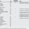
| STATEMENT | LEVEL OF EVIDENCE/GRADE OF RECOMMENDATION | REFERENCES |
|---|---|---|
1 Chung KC, Spilson SV. The frequency and epidemiology of hand and forearm fractures in the United States. J Hand Surg [Am]. 2001;26:908-915.
2 Price CT, Scott DS, Kurzner ME, Flynn JC. Malunited forearm fractures in children. J Pediatr Orthop. 1990;10:705-712.
3 Davidson JS, et al. Simple treatment for torus fractures of the distal radius. J Bone Joint Surg Br. 2001;83:1173-1175.
4 Plint AC, et al. A randomized, controlled trial of removable splinting versus casting for wrist buckle fractures in children. Pediatrics. 2006;117:691-697.
5 West S, et al. Buckle fractures of the distal radius are safely treated in a soft bandage: A randomized prospective trial of bandage versus plaster cast. J Pediatr Orthop. 2005;25:322-325.
6 Chess DG, et al. Short arm plaster cast for distal pediatric forearm fractures. J Pediatr Orthop. 1994;14:211-213.
7 Bhatia M, Housden PH. Re-displacement of paediatric forearm fractures: Role of plaster moulding and padding. Injury. 2006;37:259-268.
8 Boyer BA, et al. Position of immobilization for pediatric forearm fractures. J Pediatr Orthop. 2002;22:185-187.
9 Bochang C, Jie Y, Zhigang W, et al. Immobilisation of forearm fractures in children. Extended versus flexed elbow. J Bone Joint Surg Br. 2005;87:994-996.
10 Shaer JA, Smith B, Turco VJ. Mid-third forearm fractures in children: An unorthodox treatment. Am J Orthop. 1999;28:60-63.
11 Walker JL, Rang M. Forearm fractures in children. Cast treatment with the elbow extended. J Bone Joint Surg Br. 1991;73:299-301.
12 Bohm ER, Bubbar V, Yong Hing K, Dzus A. Above and below-the-elbow plaster casts for distal forearm fractures in children. A randomized controlled trial. J Bone Joint Surg Am. 2006;88:1-8.
13 Webb GR, Galpin RD, Armstrong DG. Comparison of short and long arm plaster casts for displaced fractures in the distal third of the forearm in children. J Bone Joint Surg Am. 2006;88:9-17.
14 Luhmann JD, et al. A randomized comparison of nitrous oxide plus hematoma block versus ketamine plus midazolam for emergency department forearm fracture reduction in children. Pediatrics. 2006;118:e1078-e1086.
15 Evans JK, et al. Analgesia for the reduction of fractures in children: A comparison of nitrous oxide with intramuscular sedation. J Pediatr Orthop. 1995;15:73-77.
16 Gregory PR, Sullivan JA. Nitrous oxide compared with intravenous regional anesthesia in pediatric forearm fracture manipulation. 1996;16:187-191.
17 Kriwanek KL, Wan J, Beaty JH, Pershad J. Axillary block for analgesia during manipulation of forearm fractures in the pediatric emergency department: A prospective randomized comparative trial. J Pediatr Orthop. 2006;26:737-740.
18 Davidson AJ, Eyres RL, Cole WG. A comparison of prilocaine and lidocaine for intravenous regional anaesthesia for forearm fracture reduction in children. Paediatr Anaesth. 2002;12:146-150.
19 Jones K, Weiner DS. The management of forearm fractures in children: A plea for conservatism. J Pediatr Orthop. 1999;19:811-815.
20 Flynn JM, Waters PM. Single-bone fixation of both-bone forearm fractures. J Pediatr Orthop. 1996;16:655-659.
21 Qidwai SA. Treatment of diaphyseal forearm fractures in children by intramedullary Kirschner wires. J Trauma. 2001;50:303-307.
22 Shoemaker SD, et al. Intramedullary Kirschner wire fixation of open or unstable forearm fractures in children. J Pediatr Orthop. 1999;19:329-337.
23 Yung PS, et al. Percutaneous transphyseal intramedullary Kirschner wire pinning: A safe and effective procedure for treatment of displaced diaphyseal forearm fracture in children. J Pediatr Orthop. 2004;24:7-12.
24 Cullen MC, et al. Complications of intramedullary fixation of pediatric forearm fractures. J Pediatr Orthop. 1998;18:14-21.
25 Kucukkaya M, et al. The application of open intramedullary fixation in the treatment of pediatric radial and ulnar shaft fractures. J Orthop Trauma. 2002;16:340-344.
26 Lascombes P, et al. Elastic stable intramedullary nailing in forearm shaft fractures in children: 85 cases. J Pediatr Orthop. 1990;10:167-171.
27 Yuan PS, Pring ME, Gaynor TP, et al. Compartment syndrome following intramedullary fixation of pediatric forearm fractures. J Pediatr Orthop. 2004;24:370-375.
28 Kapoor V, Theruvil B, Edwards SE, et al. Flexible intramedullary nailing of displaced diaphyseal forearm fractures in children. Injury. 2005;36:1221-1225.
29 Majed A, Baco AM. Nancy nail versus intramedullary-wire fixation of paediatric forearm fractures. J Pediatr Orthop B. 2007;16:129-132.
30 Calder PR, Achan P, Barry M. Diaphyseal forearm fractures in children treated with intramedullary fixation: Outcome of K-wire versus elastic stable intramedullary nail. Injury. 2003;34:278-282.
31 Fernandez FF, et al. Unstable diaphyseal fractures of both bones of the forearm in children: Plate fixation versus intramedullary nailing. Injury. 2005;36:1210-1216.
32 Van der Reis WL, et al. Intramedullary nailing versus plate fixation for unstable forearm fractures in children. J Pediatr Orthop. 1998;18:9-13.