Chapter 17 What Are the Best Diagnostic Tests for Complex Regional Pain Syndrome?
HISTORY
The French physician Claude Bernard (1813–1878) first described the condition now called complex regional pain syndrome (CRPS).1 His student, Silas Weir Mitchell, attributed it to autonomic instability and proposed the term causalgia for this diagnosis.2 Since then, a variety of other names has also been used to describe this condition, including algodystrophy, sympathalgia, post-traumatic spreading neuralgia, and reflex sympathetic dystrophy. Although reflex sympathetic dystrophy was the widely accepted diagnostic term for a long time, the role of the sympathetic nervous system in the pathophysiology of this syndrome has not been confirmed. Therefore, in a consensus workshop of the International Association for the Study of Pain (IASP) in 1995, it was recommended that the condition known as reflex sympathetic dystrophy be called complex regional pain syndrome (CRPS).
Complex Regional Pain Syndrome: Causative Factors and Natural Progression
CRPS is divided into two subtypes, I and II, on the basis of an association with peripheral nerve pathology.3 CRPS I includes cases in whom no associated nerve pathology exists and in whom CRPS develops after a noxious event.3 For example, the reported incidence of CRPS I after various fractures is 1% to 2% (7–35% after Colles’ fractures).4 CRPS II includes cases in which there are known nerve pathology, and it mostly affects large nerves such as the median or sciatic nerves.3 For example, the reported incidence of CRPS II after peripheral nerve injuries is 2% to 5%; 10% to 26% of CRPS cases are idiopathic.4
CRPS may progress through three stages.1 Stage 1 (acute) is a warm phase, and it is characterized by pain, sensory abnormalities, vasomotor dysfunction, edema, and sudomotor disturbances. Stage II (dystrophic) may be warm or cold, and it is characterized by worsening of stage I symptoms. In addition, motor and trophic (hair and nail growth) changes are first noted in stage II. Stage III (atrophic) is a cold phase in which pain and sensory disturbances decrease, but motor and trophic abnormalities worsen. In addition, atrophy of affected muscles may be noted. In some cases, the specific stages of CRPS may be difficult to identify clinically because they may blend into one another in a more continuous spectrum of manifestations. In one study, 13% of patients with acute CRPS had cold affected areas.2 In some patients, the warm phase has been noted to persist for as long as 8 to 12 years after the initial diagnosis of CRPS.4 Overall, this staging system is widely accepted in literature.1
COMPLEX REGIONAL PAIN SYNDROME: CLINICAL DIAGNOSIS
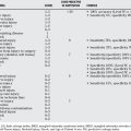
The IASP has suggested diagnostic criteria on multiple occasions,5 and these have been helpful in organizing the thought process in diagnosing CRPS. However, multiple studies have found that the IASP criteria have satisfactory sensitivity, but have poor specificity leading to a risk for overdiagnosis.6–8 Because clinical symptoms can lead to overdiagnosis, in suspicious cases, more advanced tests that have better specificity may be administered to reduce false positives of clinical diagnosis. The clinical diagnosis criteria were revised in 2003, and the new standard that has better specificity1 is described in Table 17-1. Clinical diagnosis has grade B level evidence (Table 17-2).
TABLE 17-1 Criteria for Clinical Diagnosis of Complex Regional Pain Syndrome
|
2. Patient must report at least three of the following symptoms:
|
Adapted from Harden RN, Bruehl SP: Diagnosis of complex regional pain syndrome: Signs, symptoms, and new empirically derived diagnostic criteria. Clin J Pain 22:415–419, 2006.
TABLE 17-2 Grades of Recommendation for Diagnostic Tests
| TEST | GRADE OF RECOMMENDATION* |
|---|---|
| Clinical diagnostic criteria | B |
| Radiograph | B |
| Magnetic resonance imaging | B |
| Bone scanning | B |
| Sympathetic skin response | B |
* A = good evidence (Level I studies with consistent finding) for or against recommending intervention; B = fair evidence (Level II or III studies with consistent findings) for or against recommending intervention; C = poor-quality evidence (Level IV or V with consistent findings) for or against recommending intervention; I = there is insufficient or conflicting evidence not allowing a recommendation for or against intervention.
CLINICAL DIAGNOSIS: SPECIFIC SYMPTOMS AND DIAGNOSTIC METHODS
Sensory Changes
Pain is the most frequently encountered symptom in CRPS; 81% of CRPS patients suffer severe pain.1 The nature of pain is often described as burning or stinging, which typically starts at the distal end of an extremity and propagates proximally.9
Hyperesthesia and allodynia are reported in 65% of CRPS patients,1 and they do not follow sensory nerve distribution patterns.9 This can be used to distinguish CRPS from nerve pathologies, which are limited to the affected nerve’s distribution. Mechanical allodynia can be tested by light touch or brushing over the affected area. Temperature allodynia may be assessed with warm and cold test tubes of water.
In addition, quantitative sensory testing, which is a computerized measure of temperature and vibrations sense,10 can be used for objective assessment of allodynia.11–13 Quantitative thermal testing may be done using a Marstock thermostimulator (Thermotest equipment; Somedic AB, Stockholm, Sweden).12 This equipment has an electrode that is placed on the patient’s skin. Patients have control over cooling or heating the electrode, and they are asked to raise/lower temperature until it induces pain. The thermal thresholds of the affected and unaffected region are measured and compared with each other to look for allodynia. Similarly, quantitative vibration testing can be done using a handheld vibrator (TVR model, HV-13 D; Heiwa Electronic Industrial, Osaka, Japan).12
Vasomotor Changes
Bilateral asymmetry in color, temperature, or both is a common symptom in CRPS; 87% of CRPS patients have asymmetry in color, and 79% have asymmetry in temperature.1 Classically, the affected area is warmer, redder, and edematous (80% of patients) compared with the unaffected area in early-stage CRPS.1 In 54% of patients, the affected area changes from hyperthermic to hypothermic presentation with progression of disease.9
In severe cases of CRPS, temperature asymmetry may be diagnosed by touching the affected and unaffected areas. In some cases, the temperature asymmetry is subtle and its diagnosis may require more sophisticated equipments. Simple infrared thermometers have been used for this purpose, and their sensitivity and specificity varies from 70% to 90% in literature.14 In addition, more advanced computerized thermography devices are available.13–15 For example, TIP 50 imaging unit (Bales Scientific, Walnut Creek, CA) can be used to measure the average temperature of the affected region and its contralateral normal region. The sensitivity, specificity, positive predictive value, and negative predictive value of computerized thermography depend on the cutoff asymmetry value (how different the temperature between affected and unaffected regions has to be before stating that the asymmetry is clinically significant). For example, if we choose a high cutoff value (e.g., difference of 1°C instead of 0.5°C), there would be fewer false positives, but we will not detect more patients with CRPS. One study reported that a temperature asymmetry of 0.6°C is most optimal to balance sensitivity and specificity, but if specificity is more important, then a higher cutoff (0.8°C) may be used.15 Another study found thermography to have a sensitivity of 93%, specificity of 98%, positive predictive value (PPV) of 90%, and negative predictive value (NPV) of 94%.16
Sudomotor Changes
Sudomotor changes, most notably increased sweating on the affected side, is correlated with CRPS, and this sweating asymmetry is more pronounced on exertion.9 In severe cases, asymmetry in sweating can be assessed with touch or sight. In moderate cases, dragging a smooth tool on skin can be used to approximate sweating. In addition, the following more advanced tests are available:
Motor/Trophic Changes
Motor changes are common in CRPS patients; 80% of CRPS patients report decreased range of motion, 75% report muscle weakness in the involved area, and 20% of patients suffer tremors.1 Trophic changes include brittle nails (24% of patients)1 and increased hair growth (21% of patients).1 Motor and trophic changes have to be assessed by clinical observation.
COMPLEX REGIONAL PAIN SYNDROME: ADDITIONAL TESTS
The current literature reports imaging, nuclear medicine, and sympathetic skin response (SSR) tests, which have grade B evidence (see Table 17-2) that can complement the clinical diagnosis of CRPS. The imaging techniques that may assist in the diagnosis of CRPS are plain x-ray films and magnetic resonance imaging (MRI). Radiographs are useful in the diagnosis of CRPS I after a fracture because the bone changes extend beyond the original fracture site in early stages,17 and diffuse osteoporosis with severe patchy demineralization may appear in later stages.18 In fact, the amount of bone loss in CRPS over a few months is tantamount to bone loss over 10 years of uncomplicated osteoporosis.17 One study reported radiographs to have a sensitivity of 36%, specificity of 94%, NPV of 86%, and PPV of 58% in diagnosing CRPS.18 The signs of CRPS observed on MRI are skin thickening/enhancement and joint effusion.18–20 One study found MRI to have a specificity of 98% and sensitivity of 14% in diagnosing CPRS I, 16 weeks after initial trauma.18 The literature recommends that because imaging studies have low sensitivity, they cannot be used as screening tests, but their high specificity makes them valuable as second-line tests in patients who are suspected of having CRPS after clinical observation.
In the bone scanning test, a tracer is administered to the patient, and bone imaging is done. CRPS extremities are correlated with increased uptake of tracer and accelerated blood flow to the affected limb.18 The diagnostic parameters of this test in literature are as follows: sensitivity (54–100%), specificity (85–98%), PPV (67–95%), and NPV (61–100%).21 The diagnostic accuracy is influenced by the following factors: stage of the disease, age of the patient, nature of inciting event, and location of the disease.
SSR test, which measures changes in skin conductance in response to electrical stimulation, may be useful to diagnose CRPS. The electrical stimulation is delivered using electrodes, and skin conductance is measured using electromyography. SSR is increased in the affected region of patients with early-stage CRPS.22–25 One study found that SSR was less than normal in the affected region of patients with late-stage CRPS.26 Therefore, it may be possible to differentiate stages of CRPS using SSR. No definitive data are available on the sensitivity, specificity, PPV, and NPV of SSR.
COMPLEX REGIONAL PAIN SYNDROME: A DIAGNOSTIC SCHEME
The difficulty in diagnosing CRPS is, in part, because there is no widely accepted reference standard test for this condition. In its absence, clinical observation of symptoms serves as the standard. It has high sensitivity, as we described earlier, and is useful as a screening test when CRPS is suspected. Unfortunately, clinical diagnosis lacks in specificity, which results in a relatively high number of false-positive results. This is because the symptoms used to diagnose CRPS may be seen in a variety of other illnesses. Therefore, the first thing to do after a positive clinical diagnosis is to confirm that this is CRPS and not another disease.
The diagnosis of CRPS may, in many cases, be a diagnosis of exclusion. Rheumatologic, orthopedic, neurologic, and vascular disorders can resemble the clinical features of CRPS, and it is important to differentiate among these alternative diagnoses. Unlike most rheumatic disorders, CRPS is not usually associated with increased sedimentation rates or specific antigenantibody complexes.4 Orthopedic disorders such as osteoporosis, bone bruise, stress fracture, and delayed fracture healing may also resemble CRPS,9 but they may be distinguished from it by radiography and bone scanning. Neurological disorders such as postherpetic neuralgia, plexopathy, and entrapment neuropathy may also resemble CRPS symptoms.9 However, associated venous thrombosis seen in CRPS4 does not occur in neurologic disorders. Vascular disorders, such as chronic arterial insufficiency and Raynaud’s disease, may also resemble CRPS. The former can be differentiated from CRPS because in arterial insufficiency, pulses are absent, but they are present in CRPS. Raynaud’s disease is exacerbated by cold, whereas CRPS symptoms are exacerbated by exercise.4 Therefore, cold and exercise stress tests can be used to distinguish between Raynaud’s disease and CRPS. CRPS symptoms may also be mistaken for infection. But with CRPS, there is no fever and no change in serology.4 If CRPS is still suspected after the aforementioned differential diagnosis, then the probability that the positive result is a true positive can be further clarified by administering advanced tests that have better specificity.
COMPLEX REGIONAL PAIN SYNDROME: SUMMARY
Table 17-3 provides a summary of recommendations for the treatment of CRPS.
| STATEMENT | LEVEL OF EVIDENCE/GRADE OF RECOMMENDATION | REFERENCES |
|---|---|---|
| Clinical diagnostic criteria | B | 1,9 |
| X-ray | B | 17,18 |
| MRI | B | 18–20 |
| Bone scanning | B | 21 |
| Sympathetic skin response | B | 22–26 |
1 Harden RN, Bruehl SP. Diagnosis of complex regional pain syndrome: Signs, symptoms, and new empirically derived diagnostic criteria. Clin J Pain. 2006;22:415-419.
2 Lau FH, Chung KC. Silas Weir Mitchell, MD: The physician who discovered causalgia. J Hand Surg [Am]. 2004;29:181-187.
3 Stanton-Hicks M, Janig W, Hassenbusch S, et al. Reflex sympathetic dystrophy: Changing concepts and taxonomy. Pain. 1995;63:127-133.
4 Veldman PH, Reynen HM, Arntz IE, Goris RJ. Signs and symptoms of reflex sympathetic dystrophy: Prospective study of 829 patients. Lancet. 1993;342:1012-1016.
5 IASP. Classification of chronic pain. Descriptions of chronic pain syndromes and definitions of pain terms. Prepared by the International Association for the Study of Pain, Subcommittee on Taxonomy. Pain Suppl. 1986;3:S1-S226.
6 Bruehl S, Harden RN, Galer BS, et al. External validation of IASP diagnostic criteria for Complex Regional Pain Syndrome and proposed research diagnostic criteria. International Association for the Study of Pain. Pain. 1999;81:147-154.
7 Galer BS, Bruehl S, Harden RN. IASP diagnostic criteria for complex regional pain syndrome: A preliminary empirical validation study. International Association for the Study of Pain. Clin J Pain. 1998;14:48-54.
8 Harden RN, Bruehl S, Galer BS, et al. Complex regional pain syndrome: Are the IASP diagnostic criteria valid and sufficiently comprehensive? Pain. 1999;83:211-219.
9 Vacariu G. Complex regional pain syndrome. Disabil Rehabil. 2002;24:435-442.
10 Chong PS, Cros DP. Technology literature review: Quantitative sensory testing. Muscle Nerve. 2004;29:734-747.
11 Fowler CJ, Carroll MB, Burns D, et al. A portable system for measuring cutaneous thresholds for warming and cooling. J Neurol Neurosurg Psychiatry. 1987;50:1211-1215.
12 Wahren LK, Torebjork E, Nystrom B. Quantitative sensory testing before and after regional guanethidine block in patients with neuralgia in the hand. Pain. 1991;46:23-30.
13 Gulevich SJ, Conwell TD, Lane J, et al. Stress infrared telethermography is useful in the diagnosis of complex regional pain syndrome, type I (formerly reflex sympathetic dystrophy). Clin J Pain. 1997;13:50-59.
14 Chelimsky TC, Low PA, Naessens JM, et al. Value of autonomic testing in reflex sympathetic dystrophy. Mayo Clin Proc. 1995;70:1029-1040.
15 Bruehl S, Lubenow TR, Nath H, Ivankovich O. Validation of thermography in the diagnosis of reflex sympathetic dystrophy. Clin J Pain. 1996;12:316-325.
16 Gulvich SJ, Conwell TD, Lane J, et al. Stress infrared telethermography is useful in the diagnosis of complex regional pain syndrome, Type I (formerly reflex sympathetic dystrophy). Conf Proc IEEE Eng Med Biol Soc. 2004;2:1178.
17 Masson C, Audran M, Pascaretti C, et al. Further vascular, bone and autonomic investigations in algodystrophy. Acta Orthop Belg. 1998;64:77-87.
18 Schurmann M, Zaspel J, Lohr P, et al. Imaging in early posttraumatic complex regional pain syndrome: A comparison of diagnostic methods. Clin J Pain. 2007;23:449-457.
19 Graif M, Schweitzer ME, Marks B, et al. Synovial effusion in reflex sympathetic dystrophy: An additional sign for diagnosis and staging. Skeletal Radiol. 1998;27:262-265.
20 Intenzo CM, Kim SM, Capuzzi DM. The role of nuclear medicine in the evaluation of complex regional pain syndrome type I. Clin Nucl Med. 2005;30:400-407.
21 Fournier RS, Holder LE. Reflex sympathetic dystrophy: Diagnostic controversies. Semin Nucl Med. 1998;28:116-123.
22 Cronin KD, Kirsner RL, Fitzroy VP. Diagnosis of reflex sympathetic dysfunction. Use of the skin potential response. Anaesthesia. 1982;37:848-852.
23 Aisen ML, Stallman J, Aisen PS. The sympathetic skin response in the shoulder-hand syndrome complicating tetraplegia. Paraplegia. 1995;33:602-605.
24 Clinchot DM, Lorch F. Sympathetic skin response in patients with reflex sympathetic dystrophy. Am J Phys Med Rehabil. 1996;75:252-256.
25 Bolel K, Hizmetli S, Akyuz A. Sympathetic skin responses in reflex sympathetic dystrophy. Rheumatol Int. 2006;26:788-791.
26 Rommel O, Tegenthoff M, Pern U, et al. Sympathetic skin response in patients with reflex sympathetic dystrophy. Clin Auton Res. 1995;5:205-210.


