59
Warts (verruca vulgaris)
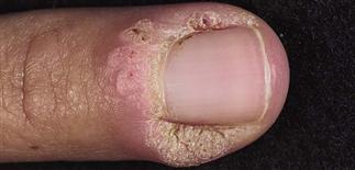
Warty hyperkeratosis is typically rough-looking and white. Proximal and lateral nail folds affected. Treatment is difficult and must be gentle enough not to scar or damage the nail matrix.
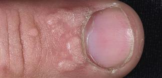
Multiple rough, flesh-colored papules grouped around the proximal nail fold. Biting this area is discouraged, as it may spread the warts to other areas, such as the lips or nails.
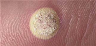
Wart treated with liquid nitrogen. A rim of normal skin is also treated. The white freezing color change should remain 15 s and be allowed to slowly thaw; often the lesion is treated twice.
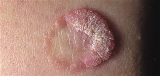
Partial resolution of a wart after liquid nitrogen treatment. The central area is resolved, although a rim of warty tissue remains. Allowing rim and center to stay white 15 s may avoid this.
DESCRIPTION
Benign epidermal overgrowths caused by human papillomavirus (HPV). Transmitted by contact, often at small skin breaks, abrasions, or other trauma.
HISTORY
• Variable onset from exposure: 1–6 months. • Duration variable; spontaneous resolution with time typical. • In children, approximately two-thirds of warts spontaneously regress within 2 years. • Warts in immunocompromised persons can be widespread, chronic.
PHYSICAL FINDINGS
• Flesh-colored papules evolve into dome-shaped, gray-brown, hyperkeratotic, discrete, rough papules, often with surface black dots. • Usually few, may be numerous. • Common sites: hands, periungual skin, elbows, knees, plantar surfaces. • Filiform warts are finger-like growths on narrow or broad base, often on the face. • HPV subtyping not routine, readily available, or necessary for common warts. • Genital HPV subtypes 16, 18, and 31 are high-risk subtypes and account for approximately 75% of invasive genital cancers. • Biopsy a non-resolving wart on hand, periungual unit, or foot to rule out squamous cell carcinoma.
TREATMENT
Multiple treatments available. No treatment is consistently highly effective. Avoid painful treatment, especially for children. No treatment is acceptable, but patients seek treatment because of unsightly appearance, fear of spread or enlargement, or discomfort. Some options follow. • Duct tape cut to wart size and applied. Leave for 6 days, remove, wash skin, gently debride. Reapply, continue cycle for 1 month if necessary. • Over-the-counter topical salicylic acid (15–40%) applied once a day. Occlusion with tape increases penetration. Treatment duration 8–12 weeks. Some options are Mediplast (40% salicylic acid), DuoFilm solution (17% salicylic acid) or patch (40% salicylic acid), and Occlusal HP solution (17% salicylic acid). • Multiple visits often necessary when treating with ablative therapy. Liquid nitrogen cryotherapy: 15-s freeze time, repeat once. Retreatment in 2–3 weeks. Side effects: pain, blister after treatment. Caution treating warts around nail. Older children may tolerate gentle cryotherapy; consider for long-standing warts or if child-friendly treatments have failed. • Imiquimod 5% cream (Aldara) has limited use in common warts; efficacy limited by poor penetration in non-mucosal skin. Treat with liquid nitrogen, then apply a salicylic acid preparation at night, and imiquimod in the morning with occlusion. Continue 6–9 weeks. • Intralesional bleomycin for refractory warts; efficacy limited. Contact immunotherapy of warts using dinitrochlorobenzene, squaric acid, and Candida antigen used by some dermatologists. • Limitations of laser surgery: pain, potential scarring. • Filiform warts easiest to treat. Local anesthesia can be used; lesion removal with snip or curette.