Vitreoretinal Lymphoma
Clinical Features:
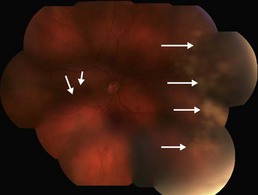
There are no clinical pathognomonic features of VRL and confirmation of the diagnosis can be difficult as it typically presents as an unspecified posterior uveitis. In certain cases, lymphoma cells can invade the subretinal space, leading to characteristic multifocal, dome-shaped yellowish subretinal deposits. These can be located within the macula, but are most striking when located in the retinal periphery (Fig. 23.2.1).
OCT Features:
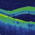
The lymphoma cells infiltrate along Bruch’s membrane and accumulate as deposits underneath the RPE. These deposits appear on OCT as medium to intense hyper-reflective dome-shaped sub-RPE elevations of varying size. They can be seen both in the macula (Fig. 23.2.2) and retinal periphery (Fig. 23.2.3).
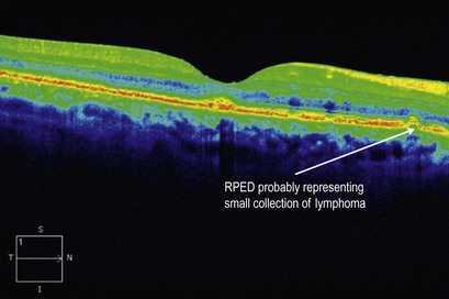
Figure 23.2.2 OCT (corresponding to Figure 23.2.1, macula) shows a small sub-RPE nodular elevation that exhibits hyper-reflectivity of medium intensity (arrow). This nodule is believed to be composed of lymphoma cells.
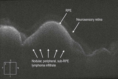
Figure 23.2.3 OCT (corresponding to Figure 23.2.1, nasal periphery) shows numerous, large sub-RPE nodular elevations, which are underneath the neurosensory retina. These lesions are intensely hyper-reflective.