Vitreomacular Adhesion and Vitreomacular Traction
OCT Diagnosis:
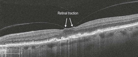
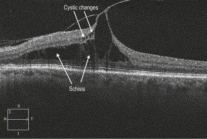
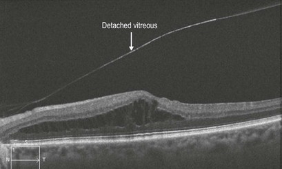
OCT is the diagnostic modality of choice for both of these entities. In fact, VMA can only be reliably diagnosed via OCT. In VMA, OCT shows vitreous separating from around the macula with persisting adhesion at the macular center, often in a concentric fashion (Fig. 10.1.1). VMT is accompanied by changes in the retina including cystic changes, macular schisis, defined as a separation between the outer nuclear and the outer plexiform layer, epiretinal membrane formation and tractional retinal detachment (Figs 10.1.2 to 10.1.4) Often the posterior hyaloid appears abnormally thickened in VMT.
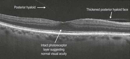
Figure 10.1.1 Vitreomacular adhesion. Note the dense posterior hyaloid face (arrows). The retina appears to be normal.