111
Vitiligo
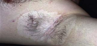
Examine the axillae, groin, and anal area in all patients suspected of having vitiligo, as these areas are commonly involved.
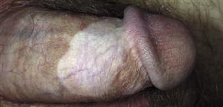
The genitalia and anal areas may be the first or only areas affected and must be examined.
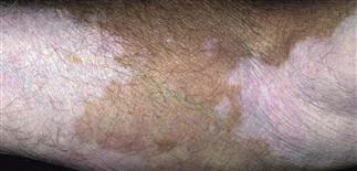
Loss of pigment may be partial or complete. Complex trichrome patterns are typical.
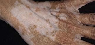
Vitiligo on the back of the hand is resistant to all forms of treatment.
DESCRIPTION
Disfiguring depigmenting disease that causes melanocyte destruction. Etiology unknown but thought to be autoimmune.
HISTORY
• One peercent of population affected; in 50%, begins before age 20. Males, females affected equally. • Positive family history in 30%. • Patients relate first onset to aftermath of emotional stress, illness, skin trauma (e.g. sunburn). Slowly progresses over years in highly variable course. Some patients have very stable disease; others progress at alarming rate. • Depigmented areas at increased risk for sunburn, subsequent skin cancers. • Can be devastating for dark-skinned people, especially if cultural implications present. Listen to patients’ concerns and make all efforts on their behalf.
PHYSICAL FINDINGS
Two clinical types. • Type A. More common pattern, involving fairly symmetric pattern of white depigmented 0.5- to 5.0-cm macules and patches with well-defined borders. Borders may have red halo of inflammation or rim of hyperpigmentation. Wood’s light accentuates hypopigmented areas. Common sites include dorsal hands and fingers, face, body folds, axillae, genitalia. Predilection for orifices, including eyes, nostrils, mouth, nipples, umbilicus, anus. Occurs at sites of trauma (Koebner phenomenon). Increased incidence of autoimmune thyroid disease. • Type B. Segmental vitiligo. Limited to one segment of body (e.g. one extremity). More common in childhood. Skin biopsy shows absence of melanocytes and sparse lymphocytic inflammation.
TREATMENT
• Broad-spectrum sunscreens that contain avobenzone, Mexoryl, titanium dioxide, or zinc oxide. • Sun-protective clothing available from Sun Precautions (http://www.sunprecautions.com), similar companies. • Concealing and camouflaging agents, such as Dermablend, Covermark, and Elizabeth Arden Concealing Cream, effective but require practice in application for good cosmesis. Suggest a local qualified aesthetician as resource for color matching. • Sunless or self-tanning lotions darken skin by staining; work best for skin phototypes 2 and 3. • Group I–V topical steroid ointments effective for limited disease. Apply twice daily for 6 weeks, stop for 2 weeks; repeat for two more cycles if no side effects. Face and neck respond better than other areas. • Tacrolimus ointment 0.1% (Protopic) effective in some cases. • Calcipotriol ointment (Vectical) may be effective in combination with Excimer laser (308 nm) therapy. •Pulse-dose systemic steroids may arrest or slow rapidly progressing cases but have potential side effects and risk. • Topical or systemic photochemotherapy with psoralen plus ultraviolet A light therapy somewhat effective. Narrowband ultraviolet B light treatment may be effective. • Punch skin grafting for limited areas of involvement may be helpful. • Patients with involvement of > 40% of body surface may choose to remove remaining normal pigment with 20% monobenzone (Benoquin cream). Apply monobenzone twice daily, 6–18 months, to chemically destroy all melanocytes in skin. Depigmentation irreversible, requiring patient to be informed, subsequent close attention to sun protection.


