Viral infections – Herpes simplex and herpes zoster
Herpes simplex
Clinical presentation
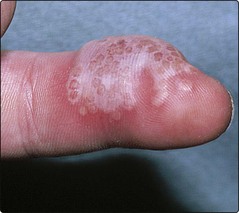
Herpetic whitlow is another presentation (Fig. 1). A painful vesicle or pustule is found on a finger in, for example, a nurse or dentist attending a patient secreting the virus. Similar direct inoculation is sometimes seen in sportsmen such as wrestlers (‘herpes gladiatorum’).
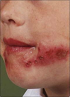
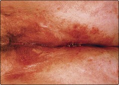
Recurrence is a hallmark of herpes simplex infection; it occurs at a similar site each time, usually on the lips, face (Fig. 2) or genitals (Fig. 3). Rarely, herpes simplex may appear in a zosteriform dermatomal distribution. The outbreak of groups of vesicles is often preceded for a few hours by tingling or burning. Crusts form within 24–48 h, and the infection fades after a week. Attacks may be precipitated by respiratory infection (hence ‘cold’ sore), sunlight or local trauma.
Complications
Complications are infrequent but can be serious. They include the following:
 Secondary bacterial infection. This is usually due to Staphylococcus aureus.
 Eczema herpeticum. Widespread herpes simplex infection is a serious and potentially fatal complication seen in patients with atopic eczema (p. 36) or Darier’s disease (p. 90).

 Chronic herpes simplex. Atypical and chronic lesions may be seen in patients with HIV infection.


 Erythema multiforme. Herpes simplex infection is the most common cause of recurrent erythema multiforme (p. 82).
Management
Mild herpetic lesions may not require any medication. The treatment of choice for recurrent mild facial or genital herpes simplex is aciclovir (Zovirax) cream (applied five times a day for 5 days), which reduces the length of the attack and the duration of viral shedding, and should preferably be started at the first indication of a recrudescence. More severe episodes warrant oral treatment with aciclovir (200 mg five times a day for 5 days), which shortens the attack. Long-term oral administration is useful in those with frequent recurrent attacks. Intravenous aciclovir may be life-saving in the immunosuppressed and in infants with eczema herpeticum. Genital herpes simplex can also be treated with oral famciclovir or valaciclovir. In those with genital herpes simplex, barrier contraception methods are advisable during intercourse, and intercourse should be avoided during symptomatic episodes.
Herpes zoster
Clinical presentation
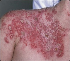
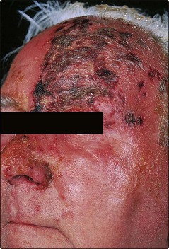
Pain, tenderness or paraesthesia in the dermatome may precede the eruption by 3–5 days. Erythema and grouped vesicles follow, scattered within the dermatomal area (Fig. 4). The vesicles become pustular and then form crusts that separate in 2–3 weeks to leave scarring. Secondary bacterial infection may occur. Herpes zoster is normally unilateral and may involve adjacent dermatomes. The thoracic dermatomes are affected in 50% of cases and, in the elderly, involvement of the ophthalmic division of the trigeminal nerve is particularly common (Fig. 5). Two-thirds of patients with herpes zoster are over 50 years of age, and it is uncommon in children. The lesions shed virus, and contacts with no previous exposure may develop chickenpox.
Complications
Serious complications may occur in herpes zoster. These include the following:


