[level-membership-for-cardiothoracic-surgery-category]
CHAPTER 20 Video-Assisted Mediastinoscopy—Video 20
Approach to Video-Assisted Mediastinoscopy
 Video-Assisted Mediastinoscopy
Step 1. Incision
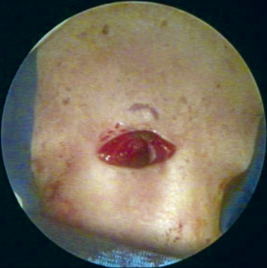
♦ Make a 2-cm incision (Figure 20-1) transversely in the base of the neck that is 1 or 2 fingerbreadths above the sternal notch.
♦ With electrocautery, cut transversely through the platysma muscle and then vertically in the midline between the strap muscles.
♦ With a closed Kelly clamp, bluntly dissect in the midline between the muscles to expose the trachea.
Step 2. Level 7 Nodes
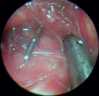
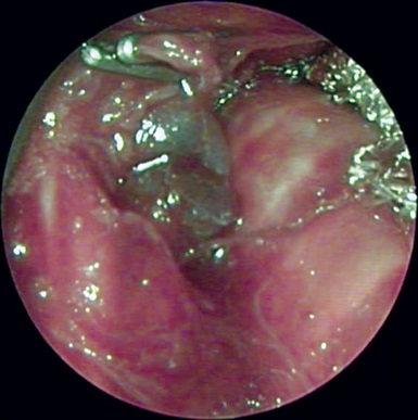
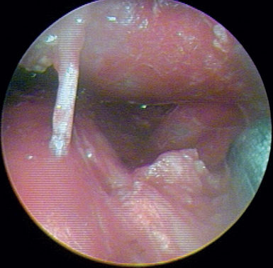
♦ An artery can usually be seen passing from the aorta to the carina. Clip the artery to reduce bleeding during removal of the subcarinal nodes (Figure 20-2).
♦ Dissect along the medial aspect of the left mainstem bronchus inferiorly to the carina. The blunt-tipped suction then sweeps the subcarinal nodes from left to right and off the surface of the esophagus.
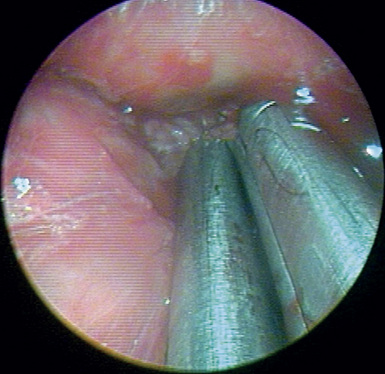
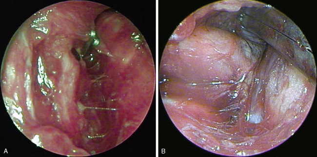
♦ Hold the subcarinal nodes to the right to allow blunt dissection between the nodes and the esophagus (Figure 20-3).
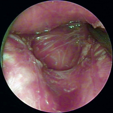
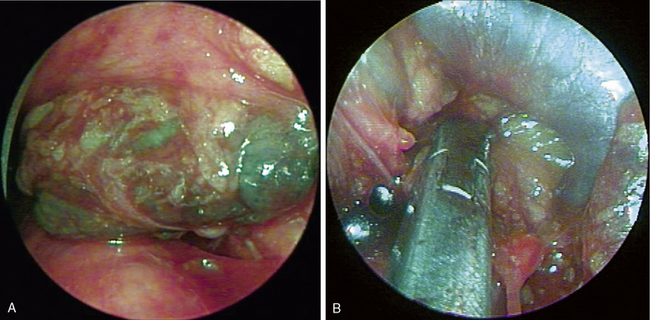
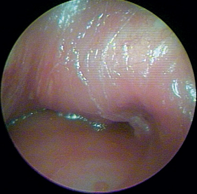
♦ Remove all the subcarinal nodes until both mainstem bronchi and the esophagus have been completely exposed (Figure 20-4).
Step 3. Left Level 10 Nodes
Step 4. Right Level 10 Nodes
Step 5. Level 4 and 2 Nodes
♦ With the biopsy forceps, grasp the fatty tissue anterior to the distal trachea, and pull posteriorly away from the superior vena cava.
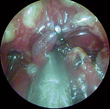
♦ There is often a small vein from the nodes to the superior vena cava. Identify and clip the vein (Figure 20-8).
[/level-membership-for-cardiothoracic-surgery-category][not-level-membership-for-cardiothoracic-surgery-category]
CHAPTER 20 Video-Assisted Mediastinoscopy—Video 20
Approach to Video-Assisted Mediastinoscopy
Video-Assisted Mediastinoscopy
Step 1. Incision
♦ Make a 2-cm incision (Figure 20-1) transversely in the base of the neck that is 1 or 2 fingerbreadths above the sternal notch.
♦ With electrocautery, cut transversely through the platysma muscle and then vertically in the midline between the strap muscles.
♦ With a closed Kelly clamp, bluntly dissect in the midline between the muscles to expose the trachea.
♦ The finger breaks through the pretracheal fascia to mobilize the lymph nodes as much as possible. This often is the safest method to dissect matted nodes away from vessels.
Buy Membership for Cardiothoracic Surgery Category to continue reading. Learn more here
[/not-level-membership-for-cardiothoracic-surgery-category]
