9 Vertebral column
The vertebral column usually consists of:
7 cervical vertebrae (movable)
12 thoracic vertebrae (movable)
4 coccygeal segments (fused to a variable extent).
A ‘typical’ vertebra
Main parts
Inferior articular processes –
projections on the inferior aspect of the vertebral arch which carry the inferior articular facets.
Cervical vertebrae
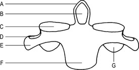
3rd to 6th (typical) (Figs 9.1 and 9.2)
Features
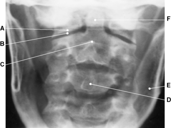
Radiographic appearances of the cervical vertebrae (Figs 9.3, 9.4, 9.5, 9.6 and Plate 6)
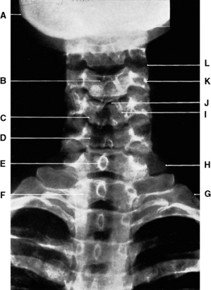
Fig. 9.3 Third to seventh cervical vertebrae: anteroposterior projection.
C–Body of 5th cervical vertebra
E–Spinous process of 7th cervical vertebra
F–Body of 1st thoracic vertebra
H–Transverse process of 7th cervical vertebra
(From Bryan 1996.)
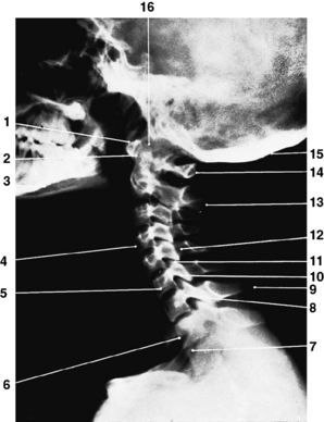
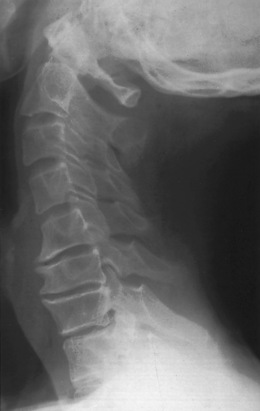
Fig. 9.4 Cervical vertebrae: lateral projection.
1 – Anterior tubercle of atlas
4 – Body of 4th cervical vertebra
6 – Body of 1st thoracic vertebra
9 – Spinous process of 7th cervical vertebra (vertebra prominens)
10 – Inferior articular process
11 – Superior articular process
14 – Posterior tubercle of atlas
(From Bryan 1996.)
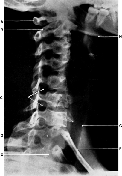
Fig. 9.5 Cervical vertebrae: oblique anteroposterior projection.
C – Right intervertebral foramina
(From Bryan 1996.)
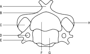
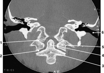
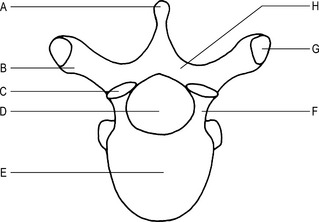
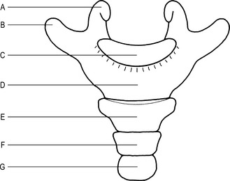
1st cervical vertebra (atlas) (Fig. 9.7)
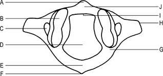
Fig. 9.7 1st cervical vertebra (superior aspect).
G–Groove for the vertebral artery
Articulations
Superior articular facets with the occipital condyles to form the atlanto-occipital joints.
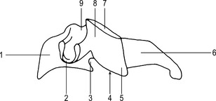
2nd cervical vertebra (axis) (Figs 9.8 and 9.9)
Articulations
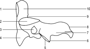
Fig. 9.9 2nd cervical vertebra (axis) (lateral aspect).
1 – Facet for the anterior arch of the atlas
6 – Inferior articular process
The body with the body of the 3rd cervical vertebra to form the intervertebral joint.
Ossification
Thoracic vertebrae
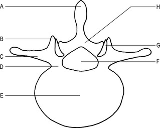
2nd to 8th (typical) (Figs 9.13 and 9.14)
Features
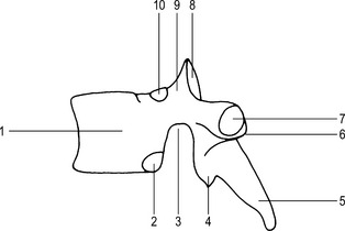
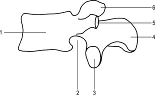
Fig. 9.14 A typical thoracic vertebra (lateral aspect).
2 – Demi-facet for the head of the rib
4 – Inferior articular process
7 – Costal facet for the tubercle of the rib
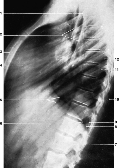
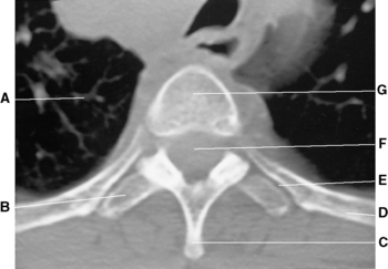
Radiographic appearances of the thoracic vertebrae (Figs 9.15, 9.16 and 9.17)
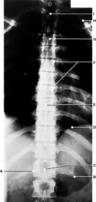
Fig. 9.15 Thoracic vertebrae: anteroposterior projection.
C–Body of 12th thoracic vertebra
I–Body of 1st thoracic vertebra.
(From Bryan 1996.)
Lumbar vertebrae
1st to 4th (typical) (Figs 9.18 and 9.19)
Features
Ossification
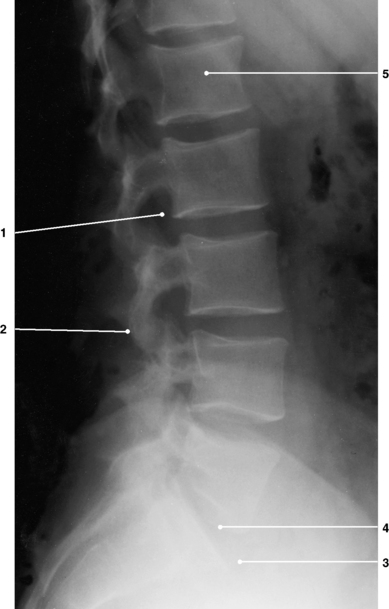
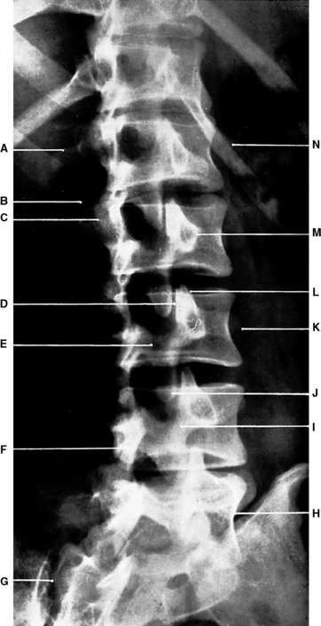
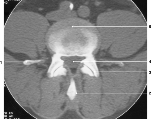
Radiographic appearances of the lumbar vertebrae (Figs 9.20, 9.21, 9.22 and 9.23)
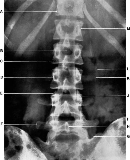
Fig. 9.20 Lumbar vertebrae: anteroposterior projection.
A–Body of 12th thoracic vertebra
F–Transverse process of 5th lumbar vertebra
H–Spinous process of 5th lumbar vertebra
M–Body of 1st lumbar vertebra.
(From Bryan 1996.)
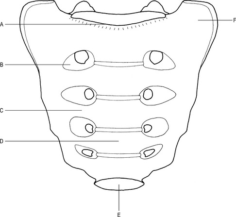
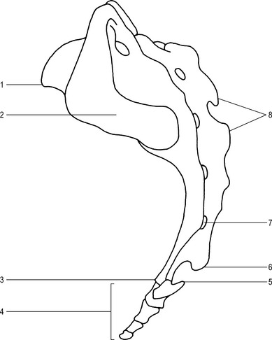
Sacrum (Figs 9.24 and 9.25)
The sacrum is composed of 5 sacral segments, which are fused together to form a single bone.
Articulations
The base articulates with the body of the 5th lumbar vertebra to form the lumbosacral joint.
The apex articulates with the base of the coccyx to form the sacrococcygeal joint.
Main parts
Base
This is formed by the 1st sacral segment and has the following features:
Pelvic surface
Pelvic sacral foramina –
4 pairs which carry the first 4 sacral spinal nerves from the sacral canal.
Dorsal surface
This faces superiorly and posteriorly, is convex and has the following features:
Medial sacral crest –
raised promontory of bone in the midline representing the fused spinous processes.
Dorsal sacral foramina –
four pairs of foramina which carry the first four dorsal sacral nerves from the sacral canal.
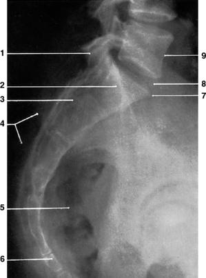
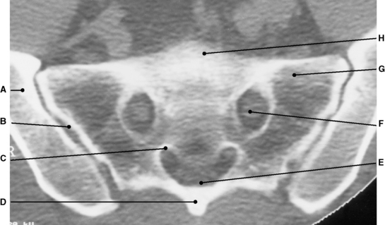
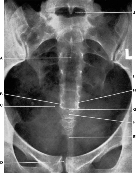
Radiographic appearances of the sacrum (Figs 9.26, 9.27, 9.28 and 9.29)
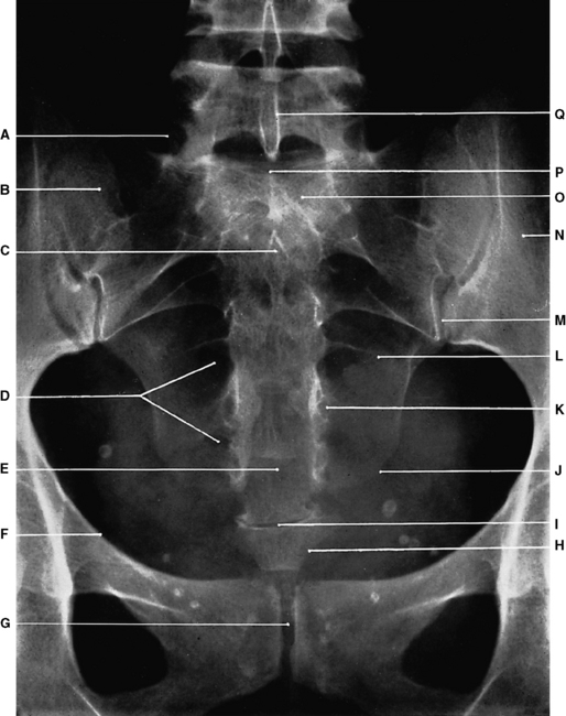
Fig. 9.26 Sacrum: anteroposterior projection.
A–Superior articular process of 1st segment
Q–Spinous process of 5th lumbar vertebra.
(From Bryan 1996.)
Coccyx (Fig. 9.30)
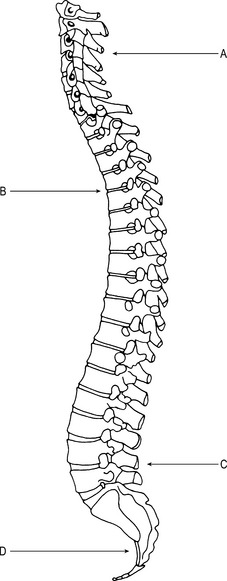
Vertebral curvatures (Fig. 9.32)
The curves of the spinal column can be divided into primary and secondary curves.
Joints of the vertebral column
Atlanto-occipital joints
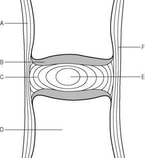
Intervertebral joints (Fig. 9.33)
Movements
Individually allow a limited degree of:
Summated over the length of the spinal column the movement is considerable.
Joints of the vertebral arches
Costovertebral joints
Fibrous capsule
Surrounds the head of the rib and the margins of the costal facets of the vertebrae.
Synovial membrane
Lines the fibrous capsule and secretes synovial fluid, which lubricates the joint.

