5 Ventricles and Cerebrospinal Fluid
The ventricular system, the remnant of the space in the middle of the embryonic neural tube (see Fig. 2-5), is an interconnected series of cavities that extends through most of the CNS.
The Brain Contains Four Ventricles
A Lateral Ventricle Curves through Each Cerebral Hemisphere
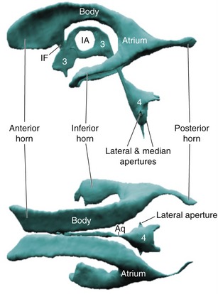
Each lateral ventricle is basically a C-shaped structure. This C shape curves from an inferior horn in the temporal lobe through a body in the parietal lobe and a bit of the frontal lobe, ending at the interventricular foramen where each lateral ventricle joins the third ventricle. Along this C-shaped course two extensions emerge—a posterior horn that extends backward into the occipital lobe and an anterior horn that extends farther into the frontal lobe (Fig. 5-1; see THB6 Figure 5-2, p. 101). The expanded area where the body and the inferior and posterior horns meet is called the atrium. Each lateral ventricle represents the cavity of an embryonic telencephalic vesicle, so telencephalic structures like the caudate nucleus and the hippocampus border much of it; the thalamus, a diencephalic derivative, also forms part of its floor (THB6 Figures 3-19 to 3-24, pp. 69-71).
Choroid Plexus Is the Source of Most CSF
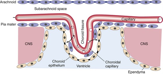
Choroid plexus is formed at certain areas where the inner lining (i.e., ependyma) and the outer covering (i.e., pia) of the CNS are directly applied to each other, with no intervening neural tissue. At these sites, the ependymal cells are specialized as a secretory epithelium called choroid epithelium; adjacent cells are joined by tight junctions, forming a diffusion barrier. Vascular connective tissue invaginates this pia/ependyma membrane, forming multiply folded choroid plexus (Fig. 5-2). This means that wherever you see choroid plexus, one side faces a ventricle and the other side faces subarachnoid space (THB6 Figure 5-8, p. 106).
CSF Is a Secretion of the Choroid Plexus
Capillaries in choroid plexus, unlike most other capillaries inside the arachnoid barrier, are permeable to plasma solutes. Plasma solutes therefore leak out, cross the pial layer, get stopped by the choroid epithelial diffusion barrier, and form the substrate for active secretion of CSF into the ventricles by the choroid epithelium. The resulting CSF is clear and colorless, low in protein, and similar (but not identical) to serum in its ionic composition.
CSF Circulates through and around the CNS, Eventually Reaching the Venous System
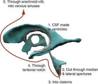
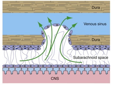
The cerebrospinal fluid secreted by the choroid plexuses moves through the ventricular system (Fig. 5-3), pushed along by newly formed CSF. It leaves the fourth ventricle through the lateral and median apertures, moves through subarachnoid space until it reaches the arachnoid villi (most of which protrude into the superior sagittal sinus), and finally joins the venous circulation (Fig. 5-4).
Imaging Techniques Allow Both CNS and CSF To Be Visualized
CT Produces Maps of X-Ray Density
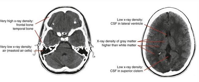
Rotating x-ray sources and detectors around someone’s head and systematically varying the center of rotation allows the construction of maps of x-ray density (THB6 Figure 5-14, p. 112). (Even though all modern tomography is a variant of computed tomography, folks usually use the term CT specifically to mean x-ray CT.) Just as with plain skull x-rays, bone is white and air is black (Fig. 5-5). White matter, because of all the lipid, is less x-ray dense than gray matter and so is darker; CSF, which is mostly water, is darker still.
MRI Produces Maps of Water Concentration
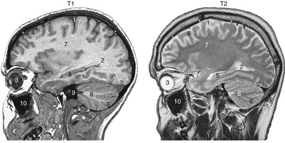
Magnetic resonance imaging (MRI) is based on similar calculations but a fundamentally different signal—the re-emission of radio waves absorbed by atoms in a strong magnetic field. The most abundant source of such signals in the CNS is protons, mostly in water but also in other molecules. Because the free water concentration and the concentrations of other molecules varies in different parts of the CNS and adjoining areas, white matter, gray matter, and CSF all look different. Different time constants can be used to produce images with different appearances (Fig. 5-6), but areas with few protons, such as bone and air-filled cavities, give off little signal.

