[level-membership-for-cardiovascular-category]
CHAPTER 77 Venous Anatomy of the Extremities
The upper and lower extremity peripheral venous systems comprise three main types of veins—superficial, deep, and perforating. The superficial and deep subsystems are defined by major and tributary veins linked by the perforating veins as well as venous networks (plexi), with the goal of directing all venous blood return into the deep system in peripheral, mid, or central extremity venous segments. Wide variability is often encountered in this anatomy, with origins for both systems beginning peripherally at the hand or foot for the upper and lower extremities, respectively. The superficial veins are located subcutaneously within the superficial fascia. The deep veins are located intramuscularly, deep to the superficial fascia, accompanying the extremity arteries (venae comitans). Perforating veins cross the muscular fascial layer, typically bridging flow from superficial to deep veins. Valves function in superficial, deep, and perforating veins to keep blood flowing antegrade and prevent blood flowing from the deep to superficial veins. Valves are more prevalent in the deep veins for both the upper and lower extremities.1–5
In general, the extremity veins are thin wall structures with little to no elastic lamina, resulting in passive distensible conduits with large compliance. The deep veins, however, inherit strength directly by having a greater degree of elastic lamina and indirectly by way of muscular contractions, countering gravity and central venous filling pressures to propagate blood flow toward the central veins and heart.5–6 The well-orchestrated anatomic physiology between the deep venous pumps, valves, and perforating veins becomes particularly apparent in the setting of elevated central venous pressures, incompetent valves, immobility, obstructed central or deep extremity veins, or a combination of these conditions.
UPPER EXTREMITY
Veins for the upper extremity direct blood flow from the hand, wrist, forearm, upper arm, and shoulder to the ipsilateral central thorax veins and ultimately the superior vena cava. In the hand, forearm, and upper arm, the superficial system functions as the principal means for venous drainage. As a result, the caliber of the superficial veins is generally larger than the deep veins. The major superficial veins of the hand, forearm, and upper arm exist as single structures and infrequently have accessory veins. The smaller deep veins occur in pairs, alongside the respective arteries. All upper extremity venous drainage transitions to the deep system at the axilla, with a single main venous channel crossing the costoclavicular space into the thoracic central venous system. Within the axillae and thorax, the major deep veins occur as single structures, accompanying the adjacent artery.7–10
Superficial Veins of the Upper Extremity
The dorsal veins include the dorsal digital veins, dorsal metacarpal veins, and dorsal venous network (plexus). Dorsal digital veins form by coalescence of dorsal distal digital venules and venous arches, and extend bilaterally along the length of dorsum of the fingers. They communicate at multiple levels via small oblique venous connections. The dorsal digital veins from the ulnar aspect of the index finger, radial aspect of the fifth finger, and both sides of the third and fourth fingers, along with intercapitular veins between the digits, drain into three superficial dorsal metacarpal veins. The metacarpal veins, in turn, feed into the dorsal venous network, which is located at the mid and base of the metacarpals (Fig. 77-1).9
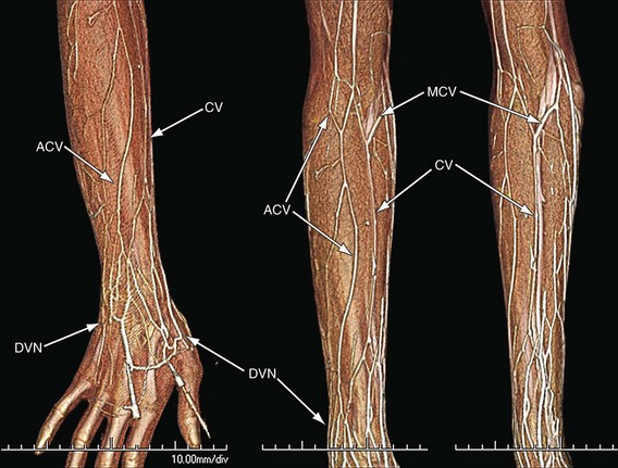
 FIGURE 77-1
Along the radial aspect of the hand, the dorsal venous network receives dorsal veins of the thumb and dorsal vein of the radial aspect of the index finger and continues with cephalad drainage, forming the cephalic vein. Along the ulnar aspect of the hand, the dorsal venous plexus receives the dorsal vein from the ulnar aspect of the fifth finger and continues with cephalad drainage, forming the basilic vein. The median portion of the network variably may drain to the mid-forearm cephalic vein via a communicating (accessory) vein (Fig. 77-2).8
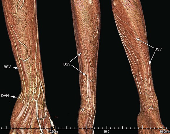
 FIGURE 77-2
The palmar veins of the hand include palmar digital veins and palmar venous plexus. Palmar digital veins parallel the dorsal digital veins, along the palmar surface. There are three routes of drainage for the palmar digital veins. Two are within the superficial system and include the palmar venous network and dorsal digital and metacarpal veins. The palmar venous network lies at the thenar and hypothenar eminences and drains primarily into the median antecubital vein of the forearm. Drainage into the dorsal digital veins and dorsal metacarpal veins occurs by way of intercapitular veins in the digital web spaces at the metacarpal heads. The third route is into the common and metacarpal palmar veins of the palmar deep system.2,3
In the forearm, main superficial venous drainage occurs via the cephalic, basilic, and median antecubital veins (Fig. 77-3). The cephalic and basilic veins ascend to the elbow along the radial and ulnar aspects of the forearm, respectively, with continuation into the upper arm. The cephalic vein, basilic vein, or both, may form the dominant drainage route into the upper arm.
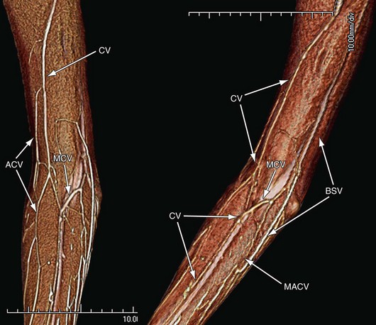
 FIGURE 77-3
The cephalic vein transitions from the dorsal to volar surfaces in the distal third of the forearm. Along its course in the forearm, the cephalic vein receives dorsal and volar tributaries. Just proximal to the elbow, the cephalic vein gives off the median basilic vein, which ascends obliquely over the volar surface, crosses the antecubital fossa, and drains into the basilic vein. The cephalic vein then crosses the ventrolateral aspect of the elbow, in a groove between the brachioradialis and biceps brachii muscles. An accessory cephalic vein is often present, with variable morphology. Most commonly, it arises from the dorsal venous plexus or from a dorsal forearm tributary network and courses along the radial aspect of the forearm, lateral to the cephalic vein. It drains into the cephalic vein below the elbow. Alternatively, the accessory cephalic vein may form a venous bridge between cephalic vein segments below and above the elbow. The accessory cephalic vein often supplies a dorsal oblique branch, forming a second level of communication between the cephalic and basilic veins within the forearm.8
In the upper arm, the basilic and cephalic veins are the major routes for superficial venous drainage, with ultimate runoff into the deep system (Figs. 77-4 and 77-5). The basilic vein is typically larger than the cephalic vein, coursing medial to the biceps brachii. The smaller cephalic vein courses lateral to the biceps brachii. The basilic vein ascends initially within the superficial fascia, but then perforates the deep fascia at the midarm level. Subsequent to this, the basilic vein courses along the medial aspect of the brachial artery until the inferior margin of the teres major muscle, at which point it joins the brachial vein (deep system) to form the axillary vein (deep system). The cephalic vein ascends completely within biceps superficial fascia, entering the infraclavicular fossa between the pectoralis major and deltoid muscles.8 It then courses medial, in the clavipectoral triangle, entering the cranial aspect of the central axillary vein with acute angulation, below the clavicle. The cephalic veins typically have a valve just proximal to its junction with the axillary vein.
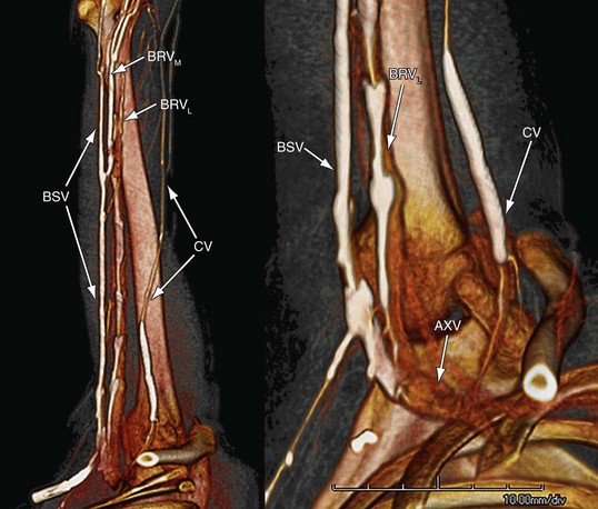
 FIGURE 77-4
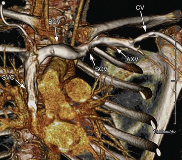
 FIGURE 77-5
Deep Veins of the Upper Extremity
In the hand, the superficial and deep palmar venous arches form the most peripheral major venous conduits of the upper extremity deep system. Usually duplicated with interconnections between each individual pair of arches, the palmar arches parallel the arterial arches and receive drainage corresponding to the respective arterial arch branches. These include common palmar digital veins for the superficial palmar venous arch and palmar metacarpal veins for the deep venous arch. The palmar metacarpal veins also communicate with the dorsal metacarpal veins via perforating veins, forming a means for the deep palmar venous arch to drain the dorsal metacarpal veins as well. The superficial palmar arch courses to the ulnar side of the wrist and drains into ulnar veins, whereas the deep palmar venous arch courses to the radial side of the wrist and drains into radial veins. Branches from the deep palmar arches and the proximal ulnar and radial veins form a carpal venous plexus, which drains into the volar and dorsal interosseous veins.4
In the forearm, the radial, ulnar, and interosseous paired deep veins ascend on either side of the respective companion arteries (Fig. 77-6). The ulnar veins are typically larger in caliber because there is more dominant drainage through the superficial palmar venous arch. Proximal to the elbow, the interosseous veins drain into the ulnar veins, which then give off tributary veins to the median cubital vein. At the elbow, the radial and ulnar veins coalesce to form the paired deep brachial veins (see Fig. 77-6).
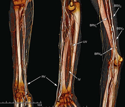
 FIGURE 77-6
In the upper arm, the paired brachial veins ascend on either side of the brachial artery, receiving small venous branch tributaries. At the level of the teres major, the brachial veins coalesce with the basilic vein to form the axillary vein. The medial brachial vein may join the basilic vein prior to this junction. The axillary vein courses to the thoracic inlet as a single vein medial and caudad to the axillary artery. It crosses the first rib into the costoclavicular space, transitioning to the subclavian vein. In addition to the cephalic vein, the axillary vein receives venous tributaries corresponding to the axillary branch arteries (see Figs. 77-4 to 77-6).
Centrally in the thorax, deep venous drainage of the upper extremities occurs by way of the subclavian and brachiocephalic veins (see Fig. 77-6). The subclavian vein extends from the first rib to the level of the clavicular head, where it joins with the internal jugular vein to form the brachiocephalic vein. The subclavian vein lies anterior and caudad to the subclavian artery and courses posterior to the scalenus anterior. Important tributaries include the external jugular, anterior jugular, and dorsal scapular veins. The right and left thoracic ducts drain into the ipsilateral subclavian veins at the respective confluences with their internal jugular veins.7,10
The right and left brachiocephalic veins join together to form the superior vena cava, which then drains into the right atrium (see Fig. 77-6). The right brachiocephalic vein has a short vertical course and lies anterior and to the right of the brachiocephalic artery. The left brachiocephalic vein has a longer, horizontal oblique course, and lies anterior to the aortic arch branch arteries. Common brachiocephalic vein tributaries include the vertebral, internal mammary, inferior thyroid, and supreme intercostal veins.7,10
LOWER EXTREMITY
The lower extremity superficial and deep venous systems in the foot, leg, and thigh direct blood flow to the ipsilateral pelvic veins and ultimately to the inferior vena cava (Figs. 77-7 and 77-8). Similar to the upper extremity, both systems have an integrated relationship, linked by major and minor perforating veins as well as by tributary veins and networks at standard and variable levels.1,5,11,12 Drainage ultimately transitions to the deep system at the groin. In distinction to the upper extremity, the deep system plays a more dominant role. In part, this is because of the greater dependence on deep intermuscular veins functioning as pumps to expel venous blood against gravity into the central venous system with each muscle contraction. The deep plantar veins of the foot and paired deep veins of the calf serve as the principle physiologic pumps to generate this flow (Figs. 77-9 and 77-10).5,6 Valves are more numerous in the lower extremity and, overall, have a greater prevalence in the deep veins and in the more peripheral lower extremity venous segments. The lower extremity superficial system draining the subcutaneous tissues is distinguished from upper extremity counterparts in that the two major superficial veins, the great and small saphenous veins, are enveloped with their own saphenous fascia layer, forming the saphenous compartment. All other superficial tributary veins course between the superficial and muscular fascia layers.11,12
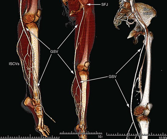
 FIGURE 77-7
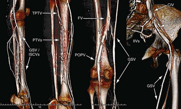
 FIGURE 77-8
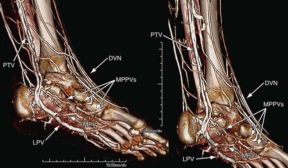
 FIGURE 77-9
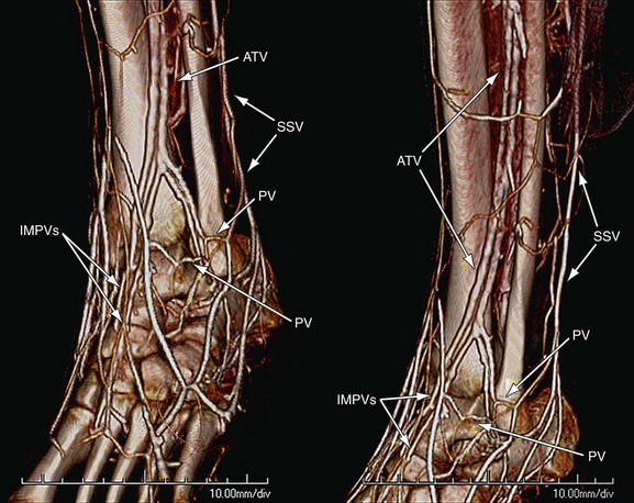
 FIGURE 77-10
Superficial Veins of the Lower Extremity
In the foot, the superficial veins can be divided into two main subsystems, those for the dorsum of the foot and those for the plantar surface. Of the two, the dorsal superficial veins function in a more dominant capacity, giving origin to the great and small saphenous veins. Beginning peripherally at the toes and extending centrally to the ankle, the superficial dorsal veins of the foot include the dorsal digital veins, dorsal metatarsal veins, dorsal venous arch, medial marginal vein, and lateral marginal vein, and dorsal venous network. All lie within the dorsal subcutaneous superficial fascia. The dorsal digital veins course along the lateral dorsal margins of the toes and converge in the webs between the toes to form the dorsal metacarpal veins. The dorsal metacarpal veins, in turn, feed into the dorsal venous arch (Fig. 77-11). The dorsal venous arch extends medially as the medial marginal vein and laterally as the lateral marginal vein. The medial marginal vein courses to the medial ankle, continuing as the great saphenous vein (Figs. 77-11 and 77-12). The lateral marginal vein courses along the lateral border of the foot to the ankle, where it continues as the small saphenous vein (Fig. 77-13). Central to the dorsal venous arch, the dorsal venous network receives small tributaries from the dorsal arch, dorsal superficial veins along the mid and basilar metatarsals and tarsals and, finally, plantar superficial veins (see Figs. 77-11 and 77-13). This network is continuous with the superficial dorsal veins of the lower leg and feeds tributaries into the medial marginal vein and the great saphenous vein.11,12
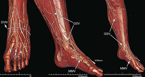
 FIGURE 77-11
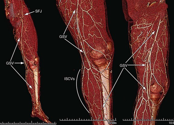
 FIGURE 77-12
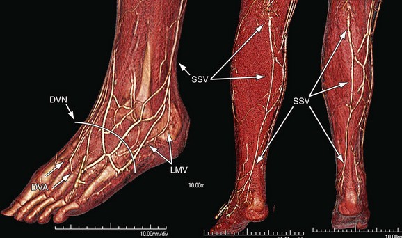
 FIGURE 77-13
The plantar superficial veins of the foot form an intricate subcutaneous network of fine veins extending from the forefoot to the hind foot. They drain into the deep plantar veins via perforating vertical veins along the intermuscular septa and into the dorsal superficial veins via medial, lateral, and interdigitary communicating veins.13
In the leg and thigh, the great and small saphenous veins are the principle, constant superficial veins. The great saphenous vein is present in both the leg and thigh, whereas the small saphenous vein most commonly only drains the calf. Communication between the great and small saphenous veins occurs in the leg via one or more intersaphenous oblique coursing veins.11,12
The great saphenous vein, continuing from its medial marginal vein origin, crosses anterior to the medial malleolus and ascends along the anteromedial aspect of the leg with the saphenous nerve (see Figs. 77-7, 77-11, and 77-12). At the knee, the great saphenous vein passes posteromedial to the medial tibial and femoral condyles and enters the thigh. In the thigh, the great saphenous vein courses anteromedially to just below the inguinal ligament, where it crosses the deep fascia at the saphenous vein opening and terminates in the anterior aspect of the femoral vein (saphenofemoral junction). Anterior and posterior accessory veins of the great saphenous vein run parallel to and are located anterior and posterior to the great saphenous vein, respectively. Both have tributary greater saphenous venous drainage in the leg and, ultimately, at the intervalvular great saphenous venous segment, just peripheral to the saphenofemoral junction. In addition to these accessory veins, a superficial accessory great saphenous vein may parallel the great saphenous vein more superficially in the leg and thigh. Tributary drainage at the intervalvular segment also includes the anterior and posterior circumflex veins of the thigh, superficial inferior epigastric veins of the lower abdominal-pelvic wall, superficial circumflex iliac veins of the lateral iliac region, and superficial external pudendal vein of the pubic and perineal regions (Fig. 77-14). The anterior and posterior circumflex veins may arise from the remnant lateral venous system, but the posterior circumflex vein may also arise from the small saphenous vein. Rather than running off into the great saphenous vein, the anterior and posterior circumflex veins may drain into the anterior and posterior accessory veins of the great saphenous vein, respectively.11,12
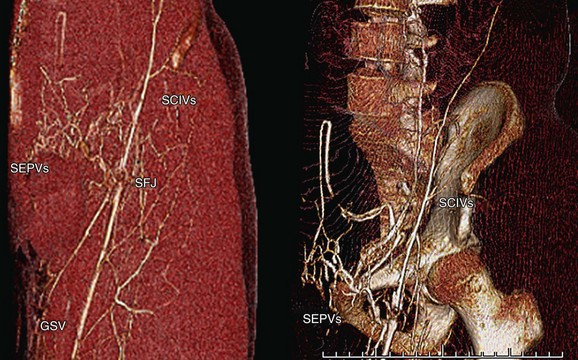
 FIGURE 77-14
The small saphenous vein, continuing from its lateral marginal vein origin, crosses posterior to the lateral malleolus and ascends with the sural nerve, in the midline of the calf (see Fig. 77-13). Most commonly, the small saphenous vein enters the popliteal fossa, crossing the deep fascia between the heads of the gastrocnemius muscles to terminate in the popliteal vein (saphenopopliteal junction) within 5 cm above the knee joint. Less commonly, the small saphenous veins may drain into the popliteal vein more than 5 cm above the knee joint, may pierce the deep fascia below the fossa, or may terminate in veins other than the popliteal vein, including the great saphenous vein (via the posterior circumflex vein), superficial communicating veins, and deep thigh muscular perforators. Rather than terminating, the small saphenous vein may have axial (deep) extension or postaxial (dorsal) extension into the thigh. A superficial accessory small saphenous vein may be present, ascending parallel to the small saphenous vein, superficial to the saphenous compartment.11,12
Deep Veins of the Lower Extremity
In the foot, the deep veins include the plantar digital veins, plantar metatarsal veins, deep plantar arch, medial plantar veins, and lateral plantar veins. Plantar digital veins are located along the plantar surfaces of the digits, receiving drainage from the plantar plexuses of each digit. They converge into four deep plantar metatarsal veins in the intermetatarsal grooves, where there is also communication to the dorsal digital veins. The deep plantar metatarsal veins drain into the plantar venous arch, paralleling the plantar arterial arch. Outflow from the plantar venous arch occurs by way of the medial and lateral deep plantar veins, which course along the medial and lateral inner plantar surfaces, respectively (see Fig. 77-9). The medial and lateral deep plantar veins function as the primary reservoir for the plantar venous pump, converging posterior to the medial malleolus to form the paired posterior tibial veins.5,6,11
The deep veins of the leg consist of three pairs of intermuscular veins and numerous intramuscular veins. The six intermuscular veins include the posterior tibial veins, peroneal veins, and anterior tibial veins (see Figs. 77-8 to 77-10). The posterior tibial and peroneal veins are located in the deep posterior compartment of the calf and the anterior tibial veins are located in the anterior compartment. From their plantar vein origins medial to the ankle, the posterior tibial veins course cephalad in the medial aspect of the deep posterior compartment, alongside the posterior tibial artery. The peroneal veins form at and drain the lateral aspect of the ankle and course in the central aspect of the deep posterior compartment, adjacent to the fibula, paired with the peroneal artery. Accessory deep venous drainage from the plantar veins may occur into the peroneal vein. The peroneal veins unite with the posterior tibial veins to form a common tibioperoneal vein. The anterior tibial veins are formed from the dorsal first intermetatarsal perforating veins, which travel with the dorsal pedis artery. Extending cephalad from the dorsal ankle level, the anterior tibial veins course in the anterior compartment lateral to the tibia along the interosseous membrane, with the corresponding anterior tibial artery. These veins drain the dorsum of the foot and anterior compartment muscles. At the inferior margin of the popliteal fossa, they cross the interosseous membrane and unite with the tibioperoneal trunk to form the popliteal vein. The popliteal vein courses cephalad across the knee joint into the thigh, traversing the adductor hiatus to become the femoral vein. In addition to draining the tibial and peroneal veins, the popliteal veins receive the sural veins and, as discussed, the short saphenous vein.5,11
Most deep intramuscular veins in the leg are minor tributaries into the paired deep intermuscular veins. The exception to this is the soleus and gastrocnemius deep veins. Both sural veins form from sinusoidal venous networks in the respective muscles of the superficial posterior compartment of the calf. The soleus veins may drain into the popliteal vein or adjacent muscular veins, whereas the gastrocnemius veins (medial gastrocnemius vein, lateral gastrocnemius, and intergemellar vein) may drain into the popliteal vein or short saphenous vein.5,11
In the thigh, the femoral and deep femoral veins comprise the major components of the deep system (see Fig. 77-8). Beginning as the continuation of the popliteal vein at the adductor hiatus, the femoral vein extends cephalad through the adductor canal, coursing alongside the femoral artery, initially posterolaterally, becoming medial to the femoral artery as it enters the femoral triangle in the groin. It receives small muscular venous tributaries primarily from the anterior and medial compartments. The deep femoral vein parallels the corresponding femoral profunda artery, coursing posterior and lateral to the femoral vein, between the anterior and medial compartments of the thigh, draining primarily posterior and lateral muscle groups. The medial and lateral circumflex veins typically drain into the femoral vein but, not uncommonly, may also drain into the deep femoral vein. In the proximal thigh, several centimeters below the inguinal ligament, the deep femoral vein joins with the femoral vein posteriorly to form the common femoral vein. After receiving the greater saphenous vein anteriorly, the common femoral vein crosses the inguinal ligament to become the external iliac vein.5,11
In the pelvis, the external iliac vein courses cephalad as a single vein, joining with the ipsilateral internal iliac vein to form the common iliac vein (see Fig. 77-8). Bilateral common iliac veins unite to form the inferior vena cava, which then traverses the abdomen and directs blood flow back to the right atrium. The right common iliac vein has a more direct path to the inferior vena cava, whereas the left crosses midline to reach the right-sided inferior vena cava. The right common iliac artery may cross the left common iliac vein, potentially resulting in anterior compression.5,11
Perforator Veins of Lower Extremity
The perforating veins in the lower extremity course obliquely across the deep fascia and communicate with unidirectional valves between the superficial and deep veins. Although these veins may have variable morphology and number, there are groups of perforating veins that maintain a consistent location. In the foot, these standard perforating veins include the dorsal foot intercapitular perforator veins and medial, lateral, and plantar foot veins, whereas in the ankle, they include the medial, lateral, and anterior perforating veins. In the leg, there are four main groups of constant perforators, corresponding to the anterior (anterior perforators), lateral (lateral perforators), deep posterior (paratibial and posterior tibial medial perforators), and superficial posterior (medial gastrocnemius, lateral gastrocnemius perforators, and intergemellar perforators) muscle compartments. The standard perforators at the knee include medial, lateral, infrapatellar, suprapatellar, and popliteal fossa perforating veins. In the thigh, the perforators are defined as medial, anterior, lateral, and posterior perforating veins. The medial perforators include the femoral canal and inguinal perforating veins. The anterior and lateral perforators are grouped according to the respective muscle compartments. The posterior perforators are divided into posteromedial thigh perforators (adductor muscles), sciatic perforators (posterior midline thigh), and posterolateral perforators (posterior muscle compartment). Perforating veins also occur in the gluteal muscles and are divided into superior, mid, and lower perforators.5,11,12


Hellinger JC, Epelman M, Rubin GD. Upper extremity computed tomographic angiography: state of the art techniques and applications in 2010. Radiol Clin North Am. 2010;48:397-421.
Uhl JF, Gillot C. Embryology and three-dimensional anatomy of the superficial venous system of the lower limbs. Phlebology. 2007;22:194-206.
1 Thomson H. The surgical anatomy of the superficial and perforating veins of the lower limb. Ann R Coll Surg Engl. 1979;61:198-205.
2 Nystrom A, Friden J, Lister GD. Superficial venous anatomy of the human palm. Scand J Plast Reconstr Surg Hand Surg. 1990;24:121-127.
3 Nystrom A, von Drasek-Asher G, Lister GD. The palmer digital venous anatomy. Scand J Plast Reconstr Surg Hand Surg. 1990;24:113-119.
4 Nystrom A, Friden J, Lister GD. Deep venous anatomy of the human palm. Scand J Plast Reconstr Surg Hand Surg. 1991;25:233-239.
5 Tretbar LL. Deep veins. Dermatol Surg. 1995;21:47-51.
6 White JV, Katz ML, Cisek P, Kreithen BA. Venous outflow of the leg: anatomy and physiologic mechanism of the plantar venous plexus. J Vasc Surg. 1996;24:19-824.
7 Godwin JD, Chen JTT. Thoracic venous anatomy. Am J Roentgenol. 1986;147:674-684.
8 Hallock GG. The cephalic vein in microsurgery. Microsurgery. 1993;14:482-486.
9 Moss SH, Schwartz KS, von Drasek-Ascher G, et al. Digital venous anatomy. J Hand Surg Am. 1985;10:473-482.
10 Chasen MH, Charnsangavej C. Venous chest anatomy: clinical implications. Eur J Radiol. 1998;27:2-14.
11 Caggiati A, Bergan JJ, Gloviczki P, et al. Nomenclature of the veins of the lower limbs: an international interdisciplinary consensus statement. J Vasc Surg. 2002;36:416-422.
12 Uhl JF, Gillot C. Embryology and three-dimensional anatomy of the superficial venous system of the lower limbs. Phlebology. 2007;22:194-206.
13 Imanishi N, Kish K, Chang H, et al. Anatomical study of cutaneous venous flow of the sole. Plast Reconstr Surg. 2007;120:1906-1910.
[/level-membership-for-cardiovascular-category][not-level-membership-for-cardiovascular-category]
CHAPTER 77 Venous Anatomy of the Extremities
The upper and lower extremity peripheral venous systems comprise three main types of veins—superficial, deep, and perforating. The superficial and deep subsystems are defined by major and tributary veins linked by the perforating veins as well as venous networks (plexi), with the goal of directing all venous blood return into the deep system in peripheral, mid, or central extremity venous segments. Wide variability is often encountered in this anatomy, with origins for both systems beginning peripherally at the hand or foot for the upper and lower extremities, respectively. The superficial veins are located subcutaneously within the superficial fascia. The deep veins are located intramuscularly, deep to the superficial fascia, accompanying the extremity arteries (venae comitans). Perforating veins cross the muscular fascial layer, typically bridging flow from superficial to deep veins. Valves function in superficial, deep, and perforating veins to keep blood flowing antegrade and prevent blood flowing from the deep to superficial veins. Valves are more prevalent in the deep veins for both the upper and lower extremities.1–5
In general, the extremity veins are thin wall structures with little to no elastic lamina, resulting in passive distensible conduits with large compliance. The deep veins, however, inherit strength directly by having a greater degree of elastic lamina and indirectly by way of muscular contractions, countering gravity and central venous filling pressures to propagate blood flow toward the central veins and heart.5–6 The well-orchestrated anatomic physiology between the deep venous pumps, valves, and perforating veins becomes particularly apparent in the setting of elevated central venous pressures, incompetent valves, immobility, obstructed central or deep extremity veins, or a combination of these conditions.
UPPER EXTREMITY
Veins for the upper extremity direct blood flow from the hand, wrist, forearm, upper arm, and shoulder to the ipsilateral central thorax veins and ultimately the superior vena cava. In the hand, forearm, and upper arm, the superficial system functions as the principal means for venous drainage. As a result, the caliber of the superficial veins is generally larger than the deep veins. The major superficial veins of the hand, forearm, and upper arm exist as single structures and infrequently have accessory veins. The smaller deep veins occur in pairs, alongside the respective arteries. All upper extremity venous drainage transitions to the deep system at the axilla, with a single main venous channel crossing the costoclavicular space into the thoracic central venous system. Within the axillae and thorax, the major deep veins occur as single structures, accompanying the adjacent artery.7–10
Superficial Veins of the Upper Extremity
The dorsal veins include the dorsal digital veins, dorsal metacarpal veins, and dorsal venous network (plexus). Dorsal digital veins form by coalescence of dorsal distal digital venules and venous arches, and extend bilaterally along the length of dorsum of the fingers. They communicate at multiple levels via small oblique venous connections. The dorsal digital veins from the ulnar aspect of the index finger, radial aspect of the fifth finger, and both sides of the third and fourth fingers, along with intercapitular veins between the digits, drain into three superficial dorsal metacarpal veins. The metacarpal veins, in turn, feed into the dorsal venous network, which is located at the mid and base of the metacarpals (Fig. 77-1).9
Along the radial aspect of the hand, the dorsal venous network receives dorsal veins of the thumb and dorsal vein of the radial aspect of the index finger and continues with cephalad drainage, forming the cephalic vein. Along the ulnar aspect of the hand, the dorsal venous plexus receives the dorsal vein from the ulnar aspect of the fifth finger and continues with cephalad drainage, forming the basilic vein. The median portion of the network variably may drain to the mid-forearm cephalic vein via a communicating (accessory) vein (Fig. 77-2).8
The palmar veins of the hand include palmar digital veins and palmar venous plexus. Palmar digital veins parallel the dorsal digital veins, along the palmar surface. There are three routes of drainage for the palmar digital veins. Two are within the superficial system and include the palmar venous network and dorsal digital and metacarpal veins. The palmar venous network lies at the thenar and hypothenar eminences and drains primarily into the median antecubital vein of the forearm. Drainage into the dorsal digital veins and dorsal metacarpal veins occurs by way of intercapitular veins in the digital web spaces at the metacarpal heads. The third route is into the common and metacarpal palmar veins of the palmar deep system.2,3
In the forearm, main superficial venous drainage occurs via the cephalic, basilic, and median antecubital veins (Fig. 77-3). The cephalic and basilic veins ascend to the elbow along the radial and ulnar aspects of the forearm, respectively, with continuation into the upper arm. The cephalic vein, basilic vein, or both, may form the dominant drainage route into the upper arm.
The cephalic vein transitions from the dorsal to volar surfaces in the distal third of the forearm. Along its course in the forearm, the cephalic vein receives dorsal and volar tributaries. Just proximal to the elbow, the cephalic vein gives off the median basilic vein, which ascends obliquely over the volar surface, crosses the antecubital fossa, and drains into the basilic vein. The cephalic vein then crosses the ventrolateral aspect of the elbow, in a groove between the brachioradialis and biceps brachii muscles. An accessory cephalic vein is often present, with variable morphology. Most commonly, it arises from the dorsal venous plexus or from a dorsal forearm tributary network and courses along the radial aspect of the forearm, lateral to the cephalic vein. It drains into the cephalic vein below the elbow. Alternatively, the accessory cephalic vein may form a venous bridge between cephalic vein segments below and above the elbow. The accessory cephalic vein often supplies a dorsal oblique branch, forming a second level of communication between the cephalic and basilic veins within the forearm.8
In the upper arm, the basilic and cephalic veins are the major routes for superficial venous drainage, with ultimate runoff into the deep system (Figs. 77-4 and 77-5). The basilic vein is typically larger than the cephalic vein, coursing medial to the biceps brachii. The smaller cephalic vein courses lateral to the biceps brachii. The basilic vein ascends initially within the superficial fascia, but then perforates the deep fascia at the midarm level. Subsequent to this, the basilic vein courses along the medial aspect of the brachial artery until the inferior margin of the teres major muscle, at which point it joins the brachial vein (deep system) to form the axillary vein (deep system). The cephalic vein ascends completely within biceps superficial fascia, entering the infraclavicular fossa between the pectoralis major and deltoid muscles.8 It then courses medial, in the clavipectoral triangle, entering the cranial aspect of the central axillary vein with acute angulation, below the clavicle. The cephalic veins typically have a valve just proximal to its junction with the axillary vein.
Deep Veins of the Upper Extremity
In the hand, the superficial and deep palmar venous arches form the most peripheral major venous conduits of the upper extremity deep system. Usually duplicated with interconnections between each individual pair of arches, the palmar arches parallel the arterial arches and receive drainage corresponding to the respective arterial arch branches. These include common palmar digital veins for the superficial palmar venous arch and palmar metacarpal veins for the deep venous arch. The palmar metacarpal veins also communicate with the dorsal metacarpal veins via perforating veins, forming a means for the deep palmar venous arch to drain the dorsal metacarpal veins as well. The superficial palmar arch courses to the ulnar side of the wrist and drains into ulnar veins, whereas the deep palmar venous arch courses to the radial side of the wrist and drains into radial veins. Branches from the deep palmar arches and the proximal ulnar and radial veins form a carpal venous plexus, which drains into the volar and dorsal interosseous veins.4
[/not-level-membership-for-cardiovascular-category]
