Venous Anatomy and Variants

Published on 13/02/2015 by admin

Filed under Cardiothoracic Surgery

Last modified 22/04/2025

Print this page
rate 1 star rate 2 star rate 3 star rate 4 star rate 5 star
Your rating: none, Average: 2.6 (27 votes)

This article have been viewed 3193 times

Chapter 6

Venous Anatomy and Variants

Venous Axial 2

image

Normal Variant

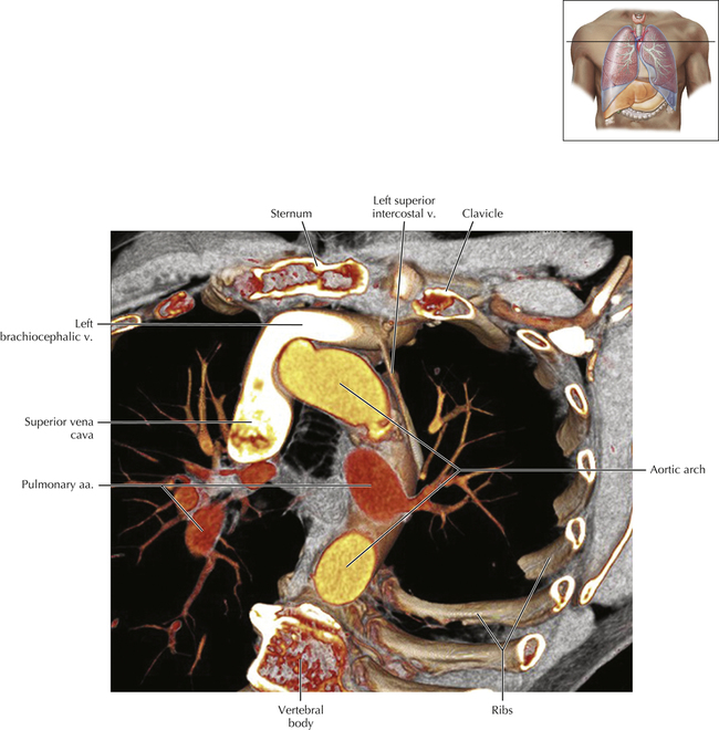
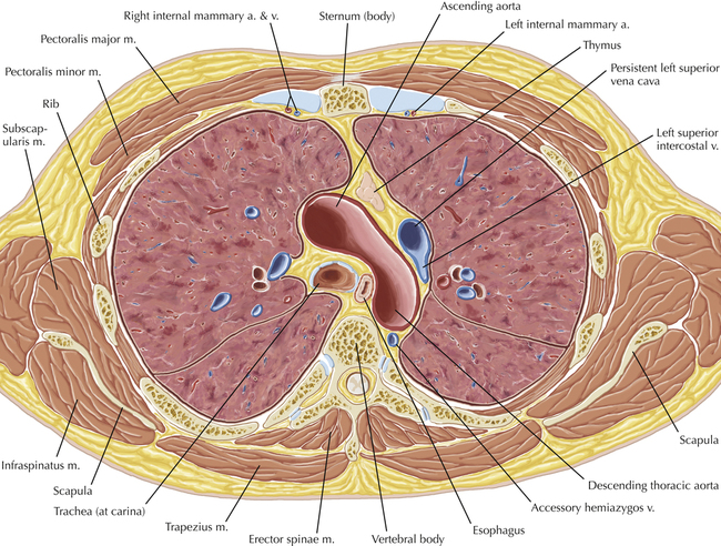
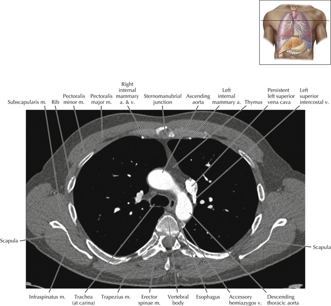
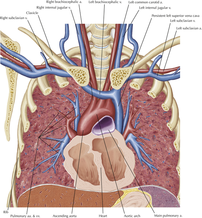
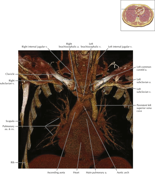
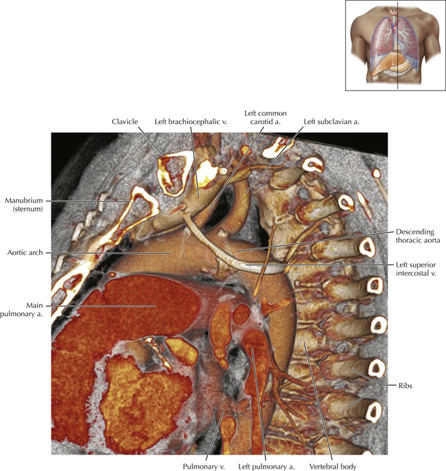
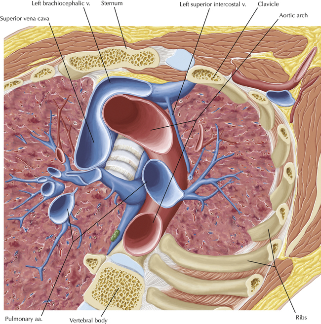
This scan, performed at the caudal surface of the aortic arch, shows a common venous anomaly affecting the thorax—the persistent left superior vena cava. The prevalence of this anomaly is estimated at nearly 0.3% of the general population, and it may occasionally be associated with other congenital cardiovascular anomalies. In its most common form, the persistent left superior vena cava forms at the confluence of the left internal jugular and subclavian veins and descends along the left aspect of the aortic arch and mediastinum, passing anterior to the left hilum, crossing the posterior wall of the left atrium, eventually emptying into the coronary sinus. Rarely, the persistent left superior vena cava empties into the left atrium, creating a right-to-left shunt. The enhanced persistent left superior vena cava is shown as it passes lateral to the aortic arch.

image

Venous Sagittal 1

image

Pathologic Process

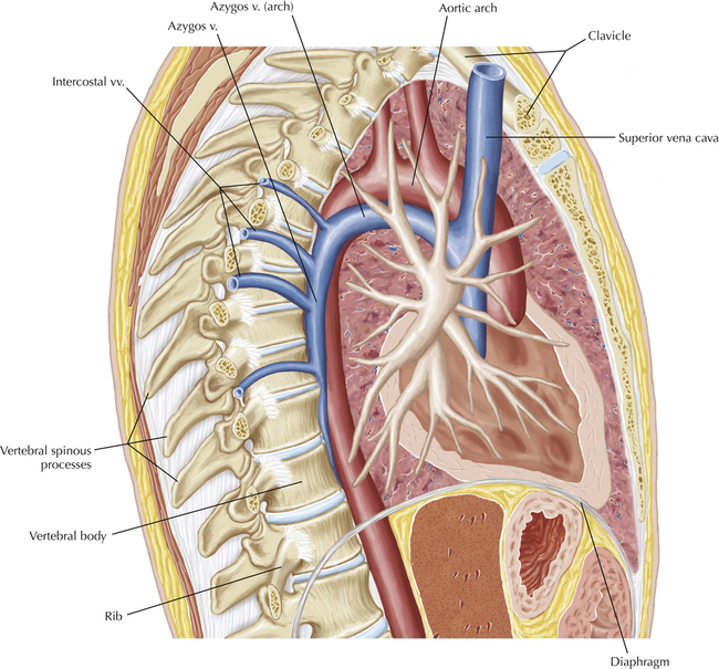
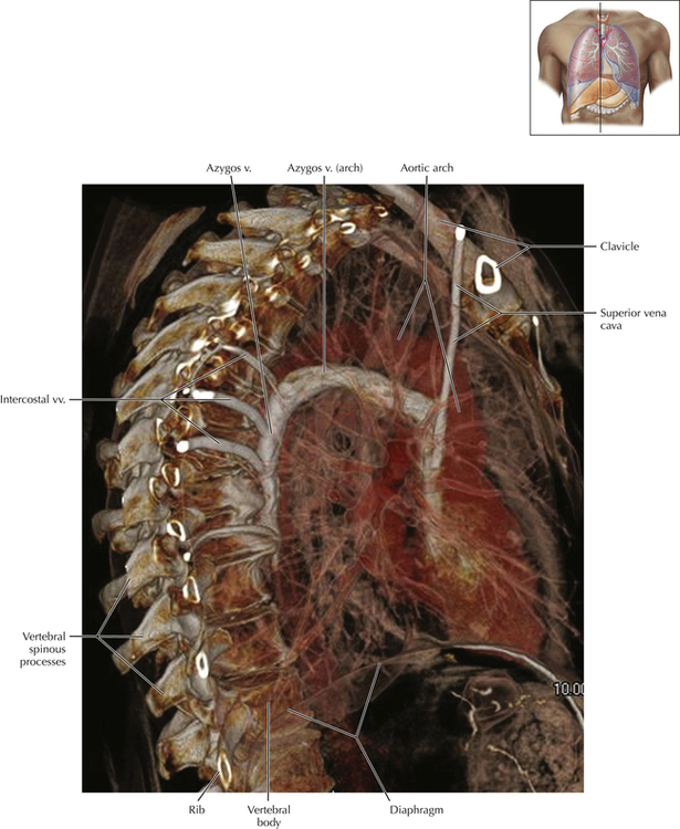
An oblique, sagittal volume rendered image performed in a patient with a right subclavian venous stenosis. This condition causes blood from the right upper extremity and chest wall to route through various chest wall and thoracic veins as the body attempts to bypass the obstructed subclavian vein. When the right upper extremity venous system is injected with intravenous contrast for thoracic CT, this situation creates a unique opportunity to demonstrate the intercostal and azygos venous systems of the thorax. The arch of the azygos vein is well visualized. The azygos vein normally courses within the posterior mediastinum on the right, adjacent to the lower eight thoracic vertebral bodies, eventually extending anteriorly, superior to the root of the right lung, to empty into the superior vena cava. The azygos vein typically receives blood flow from intercostal veins, mediastinal veins, bronchial veins, esophageal veins, and the paravertebral venous plexus. When thoracic veins become obstructed, the azygos system assumes the role of a major collateral pathway for blood flow from the upper extremities and thorax to reach the heart.

image