[level-membership-for-anesthesiology-category]
21 Veins
Veins are easily compressed with the ultrasound transducer and have thin walls that are difficult to visualize. The vein shape is highly dependent on the amount of probe compression. Often, valves can be imaged inside the vein lumen. In contrast to the pulsatile flow of arteries, veins exhibit more continuous flow. Some degree of ACV waves (from Atrial contraction, Contraction of the ventricle, and Venous filling of the atrium) and respiratory variation in flow may be present depending on how close the vein is to the thoracic cavity. Veins may have spontaneous contrast due to rouleaux formation of red blood cells in low-flow states such as congestive heart failure.1 At the usual amount of probe compression for nerve imaging, the veins are not visible in the field because the vein walls are coapted.
[/level-membership-for-anesthesiology-category][not-level-membership-for-anesthesiology-category]
21 Veins
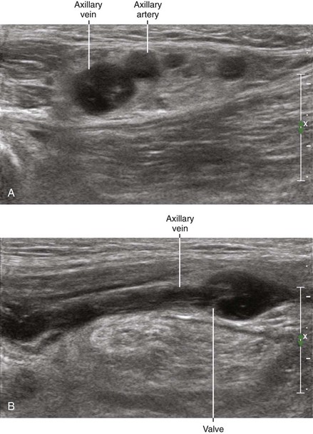
Veins are easily compressed with the ultrasound transducer and have thin walls that are difficult to visualize. The vein shape is highly dependent on the amount of probe compression. Often, valves can be imaged inside the vein lumen. In contrast to the pulsatile flow of arteries, veins exhibit more continuous flow. Some degree of ACV waves (from Atrial contraction, Contraction of the ventricle, and Venous filling of the atrium) and respiratory variation in flow may be present depending on how close the vein is to the thoracic cavity. Veins may have spontaneous contrast due to rouleaux formation of red blood cells in low-flow states such as congestive heart failure.1 At the usual amount of probe compression for nerve imaging, the veins are not visible in the field because the vein walls are coapted.
[/not-level-membership-for-anesthesiology-category]
