Ovarian Vein Thrombosis

Published on 10/03/2015 by admin

Filed under Obstetrics & Gynecology

Last modified 22/04/2025

Print this page

rate 1 star rate 2 star rate 3 star rate 4 star rate 5 star
Your rating: none, Average: 0 (0 votes)

This article have been viewed 1317 times

Ovarian Vein Thrombosis

Synonyms/Description

Septic pelvic thrombophlebitis (SPT)

Etiology

Ovarian vein thrombosis is a rare but potentially severe postpartum complication, occurring 80% to 90% of the time on the right side, and with a reported incidence of 1:600 to 1:2000 deliveries. Ovarian vein thrombosis can also occur in the immediate postoperative period after pelvic surgery or in conjunction with pelvic infection or thrombophilias such as factor V Leiden mutation. Spontaneous ovarian vein thrombosis without any of these predisposing conditions is exceedingly rare.
The ovarian veins are located in the retroperitoneum, anterior to the psoas muscle. The right ovarian vein is longer than the left, and it drains into the inferior vena cava below the right renal vein. The left drains into the left renal vein.

Ultrasound Findings

Ovarian vein thrombosis appears as a hypoechoic tubular mass cephalad to the ovary. Color Doppler typically shows an absence of flow within the mass.
The sensitivity, specificity, and accuracy of ultrasound with Doppler are reportedly 55.6%, 41.2%, and 46.2%, respectively. The sonographic evaluation is often limited by overlying bowel gas because the ovarian vein is a retroperitoneal structure; therefore CT with contrast is often used to make a more definitive diagnosis.

Differential Diagnosis

A tubular painful mass in the right lower quadrant can mimic appendicitis. The ultrasound findings may be similar, although the appendix is typically located more anteriorly in the abdomen than is the ovarian vein, which is more dorsal. Doppler is also helpful in differentiating these two diagnoses, as abundant color flow is seen in the walls of the appendix and ovarian vein thrombosis has little flow. Other possible but less likely diagnoses when dealing with a painful lower quadrant mass, especially if left-sided, include ovarian torsion, broad ligament fibroid, hydrosalpinx, and pelvic inflammatory disease. The presence of a separate ovary, location of the mass, and color Doppler pattern are very helpful to arrive at the correct diagnosis.

Clinical Aspects and Recommendations

Ovarian vein thrombosis should be considered in postpartum patients who present with abdominal or flank pain (right more frequently) and with fever, typically spiking, that is unresponsive to broad-spectrum antibiotics. These symptoms often mimic appendicitis, tubo-ovarian abscess, ovarian torsion, or broad ligament hematoma. Treatment is usually medical and may include parenteral broad-spectrum antibiotics with the addition of anticoagulant therapy.
If untreated, ovarian vein thrombosis can lead to inferior vena cava and renal vein thrombosis, pulmonary thromboembolism, and sepsis.

Figures

image

Figure O4-1 A and B, Longitudinal and transverse views of the right ovarian vein showing a hypoechoic mass with internal strandy echoes consistent with internal organizing clot. No blood flow was identified using Doppler color flow.

image

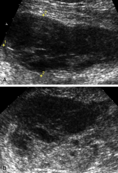
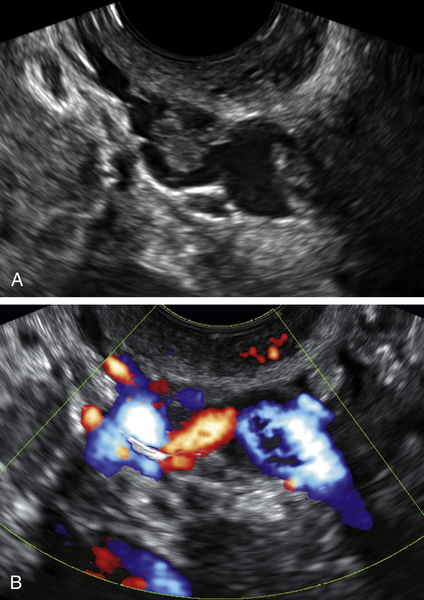
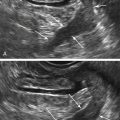
Figure O4-2 A and B, Longitudinal view of the right external iliac containing multiple clots incidentally noted on a gynecologic scan. B shows color Doppler of the vein in the same projection, showing defects within the color consistent with clot. The thrombosis was confirmed with contrast CT, and the patient was anticoagulated.

Suggested Reading

Bilgin M., Sevket O., Yildiz S., Sharifov R., Kocakoc E. Imaging of postpartum ovarian vein thrombosis. Case Rep Obstet Gynecol. 2012;2012:134603.

Dewdney S.B., Benn T., Rimel B.J., Gao F., Saad N., Vedantham S., Mutch D.G., Zighelboim I. Inferior vena cava filter placement in the gynecologic oncology patient: a 15-year institutional experience. Gynecol Oncol. 2011;121:344–346.

Sharma P., Abdi S. Ovarian vein thrombosis. Clin Radiol. 2012;67:893–898.

Stafford M., Fleming T., Khalil A. Idiopathic ovarian vein thrombosis: a rare cause of pelvic pain—case report and review of literature. Aust N Z J Obstet Gynaecol. 2010;50:299–301.