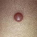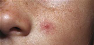
Vascular spider with prominent central feeding vessel on the chin.
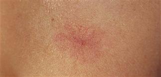
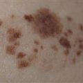
Large vascular spider.
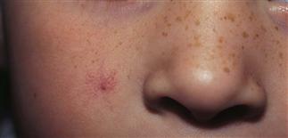
Vascular spider on the face of a child.
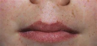
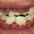
Vascular telangiectasias on the lips of a child with HHT (Osler–Weber–Rendu).
CLINICAL FEATURES
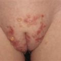
Vascular spiders (spider angioma – spider telangiectasia – nevus araneus) consist of a central arteriole with many dilated radiating telangiectasias and a surrounding flush area, varying from a few millimeters to several centimeters in diameter. Sometimes the central arteriole may be raised and prominent and may be pulsatile on diascopy. Pressure on the central vessel causes blanching. Vascular spiders occur in up to 45% of light-skinned children. They are seen most commonly on sun-exposed areas, usually the cheeks, nose, dorsa of the forearms and hands. Lesions resolve spontaneously at puberty. Children with hereditary hemorrhagic telangiectasias have multiple lesions of the skin and mucosa. A history of severe epistaxis and gastrointestinal bleeding may be obtained. Telangiectasias in collagen vascular disorders are also seen on the face and arms, but are more numerous and mat-like.
TREATMENT
Treatment is not necessary. If treatment is desired for cosmetic purposes a single treatment with the PDL is 70–80% effective.