[level-membership-for-pediatrics-category]
123 Vascular Disorders
Vascular lesions of the skin are a common pediatric problem with a wide range of clinical presentations. In 1996, the International Society for the Study of Vascular Anomalies adopted Mulliken and Glowacki’s classification (Table 123-1). There are two categories based on biologic and clinical characteristics: vascular tumors and vascular malformations. Vascular tumors are neoplasms of vascular structures that grow by hyperplasia. Vascular malformations are local anomalies in vascular development that do not demonstrate proliferation.
Table 123-1 International Society for the Study of Vascular Anomalies Classification of Vascular Anomalies
| Vascular Tumors | Vascular Malformations |
|---|---|
VM, venous malformation.
Adapted from ISSVA classification as reported in Color Atlas of Vascular Tumors & Vascular Malformations.
Vascular Tumors
Infantile Hemangiomas
Clinical Manifestations
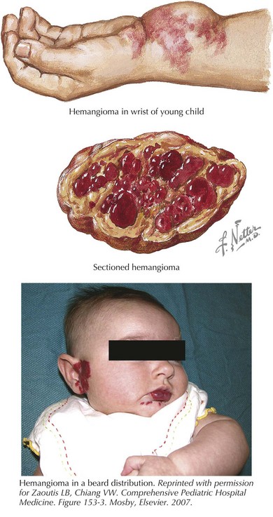
Hemangiomas are classified as superficial, deep, or mixed lesions. Superficial hemangiomas, involving the papillary dermis, are red and protuberant. Superficial hemangiomas have well-defined borders. Deep lesions invade the reticular dermis and superficial fat. They tend to have a blue-purple discoloration with normal skin texture. The margins of deep lesions may be ill defined. Most lesions have both superficial and deep components (Figure 123-1). A mature hemangioma contains characteristics of capillaries, venules, and arterioles.
Management
The most common complication of rapidly proliferating hemangiomas is ulceration. Ulceration is most common in perineal and perioral hemangiomas and can be quite painful. Ulcerated hemangiomas are also at risk for superinfection. Hemangiomas in select locations may have unique complications. Nasal tip hemangiomas cause significant disfigurement. Periocular hemangiomas disrupt visual development, and early referral to an ophthalmologist is prudent. Hemangiomas along the jaw line or neck (beard distribution) of the head and neck (see Figure 123-1) have a high association with airway lesions that may cause life-threatening airway obstruction. Spinal dysraphism is associated with midline lumbosacral lesions, and magnetic resonance imaging (MRI) is indicated. Infants with multiple cutaneous hemangiomas warrant evaluation for visceral hemangiomas.
PHACES Syndrome
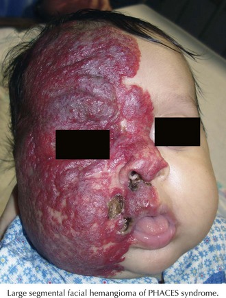
Large, segmental facial hemangiomas may be associated with PHACES (posterior fossa malformations, hemangiomas, arterial anomalies, cardiac anomalies, eye abnormalities, sternal cleft or supraumbilical raphe) syndrome (Figure 123-2). The most common associated feature of PHACES syndrome is posterior fossa malformations, particularly Dandy-Walker type and cerebellar hypoplasia. Arterial anomalies and stenosis affecting arteries of the head and neck are found in one-third of patients. Cardiac anomalies such as coarctation are also common and can be in atypical locations along the course of the aorta. Less common associations include eye anomalies and sternal clefting or supraumbilical raphe. PHACES should be considered in any child presenting with a large, segmental facial hemangioma. Suspected patients should be referred to a neurologist for complete neurologic evaluation, including MRI or magnetic resonance angiography of the brain and neck as well as cardiac evaluation. Ophthalmologic evaluation should also be considered.
Vascular Malformations: Slow-Flow Lesions
Capillary Malformations
Boon LM, Mulliken JB, Enjolras O, Vikkula M. Glomuvenous malformation (glomangioma) and venous malformation: distinct clinicopathologic and genetic entities. Arch Dermatol. 2004;140(8):971-976.
Bruckner AL, Frieden IJ. Infantile hemangiomas. J Am Acad Dermatol. 2006;55(4):671-682.
Drolet BA, Swanson EA, Frieden IJ, et al. Infantile hemangiomas: an emerging health issue linked to an increased rate of low birth weight infants. J Pediatr. 2008;153(5):712-715. 5.e1
Enjolras O, Wassef M, Chapot R. Introduction: ISSVA Classification. In: Color atlas of vascular tumors and vascular malformations. Cambridge, UK: Cambridge University Press; 2007:3-11.
Garzon MC, Huang JT, Enjolras O, et al. Vascular malformations. Part II: associated syndromes. J Am Acad Dermatol. 2007;56(4):541-564.
Guggisberg D, Hadj-Rabia S, Viney C, et al. Skin markers of occult spinal dysraphism in children: a review of 54 cases. Arch Dermatol. 2004;140(9):1109-1115.
Maguiness S, Guenther L. Kasabach-Merritt syndrome. J Cutan Med Surg. 2002;6(4):335-339.
Sans V, Dumas de la Roque E, Berge J, et al. Propranolol for severe infantile hemangiomas: follow-up report. Pediatrics. 2009;124:e423-e431.
Stier M, Glick S, Hirsch R. Laser treatment of pediatric vascular lesions: port wine stains and hemangiomas. J Am Acad Dermatol. 2008;58(2):261-285.
[/level-membership-for-pediatrics-category][not-level-membership-for-pediatrics-category]
123 Vascular Disorders
Vascular lesions of the skin are a common pediatric problem with a wide range of clinical presentations. In 1996, the International Society for the Study of Vascular Anomalies adopted Mulliken and Glowacki’s classification (Table 123-1). There are two categories based on biologic and clinical characteristics: vascular tumors and vascular malformations. Vascular tumors are neoplasms of vascular structures that grow by hyperplasia. Vascular malformations are local anomalies in vascular development that do not demonstrate proliferation.
Table 123-1 International Society for the Study of Vascular Anomalies Classification of Vascular Anomalies
| Vascular Tumors | Vascular Malformations |
|---|---|
VM, venous malformation.
Adapted from ISSVA classification as reported in Color Atlas of Vascular Tumors & Vascular Malformations.
Vascular Tumors
Infantile Hemangiomas
Clinical Manifestations
Hemangiomas are classified as superficial, deep, or mixed lesions. Superficial hemangiomas, involving the papillary dermis, are red and protuberant. Superficial hemangiomas have well-defined borders. Deep lesions invade the reticular dermis and superficial fat. They tend to have a blue-purple discoloration with normal skin texture. The margins of deep lesions may be ill defined. Most lesions have both superficial and deep components (Figure 123-1). A mature hemangioma contains characteristics of capillaries, venules, and arterioles.
Management
The most common complication of rapidly proliferating hemangiomas is ulceration. Ulceration is most common in perineal and perioral hemangiomas and can be quite painful. Ulcerated hemangiomas are also at risk for superinfection. Hemangiomas in select locations may have unique complications. Nasal tip hemangiomas cause significant disfigurement. Periocular hemangiomas disrupt visual development, and early referral to an ophthalmologist is prudent. Hemangiomas along the jaw line or neck (beard distribution) of the head and neck (see Figure 123-1
[/not-level-membership-for-pediatrics-category]
