Chapter 42 Vascular and lymphatic neoplasms
1. Which is the most common benign vascular neoplasm of childhood?
Hemangioma of infancy (HOI), which is a benign tumor of capillary endothelium. This tumor typically presents at ages 2 to 8 weeks and then goes through a rapid growth phase for the first 6 to 9 months of life. It then begins to regress, with complete regression in 50% of patients by age 5 years. Regression does not necessarily imply return of the skin to normal.
6. What is Kasabach-Merritt syndrome?
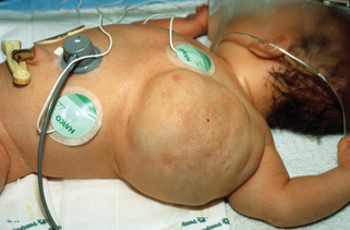
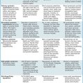
10. What is Klippel-Trénaunay syndrome?
This is overgrowth of an extremity secondary to a vascular malformation. The overgrowth invariably involves soft tissue but can also affect the bone (Fig. 42-4). The vascular malformation may be a capillary malformation (port wine stain), a venous malformation, or a combination of the two.
12. What are blue-black hyperkeratotic vascular papules?
Angiokeratomas. There are five types of angiokeratomas:
• Angiokeratoma of Mibelli: Develops in childhood or adolescence over the dorsal surface of the hands or feet
• Angiokeratoma corporis diffusum (Fabry’s disease): An X-linked recessively inherited disease that results from a deficiency of the lysosomal enzyme α-galactosidase A, leading to the accumulation of glycolipids (mainly globotriaosylceramide) in the cells from various tissues. Multiple skin lesions between the umbilicus and knees. Frequent development of hypohidrosis, paresthesias, cardiac, and renal disease
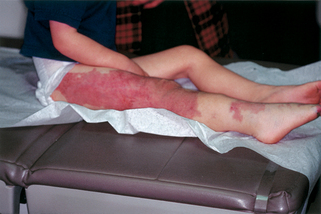
Figure 42-4. Klippel-Trénaunay syndrome. Mixed capillary-venous malformation associated with increased limb size.
Rozenfeld PA: Fabry disease: treatment and diagnosis, IUBMB Life 26:1043–1050, 2009.
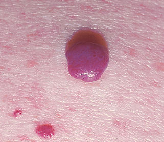
13. Where and in whom are cherry angiomas most commonly seen?
Cherry angiomas are extremely common, acquired, 1- to 5-mm, red to purple papules located primarily on the trunk and upper extremities (Fig. 42-6). They are most commonly seen in the middle-aged and elderly. In one study, 75% of the patients over age 64 had cherry angiomas.
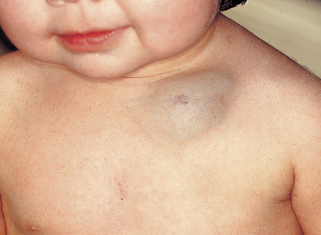
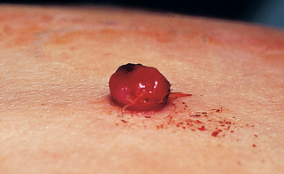
15. What is the most common presenting feature of a pyogenic granuloma?
Pyogenic granulomas are 5- to 10-mm soft red papules that bleed easily with minor trauma (Fig. 42-7). They are most common on the skin but may also occur on mucosal surfaces or, rarely, within blood vessels. Granuloma gravidarum is a variant that occurs on the gingiva during pregnancy. The pathogenesis is unknown, but approximately one third will occur after local trauma.
Lin RL, Janniger CK: Pyogenic granulomas, Cutis 74:229–233, 2004.
16. Where are you likely to find a lesion of angiolymphoid hyperplasia?
These lesions usually are found on the head and neck. They appear clinically as red-brown papules. Histologically, they demonstrate clusters of vessels with prominent endothelial cells, often accompanied by a nodular lymphocytic infiltrate that may appear as lymphoid follicles. The lesions are at times associated with a peripheral eosinophilia. The etiology is unknown.
17. What vascular tumor is associated with the Sucquet-Hoyer canal?
The Sucquet-Hoyer canal is the arterial segment of the glomus body and may give rise to glomus tumors. A solitary glomus tumor is a painful, purple nodule measuring a few millimeters in diameter. Multiple glomus tumors are inherited in an autosomal dominant fashion, may be much larger than the solitary form, and have been confused with the blue rubber bleb nevus syndrome. The cause of multiple glomus tumors are mutations in the glomulin gene located on chromosome 1p22-p21.
Anakwenze OA, Parker WL, Schiefer TK, et al: Clinical features of multiple glomus tumors, Dermatol Surg 34:884–890, 2008.
18. Matlike telangiectasias on the face, lips, tongue, ears, hands, and feet associated with internal bleeding is known as what syndrome?
Osler-Weber-Rendu syndrome, or hereditary hemorrhagic telangiectasia. Most patients present with epistaxis. Gastrointestinal telangiectasias are common, and genitourinary, pulmonary, central nervous system (CNS), and hepatic lesions may also occur. It is inherited as an autosomal dominant disease and is caused by mutations in the endoglin or activin receptor, such as kinase genes, which encode for proteins that modulate transforming growth factor–beta superfamily signaling in vascular endothelial cells.
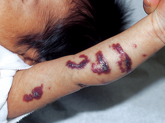
22. Name the types of lymphatic malformations.
A lymphatic malformation may be either superficial or deep. A superficial lymphatic malformation consists of superficial vesicles containing lymphatic fluid and at times blood. They have been described as looking like frog spawn. Deep lymphatic malformations are flesh-colored, soft, cystic nodules. The cysts may be either small (microcysts) or large (macrocysts).
23. Can you treat deep macrocystic lymphatic malformations?
The best treatment for this type of lesion is percutaneous injection of OK-432 (picibanil). This causes inflammation and subsequent sclerosis and shrinking of the lesion.
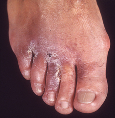
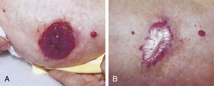
Figure 42-8. Acroangiodermatitis (pseudo-Kaposi’s sarcoma) due to underlying arteriovenous malformation.
(Courtesy of the Fitzsimons Army Medical Center teaching files.)