64
Varicella (chickenpox)
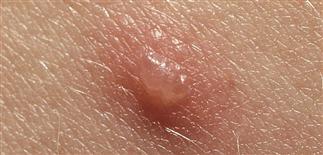
Primary lesion of chickenpox (varicella): a discrete, clear vesicle with central umbilication; it later turns cloudy, then develops a crust. There is a flare of erythema around the vesicle.
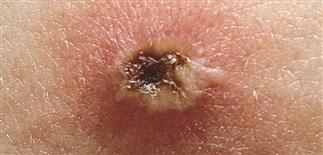
The evolving chickenpox vesicle is crusty and cloudy, and usually collapses in the middle.
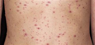
Varicella lesions are diffusely scattered. The lesions are very pruritic. Deep lesions may scar.
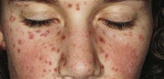
Varicella lesions in varying stages of development and widely scattered. Before the exanthem, there is typically fever, cough, and itchy eyes.
DESCRIPTION
Highly contagious infection caused by the varicella-zoster virus. Most commonly results in lifetime immunity.
HISTORY
• Transmission via airborne droplets or vesicular fluid. • Patients contagious from 2 days before rash until all lesions crusted. • Incubation period approximately 14–16 days. • In children, prodromal symptoms absent or low-grade fever, headache, malaise; fever, malaise, and generalized vesicular rash develop and last 4–7 days. Photophobia may be present. • Adolescents, adults, immunocompromised persons have more severe disease and are at risk for complications. • Moderate to intense pruritus during vesicular stage. • Complications include bacterial superinfection, pneumonia, dehydration, encephalitis, hepatitis. Most common neurologic complication is ataxia secondary to cerebellar inflammation. Roughly 15% of healthy adults develop pulmonary involvement. • Maternal infection during first 20 weeks of gestation poses risk of fetal congenital varicella syndrome.
PHYSICAL FINDINGS
• Lesions (vesicles, pustules, crusts) in all stages. • Begins on trunk and spreads to face and extremities. Extent varies. • Starts as a 2- to 4-mm red papule, then a thin-walled, clear vesicle appears. Vesicle becomes umbilicated and cloudy, breaking after 8–12 h. Crust forms as red base disappears. New lesions cease after 3 days. Crusts fall off in about 7 days. • Secondary infection or excoriation may result in scar. • Vesicles in mouth or vagina may form aphthae-like ulcers. • Pneumonia most common serious complication in healthy adults. Hepatitis most common complication in immunosuppressed patients. • Laboratory tests: Tzanck, direct fluorescent antibody test, or culture in questionable cases.
TREATMENT
Symptomatic: antipruritic lotions (e.g. Sarna). Oral antihistamines (hydroxyzine). For severe infection, i.v. acyclovir. Other antivirals, such as valacyclovir 1 g t.i.d. for 7 days and famciclovir 500 mg t.i.d. for 7 days, may have a role, although presently approved in USA for varicella-zoster infection. • Adolescents and adults. Early therapy with oral acyclovir decreases time to healing, decreases fever duration, reduces symptom severity, but does not alter viral shedding. Therapy onset after first day of illness is no value in uncomplicated cases. High morbidity rate even in otherwise healthy patients with clinically evident varicella pneumonia; i.v. acyclovir 10 mg/kg every 8 h for 7 days may help. • Immunocompromised patients. Those treated with acyclovir have decreased morbidity from visceral dissemination; modest effect on cutaneous disease. Acyclovir 10 mg/kg i.v. every 8 h for 7–10 days. Varicella-zoster immune globulin recommended for postexposure prophylaxis. Administer within 96 h post exposure.


