V
v gauge A device used to measure the total diameter of a rigid contact lens. It consists of a channel cut into a long rectangle of plastic or metal. The channel increases in width from 6.0 to 12.50 mm and a scale is printed beside it. The lens is placed with its concave surface down at the widest end of the channel and that end of the gauge is raised so that the lens slides down the channel until it stops. The diameter is then read from the scale where the lens touches the side of the channel. Syn. v-channel gauge.
validity The extent to which a measurement correctly measures what it is supposed to measure or to which extent the findings of an investigation reflect the truth. In health sciences, validity is commonly assessed by determining the sensitivity and specificity factors.
See reliability; sensitivity; specificity.
value f See f number.
value, Munsell See Munsell colour system.
value, V- See constringence.
valve of Hasner A fold of mucous membrane at the lower end of the nasolacrimal duct. If well developed, it generally prevents air from being blown back from the nose into the lacrimal sac. Syn. plica lacrimalis; valve of Bianchi.
See lacrimal apparatus.
valve of Krause A fold of mucous membrane at the junction of the lacrimal sac and the nasolacrimal duct. Syn. valve of Beraud.
See lacrimal apparatus.
valve of Rosenmuller A fold of mucous membrane found at the junction between the common canaliculus and the lacrimal sac. It is not strictly a valve because fluids can be blown back to emerge at the puncta. It is not always fully developed.
See lacrimal apparatus.
van Herick, Shaffer and Schwartz method See method, van Herick, Shaffer and Schwartz.
vancomycin See antibiotic.
varicella-zoster virus See herpes zoster ophthalmicus; herpesvirus.
varifocal lens See lens, progressive.
vasa hyaloidea propria See artery, hyaloid.
vascularization See neovascularization; pannus.
vase, Rubin’s See Rubin’s vase.
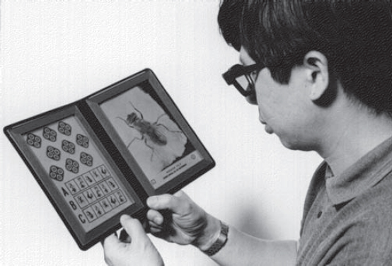
vectogram A polarized stereogram consisting of two polarized images at right angles to each other. When viewed through polarizing filters it presents one image to one eye and another image to the other eye. The Vectograph is a chart based on this principle in which almost one half of a chart is seen by one eye and almost the other half by the other eye while some lines, letters or numbers are seen binocularly to lock fusion. The Vectograph is useful for balancing refraction and to detect suppression and fixation disparity. The Titmus stereotest (Fig. V1) consists of various vectograms, including one with a stereoscopic pattern representing a housefly, to establish whether the patient has gross stereopsis (it produces approximately 3000 seconds of arc of retinal disparity at 40 cm). Children are often tested by asking them to hold one of the wings of the fly, which they will do above the plate if it is seen stereoscopically. The other vectograms of the test provide finer tests for stereoscopic acuity.
See acuity, stereoscopic visual; disparity, retinal; stereogram, random-dot; suppression; test, balancing; test, two-dimensional.
VEGF (vascular endothelial growth factor) A major protein involved in regulating the differentiation and proliferation of vascular endothelial cells thus promoting the growth of new blood vessels (angiogenesis). VEGF is essential for normal embryonic development and contributes to the maintenance and repair of tissues. There are several VEGF proteins, depending on the number of amino acids that they contain (e.g. VEGF 121, VEGF 165, VEGF 189 and VEGF 206). However, under certain circumstances (e.g. higher than normal levels of VEGF as happens in hypoxia) it may participate in cancerous processes, inflammatory processes (e.g. rheumatoid arthritis) and ocular neovascularization as in exudative (wet) age-related macular degeneration and diabetic retinopathy. Anti-VEGF drugs are used to inhibit the action of VEGF.
See anti-VEGF drugs.
veiling glare See glare, veiling.
vein A tubular vessel that carries blood towards the heart.
See artery.
anterior ciliary v. One of many veins that drains the ciliary body, the deep and superficial plexuses, the anterior conjunctival veins and the episcleral veins to empty into the vortex veins.
anterior facial v . Vein branching from the angular vein at the side of the nose and running obliquely downward and backward across the face. It crosses the mandible and joins the posterior facial vein to form the common facial vein, which opens into the internal jugular. The anterior facial vein drains the part of the eyelids anterior to the tarsus.
aqueous v . One of several veins serving as exit channels for the aqueous humour, which they carry from the canal of Schlemm to the episcleral, conjunctival and subconjunctival veins.
central retinal v. A vein formed by the junction of the superior and inferior retinal veins at about the level of the lamina cribrosa on the temporal side of the central retinal artery. After a short course within the optic nerve, it empties into the cavernous sinus, the superior ophthalmic vein and sometimes into the inferior ophthalmic vein.
See artery, central retinal; retinal vein occlusion.
conjunctival v. One of many veins that drains the tarsal conjunctiva, the fornix, and the major portion of the bulbar conjunctiva.
inferior ophthalmic v. Vein that commences as a plexus near the floor of the orbit, runs backward on the inferior rectus muscles and divides into two branches, one which runs to the pterygoid venous plexus and the other which joins the cavernous sinus, usually via the superior ophthalmic vein. The inferior ophthalmic vein receives tributaries from the lower and lateral ocular muscles, the conjunctiva, the lacrimal sac and the two inferior vortex veins.
palpebral v. One of the veins of the upper or lower eyelid that empties for the most part into the anterior facial vein as well as into the angular, supraorbital, superior and inferior ophthalmic, the lacrimal and the superficial temporal veins.
posterior ciliary v. See vein, vortex.
superior ophthalmic v. Vein that is formed near the root of the nose by a communication from the angular vein soon after it has been joined by the supraorbital vein. It passes into the orbit above the medial palpebral ligament, runs backward to the sphenoidal fissure where it usually meets the inferior ophthalmic vein, and drains into the cavernous sinus. It has many tributaries: the inferior ophthalmic vein, the anterior and posterior ethmoidal veins, the muscular vein, the lacrimal vein, the central retinal vein, the anterior ciliary vein and two of the posterior ciliary veins (the superior ones).
vortex v. One of usually four (two superior and two inferior) veins which pierce the sclera obliquely on either side of the superior and inferior recti muscles, some 6 mm behind the equator of the globe. The two superior ones open into the superior ophthalmic vein and the two inferior open into the inferior ophthalmic vein. These veins drain the posterior uveal tract. Syn. posterior ciliary vein; vena vorticosa.
See vein, anterior ciliary.
velocity of light See light, speed of.
velonoskiascopy A subjective method of detecting ametropia in which a thin rod held near the eye is moved across the pupil while the subject fixates a distant light source. The rod casts a shadow on the retina if the eye is ametropic. This shadow will appear to move with the rod in myopia and opposite to the movement of the rod in hyperopia. By moving the rod across the pupil in different meridians, astigmatism can be explored. No shadow is seen in emmetropia.
vena vorticosa See vein, vortex.
venous-stasis retinopathy See retinal vein occlusion.
ventral Relating to either the front (anterior), or to the bottom in brain orientation.
See dorsal; system, parvocellular visual.
vergence 1 . Denotes divergence of light travelling from, or convergence of light travelling from, or to an object or image. The object vergence at a refracting surface is equal to
< ?xml:namespace prefix = "mml" />

where n is the index of refraction of the first medium and l the distance between the object plane and the refracting surface in metres. The image vergence at a refracting surface is equal to
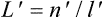
where n‘ is the index of refraction of the second medium and l‘ the distance between the image plane and the refracting surface in metres. The unit of vergence is the dioptre. 2. Disjunctive movements of the eyes such as convergence, divergence, cyclovergence, infravergence or supravergence.
See distance, image; distance, object; duction; paraxial equation, fundamental; power, refractive.
v. accommodation See accommodation, convergence.
accommodative v. See convergence, accommodative.
disparity v. See fusion, motor.
v. facility Ability of the eyes to make fusional vergence movements in a given period of time. Clinically, this is measured by introducing a relatively large prism in front of one or both eyes of a patient fixating a target until it appears single. The operation is repeated many times and the results are commonly presented in cycles per minute (one cycle indicates that single vision was reported both with the prism and after removing the prism).
See convergence, fusional; fusion, motor; lens flippers.
v. formula See paraxial equation, fundamental.
fusional v. See convergence, relative.
v. power See power, refractive.
proximal v. See convergence, proximal.
v. reflex See reflex, vergence.
relative v. See convergence, relative.
tonic v. The passive state of vergence of the eyes in the absence of a stimulus, i.e. when the eyes are in total darkness or when looking at a bright empty field. This position is maintained by the tonus of the extraocular muscles. Only at death or when paralysed do the eyes return to their anatomical position of rest and tonic vergence disappears. Syn. dark vergence; tonic convergence.
See accommodation, resting state of; position of rest, physiological; tonus.
vertical fusional v . Movement of the eyes upward until an object that was imaged on slightly disparate vertical parts of the retina falls on corresponding retinal points.
Verhoeff phi phenomenon test See movement, phi.
Verhoeff’s circles Two black concentric circles designed for use with the duochrome test and as a target for the cross-cylinder method. The thickness and overall diameter of the inner ring are equivalent to a 6/6 (20/20) Snellen letter while the thickness and overall diameter of the outer ring are equivalent to a 6/15 (20/50) Snellen letter. Syn. Verhoeff’s rings.
See chart, Snellen; test for astigmatism, cross-cylinder; test, duochrome.
Verhoeff’s rings See Verhoeff’s circles.
vernal catarrh; conjunctivitis See conjunctivitis, vernal.
vernier visual acuity See acuity, vernier visual.
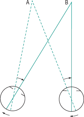
version Conjugate movements of the two eyes in the same direction, such as dextroversion, both eyes rotate to the right; laevoversion (levoversion), both eyes rotate to the left; supraversion (sursumversion), both eyes rotate upward; infraversion (deorsumversion), both eyes rotate downward: these versions bring the eyes into the secondary positions of gaze. Movements of the eyes up and to the right are called dextroelevation, up and to the left, laevoelevation, down and to the right, dextrodepression and down and to the left, laevodepression: these versions bring the eye into the tertiary positions of gaze. Version eye movements are performed by yoke muscles (Fig. V2). Syn. conjugate eye movements.
See deviation, conjugate; positions of gaze, cardinal; test, motility.
version prisms See prisms, yoke.
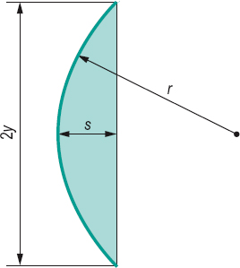
vertex The point where the optical axis intersects a reflecting or refracting surface. In a spectacle lens the back vertex is the point of intersection of the optical axis with the surface nearest to the eye, the other being the front vertex. Plural: vertices.
v. depth Distance between the posterior pole of a spectacle lens and the plane containing the posterior edge of the lens. The vertex depth s is given by the following formula
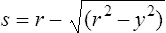
where r is the radius of curvature of the surface of the spectacle lens and y is the semi-diameter at the edge of the surface (Fig. V3). Syn. sag.
See clearance, apical; lens measure.
v. distance Distance along the line of sight between the apex of the cornea and the posterior surface of a spectacle lens. This distance normally varies between 11 mm and 15 mm.
See clearance, apical; plane, spectacle.
v. focal length The linear distance separating the principal focal point (focus) of an optical system or lens from the front or back vertices. They are called the front vertex focal length (fv) and the back vertex
focal length (f’v), respectively. In the case of a biconcave or biconvex lens the front and back vertex focal lengths are equal. In the case of a positive meniscus lens, the back vertex focal length is shorter than the front vertex focal length and vice versa in the case of a negative meniscus lens.
See power, back vertex; power, front vertex.
v. power See power, back vertex; power, front vertex.
vertexometer 1. Synonym of focimeter. 2. Synonym of distometer.
See distometer; focimeter.
vertical fusional vergence See vergence, vertical fusional.
vertigo The sensation of irregular movement in space of either oneself or of external objects. It can be experienced after vestibular stimulation.
vesicle 1. A small bladder or sac containing liquid. 2. A small elevation on the skin containing fluid, usually serous fluid. 3. Any structure that has the appearance of 1 or 2 above.
optic v’s. Hollow, spherical neuroectodermal protrusions, one on each side of the forebrain. They are derived from the optic pits after closure of the embryonic neural tube. They subsequently invaginate to form the optic cup. The surface ectoderm overlying the optic vesicles invaginates to form the lens vesicle and eventually the crystalline lens. Syn. primary optic vesicle.
See anophthalmia; cup, optic; ectoderm; pit, optic.
vestibular nystagmus See nystagmus.
vestibulo-ocular reflex See reflex, vestibulo-ocular.
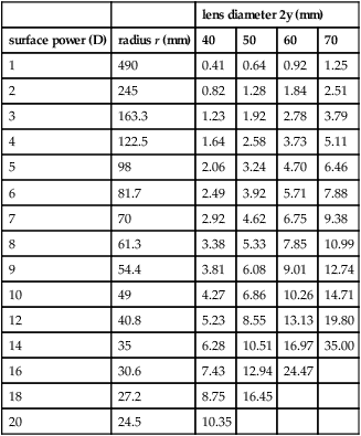
Table V1
Vertex depths of various spherical surfaces. They also represent the centre thickness of a planoconvex lens with a front surface of radius of curvature r and diameter 2y, with an edge thickness of zero. Index of refraction of the lens 1.49
| lens diameter 2y (mm) | |||||
| surface power (D) | radius r (mm) | 40 | 50 | 60 | 70 |
| 1 | 490 | 0.41 | 0.64 | 0.92 | 1.25 |
| 2 | 245 | 0.82 | 1.28 | 1.84 | 2.51 |
| 3 | 163.3 | 1.23 | 1.92 | 2.78 | 3.79 |
| 4 | 122.5 | 1.64 | 2.58 | 3.73 | 5.11 |
| 5 | 98 | 2.06 | 3.24 | 4.70 | 6.46 |
| 6 | 81.7 | 2.49 | 3.92 | 5.71 | 7.88 |
| 7 | 70 | 2.92 | 4.62 | 6.75 | 9.38 |
| 8 | 61.3 | 3.38 | 5.33 | 7.85 | 10.99 |
| 9 | 54.4 | 3.81 | 6.08 | 9.01 | 12.74 |
| 10 | 49 | 4.27 | 6.86 | 10.26 | 14.71 |
| 12 | 40.8 | 5.23 | 8.55 | 13.13 | 19.80 |
| 14 | 35 | 6.28 | 10.51 | 16.97 | 35.00 |
| 16 | 30.6 | 7.43 | 12.94 | 24.47 | |
| 18 | 27.2 | 8.75 | 16.45 | ||
| 20 | 24.5 | 10.35 | |||
vial A very small bottle. It may contain a soft contact lens, medicine or perfume.
vidarabine See antiviral agents.
videokeratoscope An electro-optical instrument for measuring the corneal topography. It produces a colour coded three-dimensional map of the shape of the cornea and of the dioptric power of the different corneal regions. These instruments are computerassisted, providing rapid, online analysis of the image and most of them are based on the corneal reflection of the Placido pattern. Topographic data are usually presented as a colour-coded map of the cornea showing regions of different power. These instruments are used to evaluate keratoconus, irregular corneal shape, contact lens fitting, monitor the cornea after keratoplasty or refractive surgery, etc. There are many commercial models including one that provides a map of the curvature and elevation of the anterior and posterior corneal surface (scanning-slit videokeratoscope).
See corneal topography; keratoscope; photokeratoscope; Topogometer.
Vieth–Müller circle See horopter, Vieth–Müller.
viewing angle See angle, visual.
viewing, eccentric Fixation in which the eye moves so as to place the image of an object outside the fovea. The object is perceived by the patient as looking ‘past’ it and not directly at it as in eccentric fixation. Eccentric viewing is often applied by people with low vision suffering from macular degeneration to improve reading a letter or a word by looking slightly above, below or to the side of it.
See fixation, eccentric; vision, low.
vignetting 1. A graduated reduction in retinal illuminance due to light reaching the pupil at very oblique angles. 2. The difference in absorption between the two portions of photochromic fused bifocal lenses when the segment is not made of photochromic glass.
See lens, photochromic; retinal illuminance.
violet One of the hues of the visible spectrum evoked by stimulation of the retina by wavelengths shorter than 450 nm and somewhat longer than 380 nm.
virtual image; object See under the nouns.
virus A submicroscopic (20–600 nm in diameter) particle (called a virion), which typically contains a protein coat (called a capsid) surrounding genetic material in the form of a double or a single strand of RNA or DNA. Viruses replicate only within cells of living hosts. They can infect cells and are the cause of various diseases. This is accomplished by releasing the viral genetic material into the host cytoplasm if it is RNA, and into the host nucleus if it is DNA, and thus inducing the production of new viral particles and newly infected cells. There are many viruses: DNA viruses as for example the adenovirus (some of which can cause epidemic conjunctivitis), herpesvirus and pox viruses, and RNA viruses as for example the picorna virus (e.g. hepatitis A), toga viruses (e.g. rubella), corona viruses (which can cause respiratory infection) and the retroviruses (e.g. HIV).
See antiviral agents; herpesvirus; gene therapy.
viscocanalostomy A type of non-penetrating filtration surgery aimed at lowering intraocular pressure by dissecting a superficial scleral flap and excising a deeper partial-thickness scleral flap below, leaving a thin membrane consisting of trabeculum and Descemet’s membrane, through which the aqueous humour diffuses. It then drains from the anterior chamber into the subconjunctival space or through Schlemm’s canal into which a high-density viscoelastic substance has been injected. The superficial flap is sutured in place at the end of the procedure. Intraocular pressure reduction is not usually as large as with trabeculectomy.
See filtration surgery.
viscosity agents See methylcellulose; wetting solution.
visibility 1. The property of being visible to the eye. 2. The range of vision through different densities of atmosphere.
visible spectrum See light.
vision (V) 1 . The appreciation of differences in the external world, such as form, colour, position, etc. resulting from the stimulation of the retina by light. 2. See acuity, unaided visual.
achromatic v. See achromatopsia.
alternating v. See lens, contact.
ambient v . Vision mediated primarily by the peripheral retina and involved in spatial orientation and recognition of motion. See vision, focal.
anomalous trichromatic v. See trichromatism, anomalous.
binocular v. (BV) Condition in which both eyes contribute towards producing a percept which may or may not be fused into a single impression. See fusion, sensory; monoblepsia; period, critical; test, bar reading; test, FRIEND; test, hole in the hand; test, Worth’s four dot; vision, Worth’s classification of binocular; zone of clear, single, binocular vision.
binocular single v. See vision, single binocular.
blue v. See chromatopsia.
blurred v. Vision characterized by poor visual acuity or in which the edges of objects are indistinct. It may be due to uncorrected or poorly corrected ametropia or presbyopia, anomalies of the ocular media (e.g. cataract, corneal opacity, haemorrhage in the vitreous), amblyopia, excess lacrimation, spasm of accommodation, optic neuritis, angle-closure glaucoma, diabetes, multiple sclerosis, migraine, etc.
central v. Vision of objects formed on the foveola or the macula.
chromatic v. See vision, colour.
colour v. (CV) Vision in which the colour sense is experienced. Syn. chromatic vision.
See theory, Hering’s of colour vision; theory, Young–Helmholtz.
daylight v. See vision, photopic.
defective colour v. See colour vision, defective.
deuteranomalous v. See deuteranomaly.
dichromatic v. See dichromatism.
distance v. (DV) Vision of objects situated either at infinity or more usually at some 5 or 6 m.
See chart, Snellen; vision, near.
diurnal v. See vision, photopic.
double v . See diplopia.
eccentric v. See fixation, eccentric; vision, peripheral.
entoptic v. See image, entoptic.
extrafoveal v. See vision, peripheral.
field of v. See field, visual.
focal v . Vision mediated by, primarily, the macular area of the retina and involved in the examination and identification of objects.
See vision, ambient.
green v . See chromatopsia.
gun barrel v . See vision, tunnel.
haploscopic v. Vision as obtained by looking in a haploscope.
indirect v. See vision, peripheral.
industrial v. The branch of optometry concerned with vision and perception by the individual at work, the evaluation of visual performance in a given occupation, the prescribing of protective ocular devices and the determination of the optimum environment (e.g. illumination) to accomplish a visual task efficiently.
intermediate v . Vision of objects situated beyond 40 cm from the eye but closer than, say, 1.5 m.
See vision, distance; vision, near.
island of v. A description of the visual field as a three-dimensional hill surrounded by a sea of darkness. Stimuli that fall within the island can be seen, whereas stimuli that fall outside the island cannot be seen. The height of the island represents the sensitivity of the eye, with the highest acuity at the top of the hill corresponding to foveal vision and declining progressively towards the periphery (when the eye is light-adapted).
low v. Vision impairment even after correction by conventional lenses, resulting from either congenital anomalies or ocular diseases such as cataract, glaucoma, age-related macular degeneration, pathological myopia, trachoma, onchocerciasis, etc. The correction and rehabilitation of patients with low vision is achieved by special aids called low vision aids (LVA) such as a telescopic lens, and appropriate counselling (e.g. about illumination and reading distance). The criteria that the health authorities normally use to classify a person as having partial sight take into consideration not only the corrected visual acuity but also the extent of visual field loss (generally less than 20°). Syn. partial sight; subnormal vision.
The World Health Organization (WHO) defines low vision as visual acuity less than 6/18 (20/60) and equal to or better than 3/60 (10/200) in the better eye with best correction.
See aids, low vision; blindness; bracketing; chart, Bailey–Lovie; chart, contrast sensitivity; clipover; deaf-blind; lamp, halogen; lens, cross-cylinder; lens, telescopic; magnification, apparent; magnification, relative distance; magnification, relative size; magnifier; rule, Kestenbaum’s; spectacles, magnifying; spectacles, pinhole; telescope, galilean; test, Pepper; typoscope; viewing, eccentric.
mesopic v. Vision at intermediate levels between photopic and scotopic vision, and corresponding to luminances ranging from about 10–3 to 10 cd/m2. Syn. twilight vision.
monochromatic v. Synonym of monochromatism.
See monochromat.
monocular v. Vision of one eye only.
multiple v. See polyopia.
near v. (NV) Vision of objects situated 25–50 cm from either the eye, or more commonly the spectacle plane.
See Jaeger test types; vision, distance.
night v.; nocturnal v. See vision, scotopic.
panoramic v. Vision of some animals whose eyes are located laterally so that the two visual fields overlap only slightly or are adjacent, thus providing vision over a much larger region of the environment than if the two lines of sight were aimed in the same direction.
peripheral v. Vision resulting from stimulation of the retina outside the fovea or macula. Syn. eccentric vision; extrafoveal vision; indirect vision.
See fusion, sensory; vision, central.
photopic v. Vision at high levels of luminance (above 10 cd/m2) and resulting from the functioning of the cones. Syn. daylight vision; diurnal vision.
See theory, duplicity; threshold, differential.
protanomalous v. See protanomaly.
red v. See chromatopsia.
v. science The scientific study of how the visual system contributes to an understanding of the environment by processing and interpreting the light stimulation to the eye. Various disciplines contribute to vision science including anatomy, biology, optics, physiology and psychology.
scotopic v. Vision at low levels of luminance, below about 10–3 cd/m2 and resulting from the functioning of the rods. Syn. night vision; nocturnal vision; scotopia.
v. screener An instrument used to measure various visual functions rapidly and inexpensively. There are various models, but most are modified stereoscopes with an internally illuminated set of targets and an optical system or variable target positioning to simulate either a near or far testing distance. Most of these instruments measure visual acuity, heterophoria, fusion, stereopsis, colour vision and visual field.
See photorefraction.
simultaneous v. See lens, contact.
single binocular v. (SBV) Condition in which both eyes contribute towards producing a single fused percept.
spatial v. See perception, depth.
stereoscopic v. See stereopsis.
telescopic v. See vision, tunnel.
v. therapy; v. training See training, visual.
tritanomalous v. See tritanomaly.
tunnel v. Vision limited to the central part of the visual field as though one were looking through a hollow tube. It may be a symptom of hysteria, malingering, the final stage of either open-angle glaucoma or retinitis pigmentosa, etc. Syn. gun barrel vision; telescopic vision.
See amblyopia, hysterical; field, visual expander.
twilight v. See vision, mesopic.
Worth’s classification of binocular v. For the purpose of visual rehabilitation, binocular vision is often classified into three grades: (1) simultaneous binocular vision (first-degree fusion or superimposition); (2) fusion (sensory fusion or second-degree fusion or flat fusion); (3) stereopsis (third-degree fusion).
See fusion, sensory; superimposition.
yellow v. See xanthopsia.
Vistech A clinical test for contrast sensitivity. It consists of a chart containing five horizontal rows, each with nine circular patches of sinusoidal gratings. The gratings are either vertical or 15° to the right or to the left. Each row has a different spatial frequency, starting from the top of the chart: 1.5, 3.0, 6.0, 12.0 and 18.0 cycles per degrees when viewed at a distance of 40 cm. The contrast level of each of the nine gratings decreases from 33% to 0% from left to right in approximately 0.2 log unit steps. The patient is asked to look along each row, identifying the orientation of the grating. The testing is carried out monocularly with optical correction, if any. The last grating of each row that is incorrectly identified is noted on an evaluation form, which is provided with the test. The end points of each of the five rows are connected to form a contrast sensitivity curve for each patient. It is then compared with normal values indicated on the form. There is also a version for testing at distance. Syn. Vision Contrast Test System (VCTS).
See chart, contrast sensitivity; test, Arden grating.
visual Relating to vision.
visual acuity; agnosia; agraphia See under the nouns.
Visual Analysis Skills Test See test, developmental and perceptual screening.
visual allesthesia; angle; area; association areas; axis; centre; cliff See under the nouns.
visual cortex See area, visual.
visual deprivation See deprivation, visual.
visual direction See line of direction.
visual display unit (VDU) The visual image appearing on the screen of a cathode ray tube.
See syndrome, computer vision.
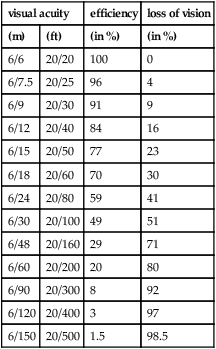
visual efficiency scale, Snell–Sterling A representation of visual efficiency as a function of visual acuity, in which are taken into account other factors such as perception, experience, etc. in estimating how much vision a person has for a given visual acuity.
visual evoked cortical potential See potential, visual evoked cortical.
visual extinction See phenomenon, extinction.
visual fatigue; field See under the nouns.
visual field, binocular See field, binocular visual.
visual field analyser See analyser, Friedmann visual field.
visual field expander See field, visual expander.
visual hallucination; illusion See under the nouns.
visual integration Term referring to the integration occurring in the brain to give us a final percept, presumably in the prefrontal cortex. Information from the dorsal (parietal or medial temporal) stream dealing with localization or movement is integrated with information from the ventral (inferotemporal) stream dealing with colour or form, so that, for example, we can see a red car moving towards us.
visual line of direction See line of direction.
visual neglect A rare phenomenon in which a patient can see all of the visual field binocularly but somehow ignores objects on one side (e.g. patient may draw a diagram omitting one side or shave only one side of the face). It is due to a lesion of the brain (e.g. a stroke), most often in the right cortex and the patient, although conscious of objects in the left visual field does not pay attention to them. The impairment occurs in the posterior parietal lobe, which receives projections from the primary visual cortex. A confrontation visual field test in which objects are presented to both sides simultaneously often facilitates detection of the condition.
See phenomenon, extinction.
visual optics; pathway; pigment; plane; point See under the nouns.
visual perseveration See palinopsia.
visual purple See rhodopsin.
visual system, magnocellular; parvocellular See under system.
visual system, sustained See system, parvocellular visual.
visual system, transient See system, magnocellular visual.
visual training See training, visual.
visualization 1. The ability to form a mental image of an object not present in the field of view. 2. Synonym for imagery. Example: visualizing the face of a person speaking on the radio.
See imagery.
visus Vision.
Table V2
Relationship between visual acuity and the Snell-Sterling visual efficiency scale (in percentage round figures)
| visual acuity | efficiency | loss of vision | |
| (m) | (ft) | (in %) | (in %) |
| 6/6 | 20/20 | 100 | 0 |
| 6/7.5 | 20/25 | 96 | 4 |
| 6/9 | 20/30 | 91 | 9 |
| 6/12 | 20/40 | 84 | 16 |
| 6/15 | 20/50 | 77 | 23 |
| 6/18 | 20/60 | 70 | 30 |
| 6/24 | 20/80 | 59 | 41 |
| 6/30 | 20/100 | 49 | 51 |
| 6/48 | 20/160 | 29 | 71 |
| 6/60 | 20/200 | 20 | 80 |
| 6/90 | 20/300 | 8 | 92 |
| 6/120 | 20/400 | 3 | 97 |
| 6/150 | 20/500 | 1.5 | 98.5 |
Visuscope A modified ophthalmoscope containing a small graticule target for the measurement of eccentric fixation. The examiner projects a shadow of the target on the patient’s retina. The patient is asked to look at the centre of the target. The position of the foveal reflex relative to the centre of the graticule target indicates whether the patient has eccentric fixation and in which direction and by how much. A modified version is the Euthyscope, in which the graticule target consists of black spots rather than a star and concentric circles as in the Visuscope. The Euthyscope is used more for eccentric fixation therapy.
See pleoptics.
vitamin A deficiency A deficiency of vitamin A (also called retinol) leads to interference with growth, atrophy of epithelial tissues resulting in keratomalacia, corneal ulcerations, xerophthalmia with Bitot’s spots, reduced resistance to infection of mucous membranes, and abnormal production and regeneration of rhodopsin resulting in night blindness. Management includes a balanced diet and may require large vitamin A supplement with a topical antibiotic to prevent secondary infections.
See carotene; hemeralopia.
vitamin B See neuritis, optic.
vitelliform macular dystrophy See disease, Best’s.
vitiligo A disease of the skin characterized by areas of depigmentation of various sizes and shapes. In the eye, it can be seen in the choroid or iris. It is often associated with syphilis or tuberculosis and forms part of the Vogt–Koyanagi–Harada syndrome.
See poliosis.
vitrectomy Removal of the whole or a portion of the vitreous humour and replacement by saline or, more commonly, silicone oil. Indications for this surgical intervention include persistent vitreous opacities (usually as a result of unabsorbed haemorrhage), severe penetrating trauma, luxation of the lens, retention of some foreign bodies which cannot be removed with a magnet, endophthalmitis, and especially advanced diabetic eye disease such as proliferative retinopathy to prevent retinal detachment because the fibrovascular network of the retina tends otherwise to adhere to the vitreous body.
See injection, intravitreal.
vitreoretinal degeneration See disease, Wagner’s; syndrome, Stickler’s.
vitreoretinopathy, familial exudative An autosomal dominant disorder involving chromosome 11q, although some cases may be X-linked. It is characterized by abrupt cessation of peripheral vessels at the equator, especially on the temporal side, resulting in vitreous degeneration, peripheral telangiectasia and fibrovascular proliferation. Complications include subretinal exudation and tractional retinal detachment. Possible treatments include photocoagulation or cryopexy.
vitreous base A dense, broad band (2 mm wide) of vitreous attachment to the peripheral retina near the ora serrata. Collagen vitreous fibrils blend anteriorly with the basal lamina of the non-pigmented epithelium of the pars plana of the ciliary body and posteriorly with the internal limiting membrane of the retina.
vitreous body See humour, vitreous.
vitreous chamber See chamber, vitreous.
vitreous detachment Separation of the vitreous body from the internal limiting membrane of the retina due to shrinkage from degenerative or inflammatory conditions, trauma, progressive myopia, old age, diabetes and in aphakic eyes in which the lens extraction was intracapsular. The most common cases are elderly individuals in whom the posterior part of the vitreous, which becomes liquid, detaches from the internal limiting membrane (called posterior vitreous detachment, PVD). Symptoms are flashes, floaters and photopsia because as the eye moves the vitreous body comes into contact with the retina. The condition is sometimes associated with retinal tears and retinal detachment.
See retinal break; syneresis.
vitreous floaters See floaters.
vitreous haemorrhage See haemorrhage, preretinal; retinopathy, diabetic.
vitreous humour See humour, vitreous.
vitreous, persistent hyperplastic primary (PHPV) A congenital, abnormal vitreous development characterized by a retrolental mass formed by remnants of the hyaloid system and tunica vasculosa lentis. The eye presents with leukocoria and there may also be cataract and congenital glaucoma. Treatment should begin as early in life as possible to avoid the risk of damage to the globe and amblyopia.
See artery, hyaloid; hyaloid remnant.
vitritis Inflammatory reaction of the vitreous or the hyaloid membrane as a result of a disease in the adjacent structures, such as the ciliary body, the choroid or the retina, which causes infiltration of cells into the vitreous. Patients complain of floaters and/or blurred vision. Note: also spelt vitreitis. Syn. hyalitis.
See uveitis, intermediate.
Vogt’s striae See striae, Vogt’s.
Vogt, palisades of See palisades of Vogt.
Vogt’s white limbal girdle A white, arc-like opacity in the cornea located concentric with the limbus in the 3 and 9 o’clock positions. Type 1 is a discontinuous band separated from the limbus by a clear zone. Type 2 is an unbroken band, made up mainly of hyaline deposits and is continuous with the sclera. This degeneration becomes more prevalent with age.
Volk lens See slit-lamp.
von Graefe’s sign See disease, Graves’; sign, von Graefe’s.
von Graefe’s test See test, diplopia.
von Hippel’s disease See disease, von Hippel’s.
von Hippel–Lindau disease See disease, von Hippel–Lindau.
von Recklinghausen’s disease See disease, von Recklinghausen’s.
V-value See constringence.


