19 Uterus and Adnexal Diseases
Anatomy of the Uterus, Adnexa, and Vagina
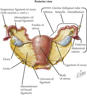
Uterus
• Derived from fusion of paired embryonic paramesonephric (müllerian) ducts: basis for divided, asymmetrical, or bifid (didelphic) uteruses
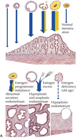
• Endometrium: highly vascular and glandular uterine lining; thickness or state varies with menstrual cycle
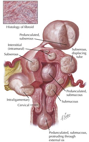
• Myometrium: dense, fibrous connective tissue and smooth muscle, derived from embryonic splanchnic mesoderm
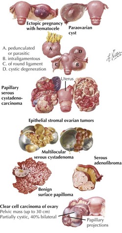
Ovaries
• Lie laterally and posterior to the broad ligament, attached near its upper borders via a peritoneal mesovarium


Vessels
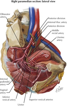
Arterial Supply
• Uterus and adnexa supplied bilaterally (on left and right sides) by three major anastomotic arteries, superior to inferior


Venous Drainage
• Uterine, ovarian, and vaginal veins interconnect in an extensive bilateral uterine plexus running within proximal broad ligaments.
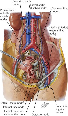
Lymphatic Drainage
• Ovarian, uterine fundus, and body lymphatics drain upward along ovarian vessels to nodes around lumbar aorta and vena cava.
• Vessels from around the uterine tube junctions drain along the round ligament into superficial inguinal nodes.
Clinical Correlates
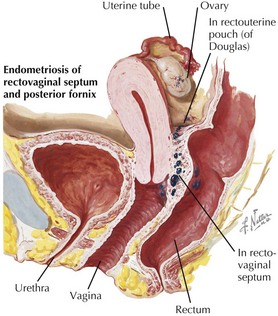
Endometriosis
• Benign foci of endometrial tissue progressively developing in pelvis—ovary, rectouterine pouch, uterine ligaments, tubes—or elsewhere in peritoneum