T-Shaped Uterus

Published on 10/03/2015 by admin

Filed under Obstetrics & Gynecology

Last modified 22/04/2025

Print this page

rate 1 star rate 2 star rate 3 star rate 4 star rate 5 star
Your rating: none, Average: 0 (0 votes)

This article have been viewed 2504 times

T-Shaped Uterus

Synonyms/Description

T-shaped uterus refers to the imaging appearance of a T rather than a triangular-shaped endometrial cavity

Etiology

Diethylstilbestrol (DES) is a synthetic estrogen that was widely prescribed to pregnant women from the late 1940s until 1970 to prevent miscarriage. An estimated 1 million to 1.5 million women received DES during their pregnancies, and this ultimately affected the reproductive organs of 35% to 69% of their female offspring. The daughters of women treated with DES developed congenital malformations of the uterus, cervix, and vagina as well as adenosis and (rarely) clear cell adenocarcinoma of the vagina. The T-shaped uterus is the most common and characteristic deformity of the uterus resulting from the prenatal exposure to DES and is highly associated with infertility and recurrent miscarriage.
Initially, the term “T-shaped uterus” was reserved for the DES daughters with the characteristic uterine cavity shape. More recently, patients with multiple D&Cs or hysteroscopic procedures can develop endometrial scarring that can be very similar in appearance and outcome to the congenital T-shaped uterus. Sometimes a patient may have recurrent miscarriages and D&Cs with a subsequent diagnosis of T-shaped uterus as a result of Asherman’s syndrome. Whether those patients had a congenital T-shaped uterus or acquired extensive scarring that distorted the endometrial cavity often cannot be determined.

Ultrasound Findings

The exact shape of the uterine cavity is usually not discernible on a standard 2-D ultrasound. The coronal view of the uterus, usually reconstructed from a 3-D volume, is necessary to evaluate the shape of the uterus and endometrial cavity. A normal uterine cavity is triangular or V-shaped, with the three apices being the two cornua and the junction of the lower uterine segment and the cervix (level of internal os). When the uterus is T-shaped, there is a waist in the sides of the triangle such that the corpus of the endometrial cavity is narrowed and takes on the shape of a T rather than a V. The outer myometrial surface of the uterus (the serosal surface) is typically unaffected.

Differential Diagnosis

The differential diagnosis of a T-shaped uterine cavity relates more to the cause, such as Asherman’s syndrome and uterine scarring versus congenital anomaly (see Scarred Uterus and Asherman’s Syndrome and also Müllerian Duct Anomalies).Occasionally a fibroid can press on the uterine cavity, creating the appearance of a T shape because of the location of the fibroid. Other congenital Müllerian duct abnormalities are usually characteristic, such as a septate or unicornuate uterus, and quite different from a T-shaped cavity.

Clinical Aspects and Recommendations

Women found to have a T-shaped endometrial cavity are best managed by specialists in infertility, hysteroscopic surgery, and high-risk obstetrics.
Fernandez and colleagues studied 97 infertile women who had T-shaped uteri, and 49.5% of them became pregnant after metroplasty. For these patients, the pregnancy rate increased from 0% to 73%, and their miscarriage rate fell from 78% to 27% (p = 0.05). For all 57 pregnancies in 48 women, the preterm delivery rate was 14%, the term delivery rate was 49%, and the live birth rate was 63%.
In another study by Katz and colleagues, which included eight patients with T-shaped uteri and recurrent miscarriage, hysteroscopic surgery resulted in four term pregnancies in three women, one ectopic pregnancy, and no abortions.

Figures

image

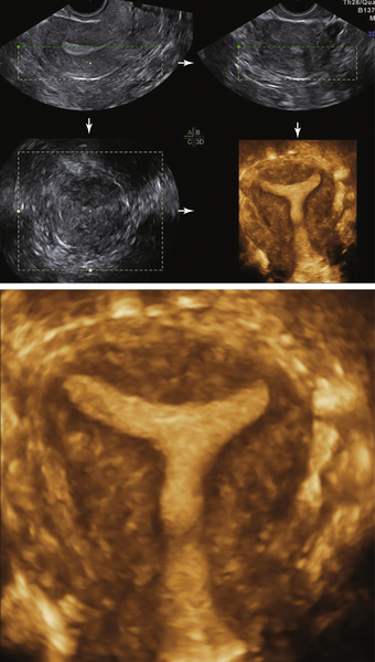
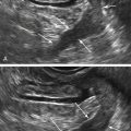
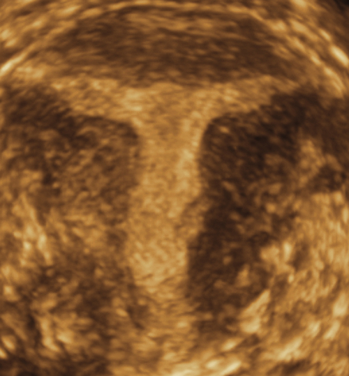
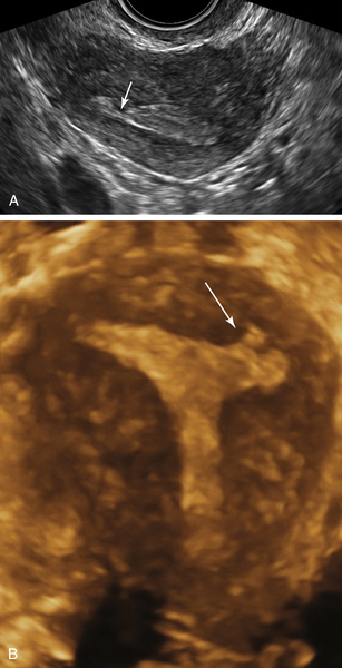
Figure T1-1 3-D image of a T-shaped uterus in a patient with recurrent miscarriage. Note the lack of triangular shape of the lateral walls of the endometrial cavity. Instead, the lateral walls are pulled inward to create a T configuration, thus narrowing the cavity. The outer uterine surface, however, is normally shaped, as is expected with this diagnosis.

image

Figure T1-2 Classic T-shaped uterus in a DES daughter who was never able to conceive.


image

Figure T1-3 A and B, 2-D and 3-D images of the uterus in a patient with multiple D&Cs. Note that although the shape is slightly T-shaped, the main abnormality is scarring (arrows) and irregularity of the left cornu with asymmetry of the shape of the uterine cavity.


Suggested Reading

Fernandez H., Garbin O., Castaigne V., Gervaise A., Levaillant J.M. Surgical approach to and reproductive outcome after surgical correction of a T-shaped uterus. Hum Reprod. 2011;26(7):1730–1734.

Katz Z., Ben-Arie A., Lurie S., Manor M., Insler V. Beneficial effect of hysteroscopic metroplasty on the reproductive outcome in a ‘T-shaped’ uterus. Gynecol Obstet Invest. 1996;41(1):41–43.

van Gils A.P., Tham R.T., Falke T.H., Peters A.A. Abnormalities of the uterus and cervix after diethylstilbestrol exposure: correlation of findings on MR and hysterosalpingography. AJR Am J Roentgenol. 1989;153(6):1235–1238.