Urticaria and Angioedema

Published on 06/06/2015 by admin

Filed under Pediatrics

Last modified 22/04/2025

Print this page

rate 1 star rate 2 star rate 3 star rate 4 star rate 5 star
Your rating: none, Average: 0 (0 votes)

This article have been viewed 2663 times

20 Urticaria and Angioedema

Urticaria, or hives, is a pruritic and episodic rash that most commonly occurs without an identifiable trigger. It occurs in approximately 25% of the population. The occurrence of urticaria increases to 50% in those affected by allergic disorders, such as asthma, allergic rhinitis, or atopic dermatitis. Acute urticaria, defined as symptoms lasting less than 6 weeks, accounts for two-thirds of all urticaria. Chronic urticaria, which can be subdivided into chronic idiopathic urticaria (CIU) and physical urticaria, is classified by symptom duration of greater than 6 weeks with symptoms on at least 2 days per week. CIU occurs in approximately 0.1% to 3% of the population and is more prevalent in females. Features of physical urticarias can be common, especially dermatographism, which occurs in almost half of the general population, including those with no history of chronic urticaria.

Chronic urticaria can have a significant social and financial impact. The hallmark of urticaria is pruritus, which contributes to difficulty sleeping and in activities of daily life. Urticaria can be a frustrating disease for patients because typically there is no identifiable trigger for exacerbations, leading to an unpredictable course. The effect on quality of life is comparable to that of coronary heart disease and atopic dermatitis. Patients with chronic urticaria also have multiple medications, medical visits, work and school absences, and use of the emergency department, all of which contribute to the economic burden of the disease.

Etiology and Pathogenesis

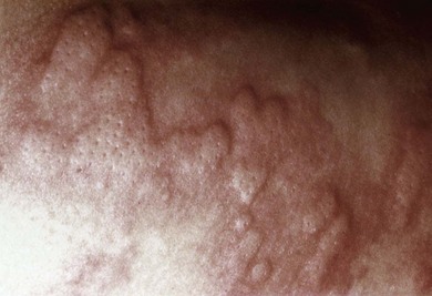
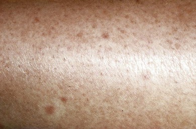
Urticaria is characterized by the waxing and waning appearance of pruritic, erythematous papules or plaques with superficial swelling of the dermis (Figure 20-1). The swelling observed with urticaria and angioedema results from the movement of plasma from small blood vessels into adjacent connective tissue. Unlike atopic dermatitis, the pruritus of urticaria is driven by histamine. Histologically, urticarial lesions consist of a lymphocyte-predominant perivascular infiltrate. In CIU, the cellular infiltrate is similar to that seen in allergen-induced late phase skin response, and the cytokine profile is both TH1 and TH2.

Chronic Idiopathic Urticaria

CIU, as the name implies, typically lacks an identifiable, consistent trigger. The episodic onset of disease is more commonly observed in adults than children. The disease duration is on average 3 to 5 years, with greater duration if severe disease, angioedema, or autoimmune features are present. The pathogenesis of CIU is still unknown, although autoantibodies and cells typically involved in IgE-mediated reactions, such as mast cells and basophils, have been implicated. About 30% to 40% of patients with CIU are classified as having autoimmune urticaria, a subgroup of CIU defined by the presence of histamine-releasing autoantibodies. The majority of these autoantibodies are directed against the α subunit of the high-affinity receptor, FcεRI; the remainder target IgE. These autoantibodies have been hypothesized to be pathogenic, although this remains controversial because they can also be found in healthy individuals, in addition to those with other autoimmune diseases, such as systemic lupus and dermatomyositis. Injection of autologous serum, also known as the autologous serum skin test (ASST), leads to a wheal-and-flare response in CIU patients, suggesting that the causative agent is in the serum. Patients with CIU have a higher prevalence of other autoantibodies such as antimicrosomal and antithyroglobulin thyroid autoantibodies and a higher frequency of certain HLA class II alleles (DR4, DQ8) associated with autoimmunity.

More recently, the role of mast cells and basophils has been investigated. Skin biopsies of patients with CIU demonstrate mast cell degranulation accompanied by increased mast cell releasibility that reverses with disease remission. Basophils, which typically are not present in the skin, are observed in both lesional and nonlesional CIU skin biopsies. Basopenia has been described in CIU and is a marker of more severe disease. More recently, basophil activation markers have been shown to be enhanced in CIU patients along with defects in basophil signaling through the IgE receptor.

Physical Urticaria

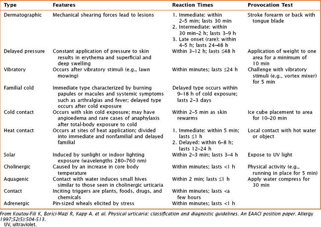
Physical urticarias have a specific trigger that directly induces hives within minutes, with hives lasting minutes to a few hours. Physical urticarias are further classified into multiple subtypes (Table 20-1), each having a different physical trigger; thus, an individual can have more than one subtype. Mast cell degranulation is included in the pathogenesis of most subtypes of physical urticaria, including dermatographic, cholinergic, cold, and solar urticaria, but serum immunoglobulin may also be involved as demonstrated by passive transfer experiments.
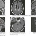
Angioedema

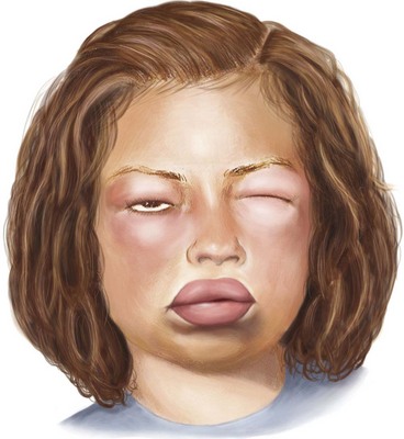
Angioedema, which is a swelling of the dermis, subcutaneous, and submucosal tissues, often coexists with urticaria but typically persists past 24 hours (Figure 20-2). Angioedema is described as painful or burning in quality. The lack of pruritus in angioedema may be caused by fewer mast cells in the lower dermis and subcutis. Idiopathic angioedema coexists with urticaria, occurring in up to 50% of those with CIU. Angiotensin-converting enzyme (ACE) inhibitors can also trigger angioedema via the bradykinin pathway.

Clinical Presentation

Urticarial lesions are intensely pruritic and can affect any location on the body. They are typically transient, lasting less than 24 hours. Unlike atopic dermatitis, excoriation is not a typical finding in urticaria, regardless of the degree of pruritus experienced. Lesions can vary in size and can be confluent. Similar to urticaria, angioedema can occur anywhere on the body, although it frequently involves face, lips, tongue, throat, and extremities, but unlike urticaria, it commonly involves mucous membranes. Cold urticaria is the exception because it may involve swelling of the tongue or palate. Urticarial lesions usually demonstrate complete resolution without skin pigment changes.

Patients with long-lasting lesions or the presence of other systemic symptoms should be evaluated for other diseases (see Box 20-1). Urticarial vasculitis is classified by painful or pruritic lesions lasting longer than 48 hours and may leave residual skin changes unrelated to excoriation. Concurrent systemic complaints or abnormal laboratory findings indicative of an inflammatory process, such as an elevated erythrocyte sedimentation rate (ESR) or C-reactive protein (CRP) or low complement levels, are seen. A skin biopsy is required to rule out urticarial vasculitis. Urticaria pigmentosa, a subset of mastocytosis, may mimic urticaria, although these lesions are typically pigmented and last longer than urticaria. The presence of fever with urticaria can occur in Schnitzler’s syndrome and Muckle-Wells syndrome. Schnitzler’s syndrome is described as recurrent urticaria with arthralgia, fever, and elevation in inflammatory markers in association with an elevation in IgM. Muckle-Wells syndrome is a periodic fever syndrome with urticaria associated with periodic, unexplained fevers.

History

In diagnosing either acute or chronic urticaria, a thorough history is the most important element along with a detailed physical examination. The history should elicit the time of onset of hives (because some patients may experience diurnal variation) and the specific days that the patient is affected because there may be an association with certain environmental exposures or stressors. A description of the lesions, including their shape, size, distribution, color, pigmentation, and the quality of pain or itch, is important in confirming the diagnosis of urticaria. To identify acute triggers, the history should elicit recent use of medications, including antibiotics, nonsteroidal antiinflammatory drugs (NSAIDs), and aspirin; food ingested shortly before symptom occurrence (especially within 2 hours of lesion appearance); implanted surgical devices; insect stings; and changes in environment. Questions regarding recent infections or to evaluate for symptoms of thyroid disease are important (see Chapter 68). For female patients, it is important to investigate if there is a variation of hives in relation to hormonal changes observed with menses or pregnancy. Physical triggers are important for defining physical urticarias. Further evaluation of life stressors may assist in understanding the timing of lesions. It is also important to evaluate how the patient is coping and what therapeutics, including nonprescription medications or dietary changes, are being used and if they are providing any relief. Some patients with chronic urticaria are on restricted diets, which are unnecessary and may lead to nutritional deficits. Medication side effects should also be evaluated.

For patients with angioedema without a history of urticaria, it is important to ask about family history of angioedema. Hereditary angioedema (HAE), an autosomal dominant disease involving a defect in C1 esterase inhibitor function, presents with isolated angioedema that can sometimes be disfiguring and life threatening if the airway is involved. It is important to question about episodic abdominal pain and abdominal surgeries in these patients because many times their sole presenting symptoms are abdominal pain and vomiting.

Evaluation and Management

Laboratory Testing

No routine laboratory testing is indicated in urticaria unless the clinician is attempting to identify the trigger or if the patient has an atypical clinical presentation. For patients with acute urticaria suspected of having a food allergy, specific skin testing and specific IgE measurements may be helpful, although the latter should only be done if skin test results are positive or if it is contraindicated. There is a high percentage of false-positive test results in skin testing to foods, so testing should occur only to foods of concern. Skin testing is difficult to perform in patients with chronic urticaria because of the high prevalence of dermatographism and delayed pressure features in this group, as well as their dependence on antihistamines. Patients with physical urticaria do not need laboratory testing. In CIU, laboratory testing acts as an adjunct to history and examination, especially in patients who fail to respond to conventional therapies or who have uncharacteristic lesions. If there are concerns for vasculitis or autoimmune disorders, an ESR and antinuclear antibody (ANA) are good screening tests. Complement levels, specifically C3 and C4, should be considered. Other laboratory screening tests, such as a complete blood count with differential, complete metabolic panel, and urinalysis, should be obtained if there are concerns for systemic disease. Patients with significant angioedema without urticaria should also have complement levels, specifically C4, and C1 inhibitor function testing; C4 levels are typically low in patients with HAE during acute episodes. Symptoms consistent with thyroid disease warrant thyroid screening with a thyroid-stimulating hormone and thyroid autoantibody testing. If there are signs, symptoms, or laboratory tests concerning for malignancy, a chest radiograph is recommended.

Even though viral infections, especially viral upper respiratory infections, are common causes of acute urticaria, testing for viruses does not change therapy or outcomes. Other infectious agents have been implicated in urticaria, and testing for these should be guided by the history and examination. Hepatitis, especially hepatitis C, has been reported to manifest as chronic urticaria; thus, screening studies for hepatitis B and C should be considered in patients with risk factors. In patients with symptoms of gastroesophageal reflux or gastritis, it may be indicated to obtain Helicobacter pylori serology because this organism is thought to trigger autoantibody production in some patients with CIU. If there is peripheral eosinophilia, the stool should be tested for ova and parasites. Bacterial and fungal cultures do not need to be ordered for urticaria because these are unusual triggers of hives.

Management

The treatment of CIU is a greater challenge than that of acute and physical urticarias. Patients may be less frustrated if they are educated on the natural history of CIU. For all types of urticaria, if there is a clear avoidable trigger, such as in confirmed food allergy or physical urticaria, it should be eliminated. In chronic urticaria, it is unlikely that foods are contributing in a significant manner, and there are no conclusive data supporting diets with avoidance of foods high in salicylates. For all patients, avoidance of aspirin and salicylate-containing medications, as well as NSAIDs, is advisable. Both aspirin and NSAIDs can aggravate urticaria and angioedema via inhibition of prostaglandin synthesis; thus, patients should switch to selective cyclooxygenase-2 inhibitors if a chronic analgesic agent is needed. For patients with angioedema, ACE inhibitors should be avoided. All patients should avoid morphine and codeine-containing products that can directly stimulate skin mast cells.

The mainstay of treatment for urticaria is symptom alleviation using antihistamines. Because of the soporific side effect of antihistamines, less sedating H1-blockers, such as loratadine and cetirizine, are preferred for more regular use, with the classic sedating H1-blockers, such as diphenhydramine and hydroxyzine, reserved for use on an as-needed basis. Different H1-blockers may be tried because one may work better per individual; for select patients with CIU, the simultaneous use of multiple H1-blockers may be necessary. H2-blockers can provide an added benefit in combination with H1-blockade and may even increase serum concentration of the H1-blocker. Doxepin is a tricyclic antidepressant with some H1- and H2-receptor antagonist properties, and it can be used for nighttime itch, with the added benefit of assisting with disease-associated depression. It should be used with caution because it is very sedating; can decrease the effectiveness of other drugs, such as cimetidine and macrolides; and is contraindicated if a patient is taking monoamine oxidase inhibitors because of the risk for developing prolonged QT syndrome.

Evidence for leukotriene receptor antagonist (LTRA) use in chronic urticaria is conflicting. Some benefit has been observed in CIU and physical urticaria (specifically cold-induced and delayed pressure subtypes), as well as acute urticaria caused by aspirin or food. The Food and Drug Administration has placed a warning indicating that LTRA drugs may alter children’s moods; thus, these medications should only be used in children without underlying mood disorders and in conjunction with other therapies, such as antihistamines, with discontinuation of the LTRA if no added benefit is observed.

For severe cases of acute urticaria, a short course of systemic corticosteroids can lead to rapid symptom alleviation. Systemic corticosteroids also have a role in severe, antihistamine-resistant CIU when rapid control is warranted, as with episodes of significant angioedema. The use of systemic corticosteroids should be judicious because of the side effect profile with prolonged use, including greater risk for osteoporosis, peptic ulcer disease, diabetes, and hypertension, to name a few. Also, it is important that the patients and their families are aware that hives may recur after the corticosteroid effect has worn off because corticosteroids do not alter the disease process. Topical corticosteroids play no role in the treatment of patients with urticaria, with the exception of perhaps some minimal benefit in localized delayed pressure urticaria.

Unlike HAE, angioedema associated with acute or chronic urticaria is rarely life threatening, but it may respond to epinephrine. Thus, an epinephrine autoinjector may provide that subset of patients with relief. There is no role for epinephrine, steroids, or antihistamines in patients with HAE.

More recently, immunomodulators have been used for the treatment of patients with severe, steroid-dependent chronic urticaria. Cyclosporine, sulfasalazine, and dapsone have demonstrated some benefit in a subset of patients. Dapsone, which inhibits neutrophil function, may be beneficial for patients with neutrophil-predominant infiltrates on skin biopsy. Isolated reports of cyclophosphamide and methotrexate use in CIU appear to demonstrate some benefit. Colchicine and hydroxychloroquine have also been suggested as therapeutics. For patients with suspected autoimmune CIU, plasmapheresis has been used to remove autoantibodies with temporary amelioration of disease. Intravenous immunoglobulin infusions have also been studied in patients with functional autoantibodies with a transient beneficial effect. Overall, these therapeutics should be used with caution and only when the patient has failed all other therapies. Most of these therapies also require frequent side effect monitoring, including laboratory testing.

Patients with CIU have a higher incidence of thyroid disease and thyroid autoantibodies compared with the general population, so only patients with underlying thyroid disease should be treated for underlying process. Euthyroid patients with thyroid autoantibodies should not be treated with thyroid hormone replacement.