[level-membership-for-dermatology-category]
Urticaria and angioedema
Urticaria (hives) is a common eruption characterized by transient, usually pruritic, wheals due to acute dermal oedema from extravascular leakage of plasma. Angioedema signifies a larger area of oedema involving the dermis and subcutis. A classification is shown in Table 1.
Table 1 Classification of urticaria and angioedema
| Allergic (IgE mediated) mast cell degranulation | SystemicSkin contact | Food, drugs, latex (aerosols)Animal saliva, pollen, latex |
| Non-allergic (non-IgE mediated) mast cell degranulation | Chronic ordinaryPhysicalPharmacological | No cause identifiable (commonest subtype)Dermographism, cholinergic, cold, solar, heat, delayed pressureAspirin, opiates, non-steroidal drugs, food additives, ACE inhibitors (p. 87) |
| Autoimmune disease | Systemic lupus erythematosus (p. 80), thyroid antibodies, anti-IgE receptor antibodies, urticarial vasculitis (p. 77) | |
| Genetic | C1 esterase inhibitor deficiency (p. 77), mastocytosis (p. 116) | |
| Other | Infection, paraneoplastic, skin contact (nettle sting) |
Aetiopathogenesis
 IgE-mediated (type I) hypersensitivity (p. 11) is the best understood mechanism; antigen cross-links immunoglobulin (Ig) E molecules on the surface of mast cells, resulting in degranulation with release of vasoactive agents.


Clinical presentation
Chronic idiopathic urticaria
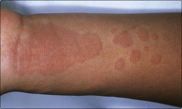
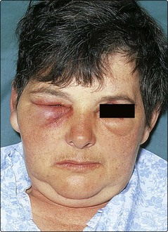
Itchy pink wheals appear as papules or plaques anywhere on the skin surface (Fig. 1). Typically, they last for less than 24 h and disappear without a trace. Wheals may be round, annular or polycyclic, and vary in diameter from a few millimetres to several centimetres. Their number ranges from a few to many appearing each day, depending on the severity of the condition. Angioedema, usually with swelling of the tongue or lips, may occur (Fig. 2). Pharmacological agents often act as provoking factors, but normally no underlying cause is found. The condition resolves spontaneously within 6 months in 50% of cases, although a minority are troubled for years.
Acute urticaria
The sudden onset of urticaria or angioedema may be due to an IgE-mediated type I reaction. The patient can often identify the offending allergen. Commonly, it is a food (e.g. egg, fish or peanuts), a drug (e.g. an antibiotic) or contact with latex (p. 125). Sometimes no cause is found.
Physical urticarias
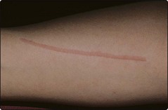
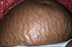
Cold, heat, sun exposure, pressure and even water can all induce urticaria at the stimulated site. Dermographism, found in 5% of normal people, describes whealing induced by firm stroking of the skin (Fig. 3). In a few individuals, it is exaggerated and symptomatic. The wheals in cholinergic urticaria are small, intensely itchy papules that appear in response to sweating, as induced by exercise, heat, emotion or spicy food. The eruption lasts for a few minutes to an hour.
Differential diagnosis
Urticaria is usually differentiated from other dermatoses, although pemphigoid (p. 78) or dermatitis herpetiformis (p. 79) occasionally present with an urticarial eruption. Toxic erythema and erythema multiforme (p. 82) may be urticated at first but, when the lesions persist for over 48 h, urticaria can be excluded. Facial erysipelas sometimes resembles angioedema but has a sharper margin and the patient is unwell with a fever.
Investigation
Underlying causes or provoking factors are better revealed by a careful history and examination than by laboratory tests. However, a full blood count, liver function tests, antinuclear antibody test, erythrocyte sedimentation rate (ESR) and urinalysis are often done to exclude systemic conditions (Table 1). Dermographism is demonstrated by firmly stroking the skin, and cold urticaria induced by holding an ice cube on the arm for up to 20 min. If C1 esterase inhibitor deficiency is suspected, C4 levels can be used as a screening test prior to C1 esterase inhibitor level assay.
Management


[/level-membership-for-dermatology-category][not-level-membership-for-dermatology-category]
Urticaria and angioedema
Urticaria (hives) is a common eruption characterized by transient, usually pruritic, wheals due to acute dermal oedema from extravascular leakage of plasma. Angioedema signifies a larger area of oedema involving the dermis and subcutis. A classification is shown in Table 1.
Table 1 Classification of urticaria and angioedema
| Allergic (IgE mediated) mast cell degranulation | SystemicSkin contact | Food, drugs, latex (aerosols)Animal saliva, pollen, latex |
| Non-allergic (non-IgE mediated) mast cell degranulation | Chronic ordinaryPhysicalPharmacological | No cause identifiable (commonest subtype)Dermographism, cholinergic, cold, solar, heat, delayed pressureAspirin, opiates, non-steroidal drugs, food additives, ACE inhibitors (p. 87) |
| Autoimmune disease | Systemic lupus erythematosus (p. 80), thyroid antibodies, anti-IgE receptor antibodies, urticarial vasculitis (p. 77) | |
| Genetic | C1 esterase inhibitor deficiency (p. 77), mastocytosis (p. 116) | |
| Other | Infection, paraneoplastic, skin contact (nettle sting) |
Aetiopathogenesis
IgE-mediated (type I) hypersensitivity (p. 11) is the best understood mechanism; antigen cross-links immunoglobulin (Ig) E molecules on the surface of mast cells, resulting in degranulation with release of vasoactive agents.
