[level-membership-for-orthopaedics-category]
4 Upper limb
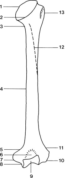
Humerus (Figs 4.1 and 4.2)
Type
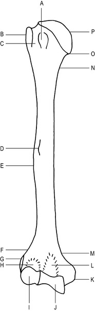
Fig. 4.1 Right humerus (anterior aspect).
A–Lesser tuberosity (tubercle)
B–Greater tuberosity (tubercle)
Main parts
Features of the upper end of the humerus
Lesser tuberosity (lesser tubercle) –
anteriorly, the tendon of the subscapularis muscle is attached.
Features of the shaft of the humerus
Deltoid tuberosity –
for the attachment of the deltoid muscle, located on the antero-lateral surface.
Trochlea –
pulley-shaped; articulates with the trochlear notch of the ulna. Has a large medial lip which forms the ‘carrying angle’ (see elbow joint).
Olecranon fossa –
posteriorly receives the olecranon process of the ulna when the elbow joint is extended.
Radiographic appearances of the humerus (Figs 4.3 and 4.4)
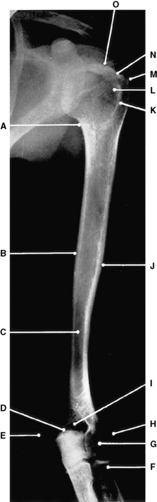
Fig. 4.3 Left humerus: anteroposterior projection.
I–Olecranon and coronoid fossae
K–Intertubercular sulcus (Bicipital groove)
(From Bryan 1996.)
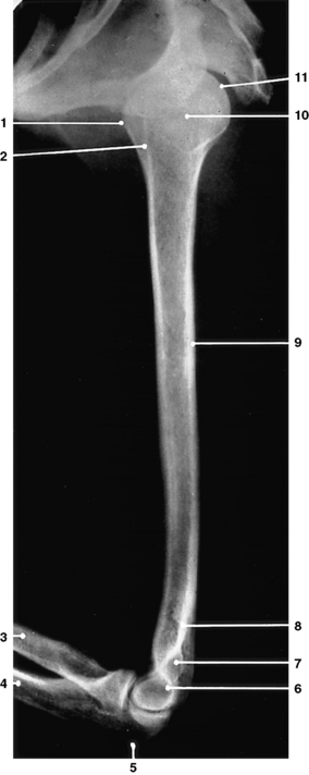
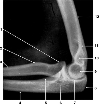
Fig. 4.4 Left humerus: lateral projection.
2 – Intertubercular sulcus (Bicipital groove)
(From Bryan 1996.)
Radius (Figs 4.5 and 4.6)
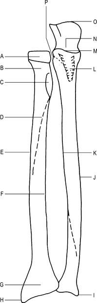
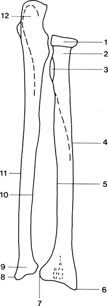
Ulna (Figs 4.5 and 4.6)
Articulations
Radial notch of the ulna with the head of the radius to form the superior radio-ulnar joint.
Head of ulna with the ulnar notch of the radius to form part of the inferior radio-ulnar joint.
Trochlear notch of the ulna with the trochlea of the humerus to form part of the elbow joint.
Main parts
Features of the upper end of the ulna
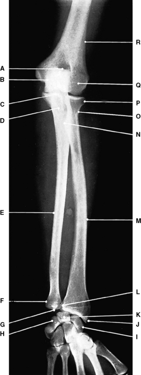
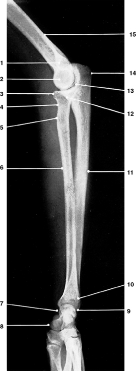
Radiographic appearances of the radius and ulna (Figs 4.7 and 4.8)
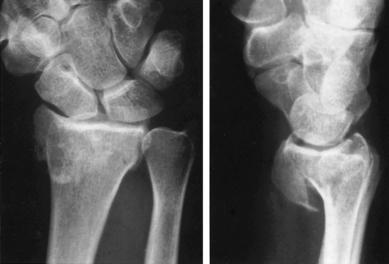
Monteggia’s fracture-dislocation (Fig. 4.9)
Fracture of the upper third of the ulna with dislocation of the radial head.

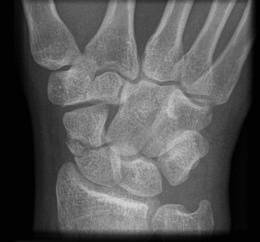
Hand (Fig. 4.11)
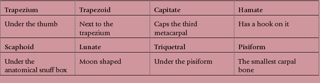
Carpal bones
Type
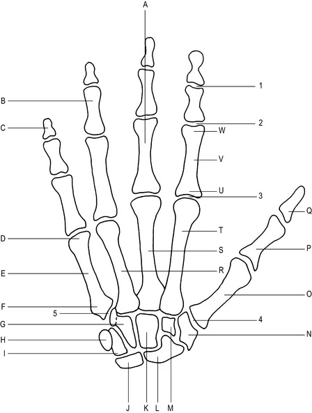
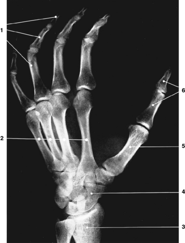
Fig. 4.11 Right hand (palmar aspect).
A–Shaft of proximal phalanx of middle finger
B–Shaft of middle phalanx of ring finger
C–Shaft of distal phalanx of little finger
P–Base of proximal phalanx of thumb
Q–Base of distal phalanx of thumb
U–Base of proximal phalanx of index finger
V–Shaft of proximal phalanx of index finger
W–Head of proximal phalanx of index finger
1–Distal interphalangeal joint of index finger (synovial hinge joint)
2–Proximal interphalangeal joint of index finger (synovial hinge joint)
3–2nd metacarpophalangeal joint (synovial ellipsoid joint)
Articulations
Scaphoid with the radius, lunate, trapezoid, trapezium.
Lunate with the radius, scaphoid, capitate, triquetral.
Triquetral with the hamate, pisiform, lunate.
Hamate with the 4th and 5th metacarpals, capitate, triquetral.
Capitate with the 2nd, 3rd and 4th metacarpals, hamate, lunate, scaphoid, trapezoid.
Trapezoid with the 2nd metacarpal, trapezium, scaphoid, capitate.
Trapezium with the 1st and 2nd metacarpals, trapezoid, scaphoid.
Ossification
Primary centres
| Capitate – 2nd month | Scaphoid – 4th–5th year |
| Hamate – 3rd month | Trapezium – 4th–5th year |
| Triquetral – 3rd year | Trapezoid – 4th–5th year |
| Lunate – 4th year | Pisiform – 9th–12th year. |
N.B. The dates at which the carpal bones ossify are subject to considerable variation.
Metacarpal bones (Fig. 4.11)
Articulations
1st metacarpal with the proximal phalanx of the thumb, and the trapezium.
2nd metacarpal with the proximal phalanx of the index finger, the trapezium, trapezoid and capitate.
3rd metacarpal with the proximal phalanx of the middle finger and the capitate.
4th metacarpal with the proximal phalanx of the ring finger, the capitate and the hamate.
5th metacarpal with the proximal phalanx of the little finger and the hamate.
Phalanges (Fig. 4.11)
Articulations
4 middle phalanges with the corresponding proximal and distal phalanges.
5 distal phalanges with the corresponding middle phalanges and the proximal phalanx of the thumb.
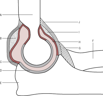
Elbow joint (Figs 4.15, 4.16 and 4.17)
Type
Synovial hinge joint, continuous with the superior radio-ulnar joint.
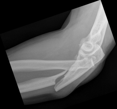
Radiographic appearances of the elbow joint (Figs 4.18 to 4.22)
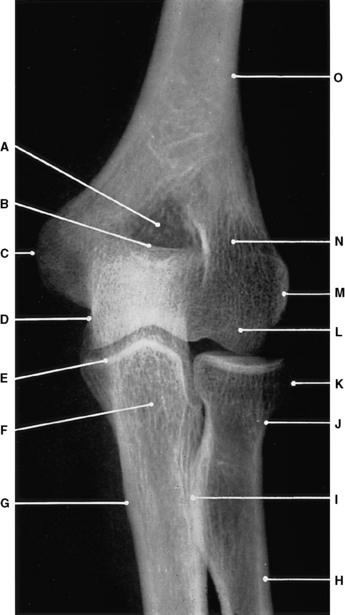
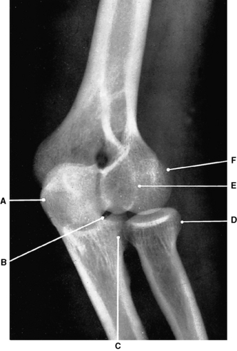
Fig. 4.18 Left elbow joint: anteroposterior projection.
A–Olecranon and coronoid fossae
(From Bryan 1996.)
Wrist joint (Figs 4.23 and 4.24)
Type
Supporting ligaments
Palmar radiocarpal ligament –
from the radius to the scaphoid, lunate and triquetral on the anterior aspect of the joint.
Palmar ulnocarpal ligament –
from the ulnar styloid process to the lunate and triquetral on the anterior aspect of the wrist.
Dorsal radiocarpal ligament –
from the radius to the scaphoid, lunate and triquetral on the posterior aspect of the wrist.
Movements
Flexion by the flexor carpi radialis and flexor carpi ulnaris muscles.
Extension by the extensor carpi radialis and the extensor carpi ulnaris muscles.
Abduction by the flexor and extensor carpi radialis muscles.
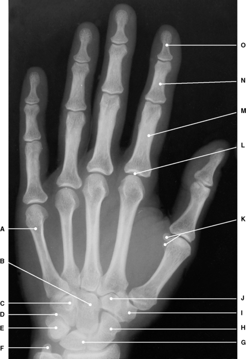
Radiographic appearances of the wrist joint (Figs 4.25 to 4.30)
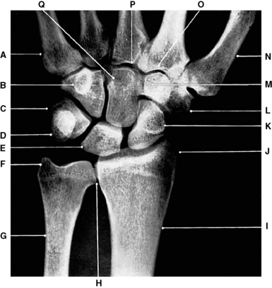
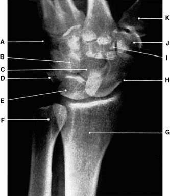
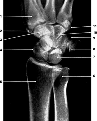
Fig. 4.25 Left wrist joint: posteroanterior projection.
P–Styloid process of 3rd metacarpal
(From Bryan 1996.)
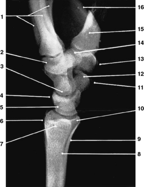
Fig. 4.26 Left wrist joint: lateral projection.
1 – 2nd to 5th metacarpal bones
10 – Styloid process of radius
(From Bryan 1996.)
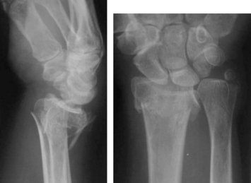
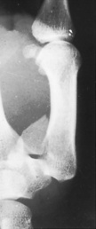
Smith’s fracture (Fig. 4.32)
Transverse fracture of the radius with anterior displacement of the distal fragment.
First carpometacarpal joint
Supporting ligaments
Movements
Flexion by the flexor pollicis, opponens pollicis and flexor pollicis longus.
Extension by the abductor pollicis longus, extensor pollicis brevis and extensor pollicis longus.
Abduction by the abductor pollicis brevis and the abductor pollicis longus.
Adduction by the adductor pollicis.
[/level-membership-for-orthopaedics-category][not-level-membership-for-orthopaedics-category]
4 Upper limb
Humerus (Figs 4.1 and 4.2)
Type
Fig. 4.1 Right humerus (anterior aspect).
A–Lesser tuberosity (tubercle)
B–Greater tuberosity (tubercle)
Main parts
Features of the upper end of the humerus
Lesser tuberosity (lesser tubercle) –
anteriorly, the tendon of the subscapularis muscle is attached.
Features of the shaft of the humerus
Deltoid tuberosity –
for the attachment of the deltoid muscle, located on the antero-lateral surface.
Trochlea –
pulley-shaped; articulates with the trochlear notch of the ulna. Has a large medial lip which forms the ‘carrying angle’ (see elbow joint).
Olecranon fossa –
posteriorly receives the olecranon process of the ulna when the elbow joint is extended.
Radiographic appearances of the humerus (Figs 4.3 and 4.4)
Fig. 4.3 Left humerus: anteroposterior projection.
I–Olecranon and coronoid fossae
K–Intertubercular sulcus (Bicipital groove)
(From Bryan 1996.)
Fig. 4.4 Left humerus: lateral projection.
2 – Intertubercular sulcus (Bicipital groove)
(From Bryan 1996.)
Radius (Figs 4.5 and 4.6)
Ulna (Figs 4.5 and 4.6)
Articulations
Radial notch of the ulna with the head of the radius to form the superior radio-ulnar joint.
Head of ulna with the ulnar notch of the radius to form part of the inferior radio-ulnar joint.
Trochlear notch of the ulna with the trochlea of the humerus to form part of the elbow joint.
