[level-membership-for-basic-science-category]
CHAPTER 47 Upper arm
SKIN AND SOFT TISSUE
SKIN
Cutaneous vascular supply
The blood supply to the skin of the upper arm can be divided into three regions with separate supply: the deltoid region, supplied by musculocutaneous perforators, and the medial and lateral regions, supplied by fasciocutaneous perforators (Cormack & Lamberty 1994; Salmon 1994) (Fig. 47.1, Fig. 47.2).
The deltoid region is supplied by the posterior circumflex humeral artery (p. 817) via musculocutaneous perforators. After the posterior circumflex humeral artery passes through the quadrangular space (p. 814) it gives off a descending branch, which runs down to the deltoid insertion and the overlying skin, and an ascending branch which passes superiorly towards the acromion, pierces the edge of deltoid and the deep fascia to fan out and supply the overlying skin.
Cutaneous innervation
The skin of the shoulder region is supplied by the supraclavicular nerves from the cervical plexus. The floor of the axilla and upper medial surface of the arm is supplied by the lateral branch of the second intercostal nerve (the intercostobrachial nerve). The lower aspect of the medial side of the upper arm is supplied by the medial cutaneous nerve of the arm. The lateral aspect of the upper arm is supplied by the upper lateral cutaneous nerve (a branch of the axillary nerve) and the lower lateral cutaneous nerve (a branch of the radial nerve). The posterior aspect is supplied by the posterior cutaneous nerve of the arm, a branch of the radial nerve (Fig. 45.15, Fig. 45.14).
SOFT TISSUE
Brachial fascia
Brachial fascia, the deep fascia of the upper arm, is continuous with the fascia covering deltoid and pectoralis major: it forms a thin, loose sheath for muscles of the upper arm, and sends septa between them. It is thin over biceps, but thicker over triceps and the humeral epicondyles, and is strengthened by fibrous aponeuroses from pectoralis major and latissimus dorsi medially and from deltoid laterally. Strong medial and lateral intermuscular septa extend from it on each side.
MUSCLES
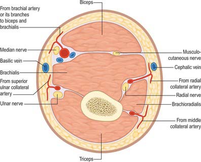
The muscles of the upper arm are coracobrachialis, which acts only on the shoulder joint; biceps and triceps, which cross both shoulder and elbow joints; and brachialis, which acts only at the elbow joint (Fig. 47.2, Fig. 47.3).
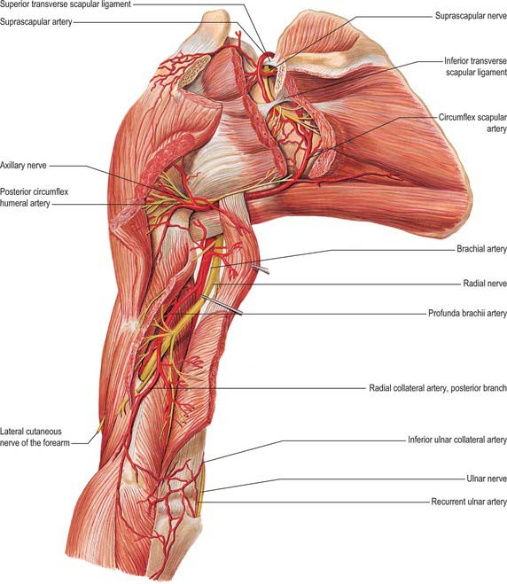
Fig. 47.3 Muscles, vessels and nerves of the left upper arm, viewed from the posterior aspect.
(From Sobotta 2006.)
ANTERIOR COMPARTMENT
Coracobrachialis
Coracobrachialis arises from the apex of the coracoid process, together with the tendon of the short head of biceps, and also by muscular fibres from the proximal 10 cm of this tendon; it ends on an impression, 3–5 cm in length, midway along the medial border of the humeral shaft between the attachments of triceps and brachialis (Fig. 46.20, Fig. 47.4). Accessory slips may be attached to the lesser tubercle, medial epicondyle or medial intermuscular septum.
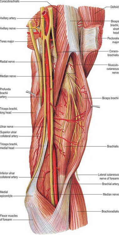
One or more branches from the axillary artery pass deep to the lateral root of the median nerve, and the musculocutaneous nerve, to reach the deep surface of the muscle. Branches from the anterior circumflex humeral artery also supply the deep surface of the muscle. The accompanying artery of the musculocutaneous nerve sends a recurrent branch to the coracoid attachment and gives off a series of branches to the muscle during its intramuscular course. Accessory branches from the thoracoacromial artery provide additional supply to the superficial part of coracobrachialis.
Biceps brachii
Biceps brachii derives its name from its two proximally attached parts or ‘heads’ (Fig. 46.20). The short head arises by a thick flattened tendon from the coracoid apex, together with coracobrachialis. The long head starts within the capsule of the shoulder joint as a long narrow tendon, running from the supraglenoid tubercle of the scapula at the apex of the glenoidal cavity, where it is continuous with the glenoidal labrum (p. 805). The tendon of the long head, enclosed in a double tubular sheath (an extension of the synovial membrane of the joint capsule), arches over the humeral head, emerges from the joint behind the transverse humeral ligament, and descends in the intertubercular sulcus, where it is retained by the transverse humeral ligament and a fibrous expansion from the tendon of pectoralis major. The two tendons lead into elongated bellies that, although closely applied, can be separated to within 7 cm or so of the elbow joint. At this joint they end in a flattened tendon, which is attached to the rough posterior area of the radial tuberosity; a bursa separates the tendon from the smooth anterior area of the tuberosity. As it approaches the radius, the tendon spirals, its anterior surface becoming lateral before being applied to the tuberosity. The tendon has a broad medial expansion, the bicipital aponeurosis, which descends medially across the brachial artery to fuse with deep fascia over the origins of the flexor muscles of the forearm (Fig. 47.5). The tendon can be split without difficulty as far as the tuberosity, whence it can be confirmed that its anterior and posterior layers receive fibres from the short and long heads, respectively.

Biceps is overlapped proximally by pectoralis major and deltoid; distally it is covered only by fasciae and skin, and it forms a conspicuous elevation on the front of the arm. Its long head passes through the shoulder joint; its short head is anterior to the joint. Distally it lies anterior to brachialis, the musculocutaneous nerve and supinator. Its medial border touches coracobrachialis, and overlaps the brachial vessels and median nerve; its lateral border is related to deltoid and brachioradialis.
Brachialis
Brachialis arises from the lower half of the front of the humerus, starting on either side of the insertion of deltoid, and extending distally to within 2.5 cm of the cubital articular surface (Fig. 46.20, Fig. 47.4). It also arises from the intermuscular septa, more from the medial than the lateral, since it is separated distally from the lateral intermuscular septum by brachioradialis and extensor carpi radialis longus. Its fibres converge to a thick, broad tendon which is attached to the ulnar tuberosity and to a rough impression on the anterior aspect of the coronoid process.
POSTERIOR COMPARTMENT
Triceps
Triceps fills most of the extensor compartment of the upper arm (Fig. 46.22, Figs 47.2–47.4). It arises by three heads (long, lateral and medial), from which it takes its name.
The long head descends between teres minor and major, dividing the wedge-shaped interval between them and the humerus into triangular and quadrangular parts (Fig. 46.22). The triangular space contains the circumflex scapular vessels; it is bounded above by teres minor, below by teres major, laterally by the long head of triceps. The quadrangular space transmits the posterior circumflex humeral vessels and the axillary nerve; it is bounded above by subscapularis, teres minor and the articular capsule, below by teres major, medially by the long head of triceps, and laterally by the humerus.
Triceps is the major extensor of the forearm at the elbow joint. The medial head is active in all forms of extension. The lateral and long heads are minimally active except in extension against resistance, as in thrusting or pushing or supporting body weight on the hands with the elbows semiflexed. When the flexed arm is extended at the shoulder joint, the long head may assist in drawing back and adducting the humerus to the thorax. The long head supports the lower part of the capsule of the shoulder joint, especially when the arm is raised. Articularis cubiti probably draws up the posterior part of the capsule of the elbow joint during extension of the forearm. In forceful supination of the semiflexed forearm, involving contraction of both supinator and biceps brachii, the triceps contracts synergistically to maintain the semiflexed position.
VASCULAR SUPPLY AND LYMPHATIC DRAINAGE
ARTERIES
Brachial artery
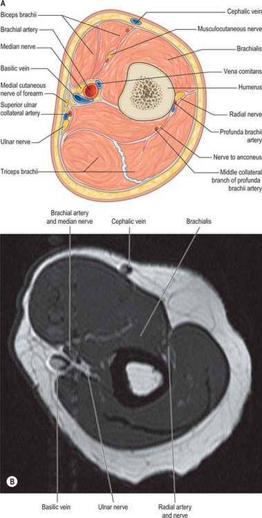
The brachial artery, a continuation of the axillary, begins at the distal (inferior) border of the tendon of teres major and ends about a centimetre distal to the elbow joint (at the level of the neck of the radius) by dividing into radial and ulnar arteries (Fig. 47.5, Fig. 47.6). At first it is medial to the humerus, but gradually spirals anterior to it until it lies midway between the humeral epicondyles. Its pulsation can be felt throughout.
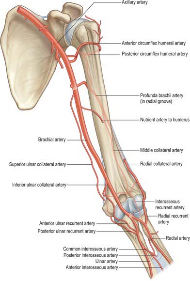
Fig. 47.6 The arterial anastomoses around the left elbow joint viewed from the anterior aspect.
(From Drake, Vogl and Mitchell 2005.)
Relations
At the elbow the brachial artery sinks deeply into the triangular intermuscular cubital fossa (p. 831).
Branches
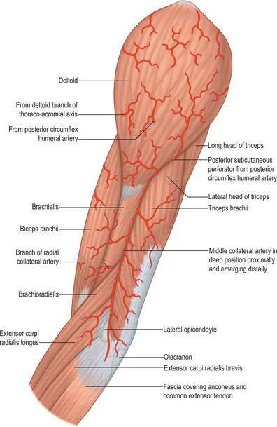
The profunda brachii (Fig. 47.2, Fig. 47.3, Fig. 47.5, Fig. 47.6) is a large branch from the posteromedial aspect of the brachial artery, distal to teres major. It follows the radial nerve closely, at first posteriorly between the long and medial heads of triceps, then in the spiral groove covered by the lateral head of triceps. It supplies muscular branches, the nutrient artery of the humerus, and finally divides into terminal radial and middle collateral branches (Fig. 47.6). The radial collateral branch pierces the lateral intermuscular septum to reach the anterior aspect of the epicondyle of the humerus in the groove between brachioradialis and brachialis, and takes part in the anastomosis around the elbow. The middle collateral branch runs posterior to the septum and epicondyle.
Middle collateral (posterior descending) branch
The middle collateral artery is the larger terminal vessel. It arises posterior to the humerus and descends down the posterior surface of the lateral intermuscular septum to the elbow (Fig. 47.4, Fig. 47.6). Proximally, the artery lies between brachialis (anteriorly) and the lateral head of triceps (posteriorly) while distally it lies between brachioradialis (anteriorly) and the lateral head of triceps (posteriorly). It may pierce the deep fascia and become cutaneous or remain deep to the fascia until anastomosing with the interosseous recurrent artery behind the lateral epicondyle; it gives off about five small fasciocutaneous perforators and often has a small branch that accompanies the nerve to anconeus.
Radial collateral (anterior descending) branch
The radial collateral artery is the continuation of the profunda (Fig. 47.6). It accompanies the radial nerve through the lateral intermuscular septum, descending between brachialis and brachioradialis anterior to the lateral epicondyle, anastomosing with the radial recurrent artery. It supplies brachialis, brachioradialis, the radial nerve and a few fasciocutaneous perforators.
The nutrient artery of the humerus arises near the mid-level of the upper arm, and enters the nutrient canal near the attachment of coracobrachialis, posterior to the deltoid tuberosity; it is directed distally.
Superior ulnar collateral artery
The superior ulnar collateral artery arises a little distal to the mid-level of the upper arm, usually from the brachial artery, but often as a branch from the profunda brachii (Figs 47.4–47.6). It accompanies the ulnar nerve, piercing the medial intermuscular septum to descend in the posterior compartment and supply the medial head of triceps. It passes between the medial epicondyle and olecranon, ending deep to flexor carpi ulnaris by anastomosing with the posterior ulnar recurrent and inferior collateral arteries. A branch sometimes passes anterior to the medial epicondyle and anastomoses with the anterior ulnar recurrent artery.
Inferior ulnar collateral (supratrochlear) artery
The inferior ulnar collateral artery begins approximately 5 cm proximal to the elbow, passes medially between the median nerve and brachialis and, piercing the medial intermuscular septum, curls round the humerus between the triceps and bone (Fig. 47.5, Fig. 47.6). It forms, by its junction with the middle collateral branch of the profunda brachii artery, an arch proximal to the olecranon fossa. As it lies on brachialis it gives off branches which descend anterior to the medial epicondyle to anastomose with the anterior ulnar recurrent artery. Behind the epicondyle a branch anastomoses with the superior ulnar collateral and posterior ulnar recurrent arteries.
VEINS
Brachial veins
These deep veins have numerous anastomoses with each other and with the superficial veins.
Superficial veins
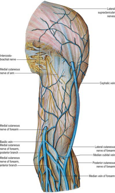
The cephalic vein ascends in front of the elbow superficial to a groove between the brachioradialis and biceps, crosses superficial to the lateral cutaneous nerve of the forearm, ascends lateral to biceps and between pectoralis major and deltoid, where it adjoins the deltoid branch of the thoracoacromial artery (Fig. 45.5, Fig. 47.7). Entering the infraclavicular fossa to pass behind the clavicular head of pectoralis major, it pierces the clavipectoral fascia, crosses the axillary artery and joins the axillary vein just below clavicular level. It may connect with the external jugular by a branch anterior to the clavicle. Sometimes the median cubital vein is large, transferring most blood from the cephalic to the basilic vein; the proximal cephalic vein is then absent or much diminished.
INNERVATION
Median nerve
The median nerve (Fig. 45.7, Fig. 47.5, Fig. 47.8) enters the arm lateral to the brachial artery. Near the insertion of coracobrachialis it crosses in front of (rarely behind) the artery, descending medial to it to the cubital fossa where it is posterior to the bicipital aponeurosis and anterior to brachialis, separated by the latter from the elbow joint.
Musculocutaneous nerve
The musculocutaneous nerve is the nerve of the anterior compartment of the arm (Fig. 47.5, Fig. 47.8). It gives a branch to the shoulder joint and then passes through coracobrachialis, which it supplies, emerging to pass between biceps and brachialis. It sends branches to both these muscles. In the cubital fossa it lies at the lateral margin of the biceps tendon where it continues as the lateral cutaneous nerve of the forearm.
The musculocutaneous nerve has frequent variations. It may run behind coracobrachialis or adhere for some distance to the median nerve and pass behind biceps. Some fibres of the median nerve may run in the musculocutaneous nerve, leaving it to join their proper trunk; less frequently the reverse occurs, and the median nerve sends a branch to the musculocutaneous. Occasionally it supplies pronator teres and may replace radial branches to the dorsal surface of the thumb.
Ulnar nerve
The ulnar nerve gives no branches in the arm (Fig. 47.5, Fig. 47.8). It runs distally through the axilla medial to the axillary artery and between it and the vein, continuing distally medial to the brachial artery as far as the midarm. Here it pierces the medial intermuscular septum, inclining medially as it descends anterior to the medial head of triceps to the interval between the medial epicondyle and the olecranon, with the superior ulnar collateral artery.
Radial nerve
The radial nerve descends behind the third part of the axillary artery and the upper part of the brachial artery, anterior to subscapularis and the tendons of latissimus dorsi and teres major (Figs 47.2–47.4, Fig. 47.8). With the profunda brachii artery it inclines dorsally, passing through the triangular space (see p. 814) below the lower border of teres major, between the long head of triceps and the humerus. Here it supplies the long head of triceps, and gives rise to the posterior cutaneous nerve of the arm which supplies the skin along the posterior surface of the upper arm. It then spirals obliquely across the back of the humerus, lying posterior to the uppermost fibres of the medial head of triceps which separate the nerve from the bone in the first part of the spiral groove. Here it gives off a muscular branch to the lateral head of triceps and a branch which passes through the medial head of triceps to anconeus. On reaching the lateral side of the humerus it pierces the lateral intermuscular septum to enter the anterior compartment; it then descends deep in a furrow between brachialis and proximally brachioradialis, then more distally extensor carpi radialis longus. Anterior to the lateral epicondyle it divides into superficial and deep terminal rami.
Cutaneous branches
Medial cutaneous nerve of arm
The medial cutaneous nerve of arm supplies the skin of the medial aspect of the arm (Fig. 47.7). It is the smallest branch of the brachial plexus, and arises from the medial cord and contains fibres from the eighth cervical and first thoracic ventral rami. It traverses the axilla, crossing anterior or posterior to the axillary vein, to which it is then medial, and communicating with the intercostobrachial nerve; it descends medial to the brachial artery and basilic vein (Fig. 47.7) to a point midway in the upper arm, where it pierces the deep fascia to supply a medial area in the distal third of the arm, extending on to its anterior and posterior aspects. Rami reach the skin anterior to the medial epicondyle, and over the olecranon. It connects with the posterior branch of the medial cutaneous nerve of the forearm. Sometimes the medial cutaneous nerve of the arm and the intercostobrachial nerve are connected in a plexiform manner in the axilla. The intercostobrachial nerve may be large and reinforced by part of the lateral cutaneous branch of the third intercostal nerve. It then replaces the medial cutaneous nerve of the arm and receives a connection representing the latter from the brachial plexus (occasionally this connection is absent).
Medial cutaneous nerve of forearm
The medial cutaneous nerve of forearm comes from the medial cord (Fig. 47.7). It is derived from the eighth cervical and first thoracic ventral rami. At first it is between the axillary artery and vein and gives off a ramus which pierces the deep fascia to supply the skin over the biceps, almost to the elbow. The nerve descends medial to the brachial artery, pierces the deep fascia with the basilic vein midway in the arm and divides into anterior and posterior branches. The larger, anterior branch usually passes in front of, occasionally behind, the median cubital vein, descending anteromedial in the forearm to supply the skin as far as the wrist and connecting with the palmar cutaneous branch of the ulnar nerve. The posterior branch descends obliquely medial to the basilic vein, anterior to the medial epicondyle, and curves round to the back of the forearm, descending on its medial border to the wrist, supplying the skin. It connects with the medial cutaneous nerve of the arm, the posterior cutaneous nerve of the forearm, and the dorsal branch of the ulnar.
Cormack GC, Lamberty BGH. The Arterial Anatomy of Skin Flaps. Edinburgh: Churchill Livingstone, 1994.
Salmon M. Anatomic studies. In: Taylor GI, Razaboni RM. Book 1: Arteries of the Muscles of the Extremities and the Trunk. Book 2: Arterial Anastomotic Pathways of the Extremities. New York: Quality Medical Publishing, 1994.
[/level-membership-for-basic-science-category][not-level-membership-for-basic-science-category]
CHAPTER 47 Upper arm
SKIN AND SOFT TISSUE
SKIN
Cutaneous vascular supply
The blood supply to the skin of the upper arm can be divided into three regions with separate supply: the deltoid region, supplied by musculocutaneous perforators, and the medial and lateral regions, supplied by fasciocutaneous perforators (Cormack & Lamberty 1994; Salmon 1994) (Fig. 47.1, Fig. 47.2).
The deltoid region is supplied by the posterior circumflex humeral artery (p. 817) via musculocutaneous perforators. After the posterior circumflex humeral artery passes through the quadrangular space (p. 814) it gives off a descending branch, which runs down to the deltoid insertion and the overlying skin, and an ascending branch which passes superiorly towards the acromion, pierces the edge of deltoid and the deep fascia to fan out and supply the overlying skin.
Cutaneous innervation
The skin of the shoulder region is supplied by the supraclavicular nerves from the cervical plexus. The floor of the axilla and upper medial surface of the arm is supplied by the lateral branch of the second intercostal nerve (the intercostobrachial nerve). The lower aspect of the medial side of the upper arm is supplied by the medial cutaneous nerve of the arm. The lateral aspect of the upper arm is supplied by the upper lateral cutaneous nerve (a branch of the axillary nerve) and the lower lateral cutaneous nerve (a branch of the radial nerve). The posterior aspect is supplied by the posterior cutaneous nerve of the arm, a branch of the radial nerve (Fig. 45.15, Fig. 45.14).
SOFT TISSUE
Brachial fascia
Brachial fascia, the deep fascia of the upper arm, is continuous with the fascia covering deltoid and pectoralis major: it forms a thin, loose sheath for muscles of the upper arm, and sends septa between them. It is thin over biceps, but thicker over triceps and the humeral epicondyles, and is strengthened by fibrous aponeuroses from pectoralis major and latissimus dorsi medially and from deltoid laterally. Strong medial and lateral intermuscular septa extend from it on each side.
MUSCLES
The muscles of the upper arm are coracobrachialis, which acts only on the shoulder joint; biceps and triceps, which cross both shoulder and elbow joints; and brachialis, which acts only at the elbow joint (Fig. 47.2, Fig. 47.3).
Fig. 47.3 Muscles, vessels and nerves of the left upper arm, viewed from the posterior aspect.
(From Sobotta 2006.)
ANTERIOR COMPARTMENT
Coracobrachialis
Coracobrachialis arises from the apex of the coracoid process, together with the tendon of the short head of biceps, and also by muscular fibres from the proximal 10 cm of this tendon; it ends on an impression, 3–5 cm in length, midway along the medial border of the humeral shaft between the attachments of triceps and brachialis (Fig. 46.20, Fig. 47.4). Accessory slips may be attached to the lesser tubercle, medial epicondyle or medial intermuscular septum.
One or more branches from the axillary artery pass deep to the lateral root of the median nerve, and the musculocutaneous nerve, to reach the deep surface of the muscle. Branches from the anterior circumflex humeral artery also supply the deep surface of the muscle. The accompanying artery of the musculocutaneous nerve sends a recurrent branch to the coracoid attachment and gives off a series of branches to the muscle during its intramuscular course. Accessory branches from the thoracoacromial artery provide additional supply to the superficial part of coracobrachialis.
Biceps brachii
Biceps brachii derives its name from its two proximally attached parts or ‘heads’ (Fig. 46.20). The short head arises by a thick flattened tendon from the coracoid apex, together with coracobrachialis. The long head starts within the capsule of the shoulder joint as a long narrow tendon, running from the supraglenoid tubercle of the scapula at the apex of the glenoidal cavity, where it is continuous with the glenoidal labrum (p. 805). The tendon of the long head, enclosed in a double tubular sheath (an extension of the synovial membrane of the joint capsule), arches over the humeral head, emerges from the joint behind the transverse humeral ligament, and descends in the intertubercular sulcus, where it is retained by the transverse humeral ligament and a fibrous expansion from the tendon of pectoralis major. The two tendons lead into elongated bellies that, although closely applied, can be separated to within 7 cm or so of the elbow joint. At this joint they end in a flattened tendon, which is attached to the rough posterior area of the radial tuberosity; a bursa separates the tendon from the smooth anterior area of the tuberosity. As it approaches the radius, the tendon spirals, its anterior surface becoming lateral before being applied to the tuberosity. The tendon has a broad medial expansion, the bicipital aponeurosis, which descends medially across the brachial artery to fuse with deep fascia over the origins of the flexor muscles of the forearm (Fig. 47.5). The tendon can be split without difficulty as far as the tuberosity, whence it can be confirmed that its anterior and posterior layers receive fibres from the short and long heads, respectively.
Biceps is overlapped proximally by pectoralis major and deltoid; distally it is covered only by fasciae and skin, and it forms a conspicuous elevation on the front of the arm. Its long head passes through the shoulder joint; its short head is anterior to the joint. Distally it lies anterior to brachialis, the musculocutaneous nerve and supinator. Its medial border touches coracobrachialis, and overlaps the brachial vessels and median nerve; its lateral border is related to deltoid and brachioradialis.
