Published on 06/02/2015 by admin
Filed under Anesthesiology
Last modified 22/04/2025
This article have been viewed 1212 times
9 Ultrasound Transducers
Ultrasound transducers consist of arrays of piezoelectric crystals that produce high-frequency sound waves in response to an electrical signal. These crystals interconvert electrical and mechanical energy, allowing for both transmission and reception of sound waves. The piezoelectric element vibrates to produce ultrasound. Piezoelectric crystals change shape under the influence of an electric field. The thickness of the crystal and the propagation speed within determine the frequency. With some transducers, the sonographer can select different crystals within the assembly to produce a different frequency.
The first ultrasound transducers were made using natural piezoelectric crystals (quartz, Rochelle salts, tourmaline). Modern transducers use synthetic crystals, such as PZT (lead zirconate titanate), that have high density, velocity, and acoustic impedance.
Linear arrays typically produce a rectangular image format. The piezoelectric crystals are arranged in a straight line. Curvilinear arrays produce images in sector format (that do not originate from a single point). The range of angles with curved arrays (typically, 0-60 degrees) is much larger than with beam steering for spatial compound imaging (typically, 0-20 degrees).
Most regional blocks are performed with linear transducers because the high scan line density produces the resolution necessary for direct nerve imaging. Small curved probes are useful for infraclavicular and suprascapular nerve blocks because working room is limited. With curved probes, inaccurate estimation of needle tip location can occur despite complete line-up due to the different angles at which the ultrasound beam hits the needle.
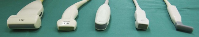
FIGURE 9-1 Ultrasound transducers for regional blocks. The photograph includes (left to right) broad linear, small footprint linear, curved, sector, and hockey-stick transducers.
Atlas of Ultrasound-Guided Regional Anesthesia 2e
WhatsApp us