Foot Ulcers
Ulcers on the feet are common. The majority of them are of vascular or neuropathic origin.
Causes
Vascular
Small Vessel Disease
Neuropathic
Peripheral Nerve Lesions
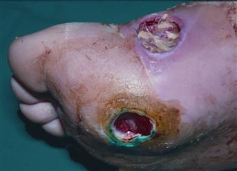
Figure 24 Chronic penetrating ulcers in a 60-year-old man with maturity-onset diabetes.
He had no evidence of major vessel disease but had signs of neuropathy. The deep ulcer beneath the head of the first metatarsal is characteristically surrounded with a thick keratin margin and the ulcer on the medial side of the foot has an exposed tendon in its base.
History
Vascular
Ischaemic ulcers are common in the elderly. They are painful and do not bleed much. They show no signs of healing. There is often a history of intermittent claudication and rest pain. Check for a history of diabetes. In the younger patient, diabetes, Buerger’s disease or Raynaud’s disease may be responsible. Check for a history of cardiac disease, which may suggest embolism leading to ischaemic ulceration. Proximal arterial disease, e.g. aneurysm, may also cause emboli.
Neuropathic
These are painless ulcers occurring over pressure points. Patients may give a history of neuropathy. They may describe a feeling, as though they are walking on cotton wool. Check for a history of diabetes and peripheral nerve lesions. Spinal cord lesions may also be present. In diabetes, ulceration may be associated with both ischaemia and neuropathy.
Neoplastic
Malignant ulcers may occur on the foot. Squamous cell carcinoma is rare but malignant melanoma is not infrequent, especially on the sole of the foot. The patient may have noticed the pigmented lesion, which has changed, with bleeding, itching and ulceration. The patient may also have noticed a lump in the groin, suggesting lymph node secondaries.
Traumatic
Foot ulcers may be caused by minor trauma, e.g. ill-fitting shoes. However, there is usually a history of an underlying predisposing condition, e.g. poor circulation, steroid therapy, neuropathy.
Infective
Pure infective ulcers on the foot are rare. Infections may occur with Nocardia species in tropical countries causing Madura foot. Check for a history of foreign travel.
Examination
Vascular
Ischaemic ulcers are found on the tips of the toes and over the pressure areas. The edge is usually punched out and healing does not occur. The base may contain slough or dead tissue. Occasionally tendons are seen in the base of the ulcer. Pulses may be absent. Check for atrial fibrillation, which may be associated with embolism as a cause of ischaemic ulceration.
Neuropathic
Neuropathic ulcers are deep, penetrating ulcers. They occur over pressure areas but the surrounding tissues are healthy and have good circulation. The ulcers themselves are painless. Examine the surrounding tissues for blunting of sensation, e.g. absence of pinprick sensation. Pulses are usually present. Carry out a full neurological examination looking for peripheral nerve injuries or evidence of spinal cord lesions.
Neoplastic
Squamous cell carcinomas have an everted edge, often with necrotic material in the base of the ulcer. The edge of the ulcer is hard. Check for inguinal lymphadenopathy. Ulcers associated with malignant melanoma tend to vary from brown to black, although they may be amelanotic. Bleeding and infection may make the surface of the tumour appear wet, soft and boggy. Check for inguinal lymphadenopathy and hepatomegaly.
Traumatic
Traumatic ulcers tend to occur either at pressure points, due to ill-fitting footwear, or at a site of injury. They usually have sloping edges and granulation tissue developing in the base. Always check the circulation, as most traumatic foot ulcers readily heal unless there is an abnormality of the circulation.
Infective
Infective ulcers by themselves are rare. Secondary infection may occur on any type of ulcer. With Madura foot, there may be ulceration and bone destruction with little systemic illness.