Facial Ulcers
Most facial ulcers are serious. Malignant facial ulcers are common in patients who work outdoors and are exposed to ultraviolet light. Lesions of the lips have been dealt with elsewhere (p. 316) and will not be included here.
Causes
Traumatic
Neoplastic
• Basal cell carcinoma (rodent ulcer) (Fig. 20) • (rare in tropical Africa)
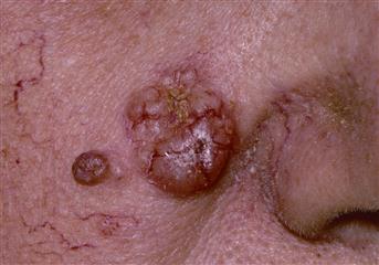
Figure 20 The larger nodular lesion on the cheek is a typical basal cell carcinoma.
Note the pearly appearance of the lesion which is beginning to ulcerate. The smaller lesion lateral to the basal cell carcinoma is a benign naevus.
Infective
History
A history of trauma should be sought. This may be as minor as scratching a spot. Self-inflicted injury resulting in dermatitis artefacta may be suspected when other causes have been excluded. Has the patient had radiotherapy in the past to suggest irradiation causes? Anaesthetised skin is easily traumatised. In the case of the face, anaesthesia may have arisen following surgery to the trigeminal ganglion for trigeminal neuralgia. Syringobulbia is a rare cause of facial anaesthesia. Check for a history of bites, insect, animal or human, which may become infected and ulcerate.
Malignant ulcers are usually on exposed areas of the face. Check the patient’s occupation. Malignancy is more common in outdoor workers exposed to ultraviolet light. Most rodent ulcers occur above a line drawn from the angle of the mouth to the lobe of the ear. Their incidence increases with age. Malignant ulcers are usually painless, unless they become infected. They may be pigmented and there may be a history of a change in a pre-existing mole to suggest malignant melanoma. These changes include: change in size; change in colour, with deepening pigmentation; bleeding or ulceration; itching; inflammatory ‘halo’; satellite nodules; palpable regional lymph nodes.
Infection may be due to herpes simplex, which may spread from around the lips or nose. The patient will complain of an itchy, burning red area, on which vesicles form and then crust over and ulcerate. Herpes zoster may arise in the distribution of the trigeminal nerve. Pain precedes malaise and fever by a few days. Vesicles develop later and may ulcerate. Syphilis is rare and may give rise to either a chancre or gumma (see Lip lesions, p. 316). Leishmaniasis is spread by sandflies and there would be a history of travel to India, Africa, the Middle East or the Mediterranean. There would be a history of bite leaving an itchy papule, leading to an ulcer. Keratoacanthoma may be due to a virus. It resembles a squamous cell carcinoma, from which it needs to be carefully distinguished. It occurs in adults as a rapidly growing lump with a central core filled with keratin. It usually takes 2–3 weeks to grow and often resolves spontaneously over several months. A history of inflammatory bowel disease will suggest pyoderma gangrenosum.
Examination
Benign ulcers
These usually have a sloping edge. Check the face for normal sensation to pain and temperature.
Malignant ulcers
Basal cell carcinomas (rodent ulcers) usually have a raised, rolled, pearly margin. They may become very large and erode deeply and locally. They do not metastasise and therefore local lymph nodes should not be enlarged unless the lesion becomes infected. Squamous cell carcinoma presents as an ulcerated lesion with an everted edge. Metastasis occurs to local lymph nodes and these may be palpable. Care should be taken to distinguish a squamous cell carcinoma from a keratoacanthoma, which is a benign, fast-growing, self-limiting papule surmounted by a keratin plug (it resembles a small volcano with a crater). Malignant melanomas vary in colour from pinkish-brown to black. They may develop a purplish hue due to a rich blood supply. There may be a brownish-pink ‘halo’ around the lesion or there may be ‘satellite’ nodules. The local lymph nodes may be enlarged. Early metastases may occur to the liver and therefore patients should be examined for hepatomegaly.
Infective
The characteristic lesions of herpes simplex will be seen around the lips and nose. In immunosuppressed patients, these lesions may coalesce and become infected. Ophthalmic herpes zoster will be recognised by its characteristic distribution in the ophthalmic division of the trigeminal nerve. It may affect the cornea. A syphilitic chancre begins as a macule, becoming a painless, hard ulcer, which is very infectious. It develops rapidly and is associated with enlargement of the local lymph nodes. A gumma occurring on the face is uncommon. It appears as a punched-out ulcer with a wash-leather base. Cutaneous leishmaniasis (oriental sore) develops at the site of a sandfly bite, commencing as an itchy papule, from which the crusts may separate, leaving an ulcer with deep perpendicular edges. Pyoderma gangrenosum presents with a nodule or pustule, which ulcerates with tender, reddish, blue necrotic edges.
General Investigations
■ FBC, ESR
WCC ↑ infected ulcer. ESR ↑ malignancy, syphilis.
■ LFTs
Alkaline phosphatase may be raised with liver secondaries, e.g. malignant melanoma.
■ Swab
C&S from infected ulcer. Dark-ground illumination microscopy for Treponema pallidum.
■ Biopsy
Benign versus malignant. Keratoacanthoma versus squamous cell carcinoma. In the case of malignant melanoma, excision biopsy is required.
Specific Investigations
■ Aspiration of lesion
Leishmaniasis – protozoal organism may be seen by microscopy in aspirates from fluid at the edge of the ulcer.
■ Viral culture
Herpes simplex, herpes zoster (rarely needed – diagnosis usually obvious clinically).
■ Antibody titres
Herpes simplex, herpes zoster (rarely needed – diagnosis usually obvious clinically).