TUMORS OF THE BRAIN

Published on 10/04/2015 by admin

Filed under Neurology

Last modified 22/04/2025

Print this page

rate 1 star rate 2 star rate 3 star rate 4 star rate 5 star
Your rating: none, Average: 0 (0 votes)

This article have been viewed 2312 times

CHAPTER 98 TUMORS OF THE BRAIN

Tumors of the brain are regarded as one of the most devastating group of neurological diseases—they are associated with significant neurological morbidity, they lead to progressive physical, cognitive and emotional dysfunction and are frequently fatal. The term brain tumor is used to describe both primary tumors that originate from the brain, cranial nerves, pituitary gland. or meninges and secondary tumors (metastases) that arise from organs outside the nervous system. These tumors present in many different ways dependent on their location, their rate of growth, and their effect on healthy neural tissue. Diagnosis requires careful history and examination, imaging, and histological examination, and management is best determined in a multidisciplinary team environment comprising neurologists, neurosurgeons, oncologists, neuropathologists, neuroradiologists, and clinical nurse specialists.

Despite the dramatic advances in neurosurgical technology, imaging, neuroanesthesia, radiotherapy techniques, and new drug development, the prognosis for many brain tumors, particularly malignant gliomas, remains bleak. The lack of a “cure” for the majority of these patients requires increasing emphasis on quality of life issues, and assessment of these is a standard feature of modern brain tumor trials.

This chapter aims to cover the key points required to manage patients with brain tumors effectively and sensitively, with specific emphasis on diagnostic and treatment considerations.

EPIDEMIOLOGY

Intracranial tumors are the eighth most common neoplasm in adults (approximately 5% of all primary neoplasms) and the most common solid tumor in children. They are the second leading cause of death from neurological disease in the United Kingdom (second only to stroke) and account for 2% of all cancer deaths in adults.

The incidence of primary brain tumors is considerably higher than tumor registry figures suggest. Based on a study from the southwest of England ascertaining data mainly from radiology records, the crude annual incidence for primary tumors was found to be 21 in 100,000.1 The annual incidence in the United States as ascertained from the Central Brain Tumor Registry is lower at 6.7 in 100,000 persons.2 There is increasing evidence that the incidence of gliomas and lymphomas is increasing, particularly in elderly patients, although this is more likely to be due to increased case ascertainment, with the increasing availability of modern imaging techniques.3

Brain tumors can present at any age. In children, they are the most common solid tumor and mainly occur in the posterior fossa, such as medulloblastoma, ependymoma, and juvenile pilocytic astrocytoma. In contrast, adults more commonly present with supratentorial tumors, particularly gliomas and meningiomas, accounting for over 75% of brain tumors. The most frequent tumors of middle life (third and fourth decades) are astrocytomas, meningiomas, pituitary adenomas, and vestibular schwannomas, whereas glioblastoma multiforme and metastases are more frequent in the fifth and six decades of life. There is a strong female preponderance of meningiomas, particularly in the spinal canal, whereas gliomas occur slightly more frequently in men. Germ cell tumors, particularly of the pineal region, occur frequently in adolescent males.

ETIOLOGY

Numerous epidemiological studies have been carried out to investigate etiological factors, but no clear risk factors have emerged apart from therapeutic ionizing irradiation. Cranial radiotherapy, even at low doses, has been shown to increase the relative risk of meningiomas by a factor of 10 and gliomas by a factor of 3.4 Other radiotherapy-induced tumors include cranial osteosarcomas, soft tissue sarcomas, schwannomas, and peripheral nerve sheath tumors. They have been described following radiotherapy for tinea capitis, craniopharyngioma, and pituitary adenomas and prophylactic cranial irradiation for acute lymphoblastic leukemia. Second tumors tend to lie within the radiation field, usually in lower dose regions, and develop from a few years to many decades after irradiation. The reported median time to the development of gliomas is 7 years. Sarcomas develop with a longer lag time and meningiomas may be seen 30 or 40 years later. The histology is identical to spontaneous tumors, although meningiomas are more likely to contain atypical features and have a worse prognosis.

No other environmental exposure has been clearly identified as a risk factor. There is widespread concern about the possible risks of cellular telephones, but case-control studies have not shown any increased risk in respect of any subtype of brain tumor using measures of the type of telephone, duration and frequency of use, and cumulative hours of use.5 So far, the consensus of opinion based on four studies is that mobile telephone use does not increase the risk of developing a brain tumor. However, with the exponential increase in the ownership and duration of use of these hand-held devices, it is important to continue surveillance of brain tumor trends in order to detect a latent period of several decades for the development of a tumor.

Genetic causes of brain tumors are rare but important. Occasionally, brain tumors occur in successive generations without any other tumor predisposition. More commonly, they are associated with neurocutaneous syndromes such as neurofibromatosis (optic nerve glioma, meningioma, vestibular schwannoma) and tuberose sclerosis (subependymal giant cell astrocytoma), von Hippel-Lindau syndrome (hemangioblastoma), and familial tumor syndromes such as Li-Fraumeni syndrome (glioma) and Cowden disease (dysplastic cerebellar gangliocytoma or Lhermitte-Duclos disease).

PATHOGENESIS

Brain tumors arise from an accumulation of mutations in genes that normally regulate the pathways of cell proliferation and differentiation. As in all human cancers, oncogenes and tumor suppressor genes are involved in the pathogenesis of brain tumors. One of the more frequent aberrations in human cancers are deletions or mutations of the TP53 gene, located on chromosome 17p, which encodes a 53-kDa protein, p53. These are found in approximately 40% of astrocytic tumors. This protein influences multiple aspects of cell cycle control as well as DNA repair after radiation damage and the induction of apoptosis. Other important genetic aberrations seen in malignant gliomas include amplification and mutations of the epidermal growth factor receptor (EGFR) gene and deletions and mutations of PTEN (phosphatase and tensin homology). Studies of astrocytic tumors show that the accumulation of predictable genetic alterations is associated with increasing malignant progression.

The mammalian cell cycle is divided into four phases: G1, S, G2, and M. Unrestricted cell multiplication is one of the hallmarks of cancer and is controlled in the normal cell by a complex series of positive and negative regulators that constitute the cell cycle checkpoints. Important proteins involved in these checkpoints include p53, MDM2, p14ARF, and p21, which regulate the progression of cells through the G1 cell cycle phase. Disruption of these protein complexes by gene deletions or mutations is found in varying proportions of gliomas, such as MDM2 gene amplification (in 10% to 15% of anaplastic astrocytomas) and glioblastoma multiformes lacking TP53 mutations. MDM2 is a cellular protein, which binds to and inactivates p53 and thus acts as an oncogene promoting glioma growth.

Taken overall, the various gene mutations mentioned above can all lead to a slight growth advantage, which can be further amplified by a gradual accumulation of further mutations, particularly when associated with loss of heterozygosity of the other allele. Current theories of malignant transformation postulate that this process is a sequence of multiple genetic alterations, each of which contributes to some expression of the tumor’s malignant characteristics. The various cellular pathways involved in proliferation, growth control, apoptosis, DNA repair, and genomic stability may differ from one tumor to another, but the phenotypes of the resulting tumors are similar.

CLINICAL FEATURES

Brain tumors can present in many different ways, and with the increased accessibility to high quality neuroimaging, they are being detected at a much earlier stage of their natural history than was the case previously. In some respects, this has made management decisions more difficult, particularly when a tumor is found that is in an inoperable location and is causing very few symptoms. Asymptomatic tumors are sometimes detected when patients are scanned for unrelated conditions and in many cases may be left alone.

The symptoms of brain tumors can be either focal or generalized and are most conveniently classified into five clinical syndromes that may coexist in the same patient or be present at different stages of the disease course:

The precise combination of clinical features varies on the location, histology, and rate of growth of the tumor. For instance, a patient with a low-grade glioma typically presents with a seizure disorder that may remain static for many years, whereas a patient with a malignant glioma may present with a rapidly progressive neurological deficit and raised intracranial pressure and be dead within a few weeks.

Raised Intracranial Pressure

As a brain tumor grows, there is displacement of cerebrospinal fluid into the spinal compartment and a reduction of blood volume. Eventually, the intracranial pressure rises because the skull behaves as a rigid box. Headache is the most common symptom of brain tumors, occurring in 23% of patients at initial presentation and 46% by the time of hospital admission. Headache alone, however, is an extremely rare presenting symptom, occurring in only 1.9% of patients.6 Because headache is such a common symptom in the population as a whole, it accounts for a disproportionate number of referrals of patients to neurology clinics concerned about the possibility of a brain tumor. Most brain tumor headaches are intermittent and nonspecific and may be indistinguishable from tension headaches.7 They may occasionally indicate the side of the tumor. Certain features of a headache are suggestive but not pathognomic of raised intracranial pressure. These include headaches that wake the patient at night or are worse on waking and improve shortly after rising, as well as headache associated with visual obscurations (transient fogging associated with changes in posture). Supratentorial tumors typically produce frontal headaches, whereas posterior fossa tumors usually result in occipital headache or neck pain. Nausea and vomiting may be a feature of raised pressure but may also occur as an early symptom of fourth ventricular tumors.

Brain tumors cause increased intracranial pressure by a variety of different mechanisms. They may have grown so large in a relatively short space of time that they cause stretching of pain-sensitive intracranial structures by a direct mass effect or by an effect on the microvasculature leading to cerebral edema. Smaller tumors, particularly those located in the posterior fossa, may cause headaches by obstructing cerebrospinal fluid circulation and producing obstructive hydrocephalus. Tumors may also cause raised intracranial pressure by producing large cysts. Occasionally, meningeal-based tumors cause localized headache through stretching of overlying dura. As a general rule, headaches with migrainous features are rarely due to an underlying tumor, although occasionally occipital tumors produce occipital seizures that are similar in many respects to migraine.

Untreated intracranial pressure leads to gradual deterioration in cognition, intermittent drowsiness, and eventually coma. Brainstem compression is the usual mode of death in patients with progressive brain herniation. The exact structures that are involved depend on the type of herniation—the most common types are uncal herniation, where the medial temporal lobe herniates across the tentorium, giving rise to an ipsilateral third nerve palsy, and tonsillar herniation, where the cerebellar tonsils are pushed down into the foramen magnum, leading to coma and death.

Progressive Neurological and Cranial Nerve Deficits

Focal neurological symptoms due to brain tumor are typically subacute and progressive with over 50% of patients having focal signs by the time of diagnosis. However, they may present acutely with strokelike symptoms, particularly if there is intratumoral hemorrhage, or even as a reversible event similar to a transient ischemic attack. Cortical tumors produce contralateral weakness, sensory loss, dysphasia, dyspraxia, and visual field loss depending on their location. A progressive hemianopia is often not detected by the patient, who may simply complain of bumping into objects or having a number of unexplained scrapes with parked vehicles or other stationary objects. Nondominant parietal tumors may present with topographical disorientation. Posterior fossa tumors cause gait ataxia, usually associated with headache and vomiting, whereas tumors in the cerebellopontine angle present with progressive unilateral deafness followed by ipsilateral facial sensory loss. Tumors in the fourth ventricle may present with effortless vomiting without nausea, a symptom that may predate more classic “posterior fossa” symptoms by many months or even years. Pituitary tumors compressing the optic chiasm cause a bitemporal quadrantanopia progressing onto hemianopia or pituitary apoplexy if there is hemorrhage or infarction. Subfrontal meningiomas may present with anosmia due to compression of the olfactory nerves in the anterior cranial fossa but may grow to a large size before clinical symptoms become apparent.

Seizure Disorder

Brain tumors account for about 5% of epilepsy cases, although they are overrepresented in cases of intractable epilepsy. Seizures are the presenting symptom in 25% to 30% of patients with gliomas and are present at some stage of the illness in 40% to 60% of patients. Approximately one half the patients have focal seizures, and the other one half have secondarily generalized seizures. Seizures occur in over 90% of cases of low-grade gliomas and frequently remain the only complaint for many years. Conversely, malignant gliomas have a lower frequency of seizures presumably because of their more rapid growth and destructive characteristics. Seizures are also common presenting symptoms for meningiomas (40% to 60%) and metastases (15% to 20%).

Temporal and frontal tumors are more likely to cause seizures than are occipital or parietal tumors, particularly when cortically based. The characteristics of the seizure depend on the location of the tumor. Frontal lobe tumors cause typically brief, frequent, and nocturnal seizures, which tend to spread rapidly and may become generalized. Common manifestations of a frontal lobe seizure include bicycling movements of the legs at night, turning of the head and eyes to the side away from the tumor (frontal adversive seizure), speech arrest, and hemiclonic spasms with a jacksonian march (posterior frontal tumors) in clear consciousness. In contrast, mesial temporal tumors can begin with olfactory or gustatory hallucinations, an epigastric rising sensation, or psychic experiences such as déjà vu or depersonalization. Once the seizures progress to a loss of awareness, the patients may stare blankly, speak unintelligibly, or exhibit lip smacking, picking at clothing, or other automatisms. Secondary generalized tonic-clonic seizures may follow on from partial seizures, more frequently in untreated patients. The presence of seizures is a favorable prognostic factor for survival, possibly due to lead-time bias in diagnosis and possibly due to the slow growth of epileptogenic tumors compared with more high-grade destructive tumors. In a study of patients with low-grade astrocytomas, the 5-year survival for patients with epilepsy as the only sign of tumor was 63% compared with 27% among the whole group.8

DIAGNOSIS

The diagnosis of a brain tumor is made by a combination of contrast-enhanced computed tomography scanning/magnetic resonance imaging and pathological classification of either a biopsy or resection specimen. Over the past decade or so, there have been a number of newer techniques introduced to complement conventional structural imaging, including proton magnetic resonance spectroscopy, functional metabolic imaging (single photon and positron emission tomography), and advanced magnetic resonance techniques, such as perfusion imaging (measuring blood flow and blood volume), diffusion weighted imaging (measuring cellularity), and diffusion tensor imaging (assessing integrity of white matter pathways). These are being gradually integrated into the routine preoperative evaluation of a brain tumor but add little to the conventional sequences in terms of refining diagnostic certainty.9

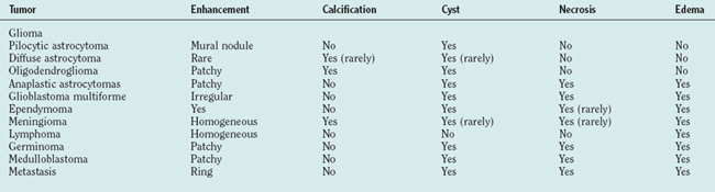
Just as there is clinicopathological correlation between World Health Organization (WHO) grade and prognosis, there is also a radiological/pathological correlation, specifically with respect to the degree of contrast enhancement seen within the tumor. Most grade II gliomas do not enhance, unlike grades III and IV, where there is usually irregular ring enhancement and, in the case of grade IV tumors, central necrosis. However, as mentioned, certain grade I gliomas, particularly juvenile pilocytic astrocytomas, also enhance, and this can occasionally give rise to diagnostic confusion, particularly in adults, in whom juvenile pilocytic astrocytomas are much less common than malignant gliomas. The key imaging characteristics of common types of tumors are summarized in Table 98-1; see also Figure 98-3 for radiological/pathological correlations.

image

Figure 98-3 Radiological and pathological correlations of common central nervous system tumors. Each row of figures shows imaging appearances (magnetic resonance imaging or computed tomography) (left), macroscopic aspect on formalin fixed postmortem brains with a tumor at similar locations (center), and representative histological sections (right), stained with hematoxylin and eosin, which stains nuclei dark blue and cytoplasm and extracellular matrix pink.

Low-grade astrocytoma: Magnetic resonance imaging (T1-weighted with contrast) shows involvement of the temporal and frontal lobes and considerable mass effect with midline shift. Gross section of a brain with a similar tumor shows effacement of cortical structures, basal ganglia, and midline shift. Histology shows a tumor with low density of tumor cells, ample fibrillary background, and thin vessels.

Glioblastoma multiforme: magnetic resonance imaging (T1-weighted with contrast) shows ring enhancement indicating neovascularization and vascular permeability as well as central necrosis. The brain section shows a hemorrhagic tumor in the left temporal and frontal lobes. There is considerable midline shift and hemorrhage in the corpus callosum and fornix, indicating tumor extension over the midline. Histologically, the tumor is characterized by a very high density of cells (dark nuclei with sparse cytoplasm), and a typical feature is necrosis with pseudopallsading arrangement of tumor cells.

Oligodendroglioma: magnetic resonance imaging (coronal FLAIR sequence) shows involvement of parietal cortex and subcortical white matter with expansion of the tumor outside of the brain and erosion of the inner skull table. A brain section of a similar tumor shows a relatively well-demarcated cystic, partially hemorrhagic space occupying lesion. Classic histological features of oligodendroglioma are cells with clear appearance (“fried-egg” cells), central nuclei, and a network of thin, branching vessels (not shown).

Medulloblastoma: computed tomography scan with contrast shows an enhancing midline mass lesion arising from the cerebellar hemisphere. Note the hydrocephalus as a result from compression of the fourth ventricle. A sagittal section shows a large tumor in the posterior fossa that displaces the cerebellar vermis and compresses the brainstem. Typical histological features are wedge-shaped nuclei that are arranged to rosettes with neuropil-like matrix in their center.

Meningioma: computed tomography scan with contrast shows a typical strongly enhancing tumor that is dural based. Meningiomas are often associated with considerable edema, and in this case, there is very significant displacement of brain tissue, which becomes symptomatic relatively late, due to the slow growth of these tumors. Postmortem finding of an asymptomatic meningioma impressing the temporal lobe in situ (upper section) and after (lower section) removal, leaving a cavity. Histologically, meningiomas show variable features. A typical feature shown here is the concentric arrangement of tumor cells, which can become calcified.

(Courtesy of Professor Sebastian Brandner.)

PATHOLOGY

There are over 160 types of primary brain tumors arising from neuroepithelial tissue within the brain, the meninges covering the brain, the sellar region, and the cranial nerves.

The WHO published a landmark classification in 1993 and, in 2000, further refined their classification system (Table 98-2).10 The key to the WHO classification is the stratification of tumors according to their biological activity so that the lower the WHO grade, the better the overall prognosis. As a general rule, the category of grade I tumors is reserved for neoplasms that have a stable histology and that are potentially curable by surgical removal alone. In contrast, tumors that appear histologically “benign,” yet are known to progressively transform over time into higher grade lesions, are categorized as grade II neoplasms. Those tumors with anaplastic histology are regarded as grade III high-grade neoplasms, and the most malignant phenotype is classified as grade IV. The peak of age of incidence is proportional to the most common histological grade; that is, grade I tumors usually present in childhood, grade II in young adulthood, grade III in middle age, and grade IV in older age. The exception to this rule is the grade IV primitive neuroectodermal tumors, which occur most frequently in childhood.

TABLE 98-2 World Health Organization Classification and Grading of Tumors of the Nervous System

Tumors of Neuroepithelial Tissue WHO Grade
Astrocytic tumors
Pilocytic astrocytoma I
Pleomorphic xanthoastrocytoma I, II
Subependymal giant cell astrocytoma I
Desmoplastic infantile astrocytoma I
Diffuse astrocytoma II
Anaplastic astrocytoma III
Glioblastoma multiforme IV
Gliosarcoma IV
Oligodendroglial tumors
Oligodendroglioma II
Anaplastic oligodendroglioma III
Mixed astrocytic and oligodendroglial tumors
Oligoastrocytoma II
Anaplastic oligoastrocytomas III
Ependymal tumors
Subependymoma I
Myxopapillary ependymoma I
Ependymoma II
Anaplastic ependymoma III
Choroid plexus tumors
Choroid plexus papilloma I
Choroid plexus carcinoma IV
Neuronal and mixed neuronal-glial tumors
Gangliocytoma I
Ganglioglioma I, II
Desmoplastic infantile ganglioglioma I
Dysembryoplastic neuroepithelial tumor I
Central neurocytoma I
Pineal parenchymal tumors
Pineocytoma I
Pineoblastoma IV
Embryonal tumors
Primitive neuroectodermal tumors IV
Medulloblastoma IV
Meningeal tumors
Meningioma I
Atypical meningioma II
Anaplastic meningioma III
Hemangiopericytoma
Melanocytic tumor of the meninges
Tumors of vascular origin
Cavernous angioma
Hemangioblastoma
Germ cell tumors
Germinoma
Embryonal carcinoma
Yolk sac tumor (endodermal sinus tumor)
Choriocarcinoma
Teratoma
Tumors of the sellar region
Pituitary adenoma
Pituitary carcinoma
Craniopharyngioma
Primary central nervous system lymphomas
Peripheral nerve sheath tumors
Vestibular schwannoma
Trigeminal schwannoma
Facial nerve schwannoma
Malignant peripheral nerve sheath tumor
Metastatic tumors

Neuroepithelial tumors (predominantly gliomas) account for approximately 50% to 60% of all primary brain tumors. The other common types are meningiomas (20%) and pituitary adenomas (15%). Rarer tumors include primary central nervous system lymphomas, neuronal tumors, and germ cell tumors. Metastatic tumors are more common than primary brain tumors and usually originate from lung (50%), breast (15%), melanoma (10%), and unknown (15%). In children, the most common tumors are low-grade astrocytomas, ependymomas, suprasellar tumors, medulloblastomas, and other primitive neuroectodermal tumors. There is increasing use of molecular classification in addition to morphology, but at present, this is not an integral part of the standard neuropathological report.

The grading system is based on a number of histological features including the degree of cellular atypia and pleomorphism, the presence of vascular proliferation and necrosis, and the cellular proliferation rate and correlates with the degree of malignancy, the likelihood of metastatic spread and recurrence, and ultimately survival.

Gliomas

Gliomas are the most common type of primary brain tumor and are so called because they share morphological and immunohistochemical features with astrocytes and oligodendroglial and ependymal cells. Astrocytic neoplasms are the most common of the three and include tumors of all WHO grades. Low-grade gliomas can be subdivided into WHO grade I tumors (e.g., pilocytic astrocytomas, pleomorphic xanthoastrocytomas, and subependymal giant cell astrocytomas [usually but not invariably associated with tuberose sclerosis]) and WHO grade II tumors (e.g., diffuse and gemistocytic astrocytomas, oligodendrogliomas, and oligoastrocytomas [which contain elements of both astrocytic and oligodendroglial lineage, otherwise known as mixed gliomas]). These two grades should be regarded as distinctive groups in that grade I tumors never progress into grade II tumors, unlike grade II tumors, which frequently transform into grade III and grade IV tumors (as discussed later). Therefore, grade II tumors should be thought of as part of a biological continuum that extends through to grade IV tumors.

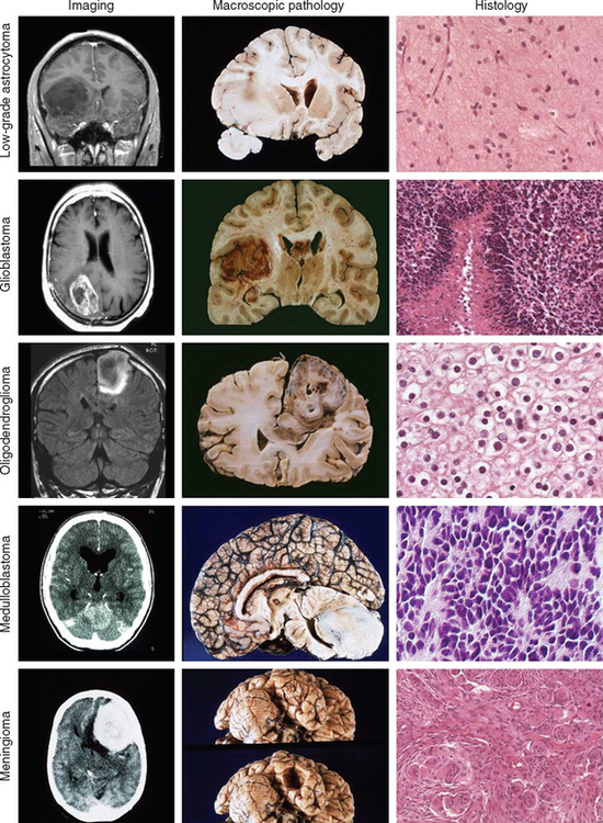
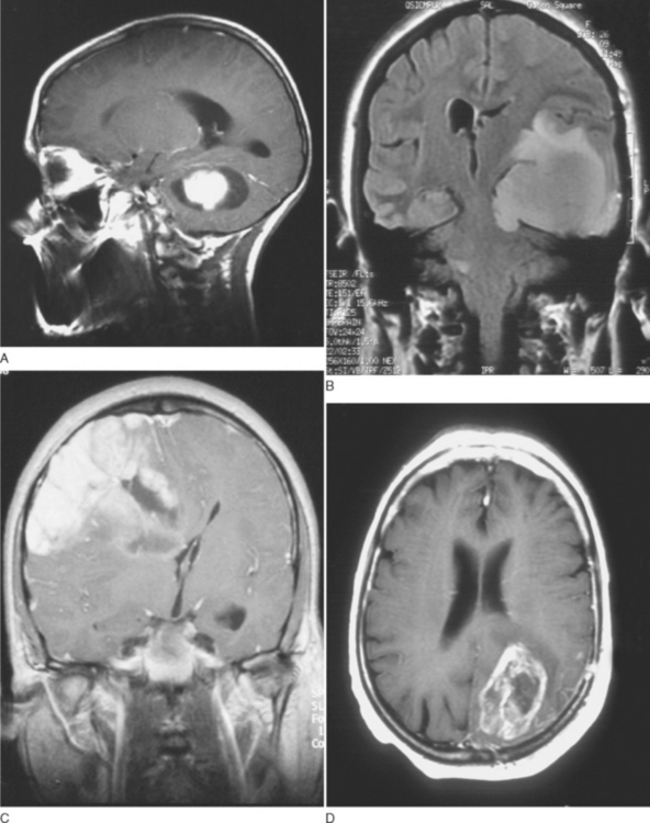
Grade I gliomas are usually well circumscribed and potentially curable by surgical resection alone. The most common type is the juvenile pilocytic astrocytoma, so called because it presents in childhood and is characterized by the presence of astrocytes with hairlike (pilocytic) processes. The tumor has a narrow zone of microscopic infiltration and appears radiologically as an enhancing mural nodule, which is supplied by capillaries that lack a complete blood-brain barrier surrounded by a cyst. This leads to the contrast enhancement seen on computed tomography scans or magnetic resonance images (Fig. 98-1).

Other grade I tumors may contain neuronal elements such as ganglioglioma and are known as mixed glioneuronal tumors. They can occur anywhere in the brain but have a predilection for the temporal lobes and may give rise to intractable temporal lobe epilepsy.

Grade II astrocytomas also grow slowly and act in a “benign” manner, usually presenting with seizures but, in contrast to grade I tumors, they are diffusely infiltrative and recur after surgery, usually as a higher-grade glioma.

The macroscopic and microscopic appearances are of diffuse spread into normal brain parenchyma, making it impossible for the surgeon to distinguish macroscopically between tumor and normal brain tissue. The astrocytic morphology may be fibrillary or protoplasmic, but these distinctions are not important in clinical practice. The gemistocytic variant, however, tends to behave in a more aggressive manner. Astrocytomas are hypercellular compared with normal white matter, and the tumor cells show hyperchromasia and pleomorphism, with striking irregularity of the nuclei. The normal architecture is commonly disrupted into a microcystic pattern. Mitoses are absent or solitary. The other main cellular type of low-grade glioma is the oligodendroglioma characterized by the presence of uniform round nuclei with small nucleoli and perinuclear halos. These tumors are often calcified and may have areas of focal increased cellularity. Some grade II gliomas have intermixtures of astrocytic and oligodendroglial cellular elements and are appropriately called oligoastrocytomas. All three types have a propensity to undergo anaplastic change and turn into a high-grade glioma. Time to malignant transformation is variable and at present cannot be predicted on the basis of histology or genetics alone, although it may be shorter in older patients.

High-grade or malignant gliomas encompass grade III (anaplastic) tumors and grade IV gliomas. These tumors are characterized histologically by increasing cellular density and pleomorphism, mitotic activity, and proliferation indices. Grade III tumors are defined by the presence of frequent mitoses, whereas the presence of vascular proliferation and/or necrosis is the hallmark of a grade IV tumor (glioblastoma multiforme). Tumors with higher-grade histology behave in a more destructive manner and present more commonly with symptoms of raised intracranial pressure, focal neurological deficit, or mental state changes. These tumors are generally incurable.

GENETICS

The molecular classification of gliomas has become increasingly important in therapeutic decision making, particularly the distinction between tumors of astrocytic lineage and oligodendroglial lineage, with the latter being characterized by losses of part of chromosomes 1 and 19.

The only molecular genetic alteration consistently observed in patients with low-grade astrocytomas is mutation of the TP53 gene, located on chromosome 17, which occurs in 50% to 60% of patients with astrocytomas. Loss of normal p53 function promotes growth and malignant transformation and is therefore believed to be one of the most important gene alterations associated with the development of malignant gliomas. Loss of p53 is also seen in anaplastic astrocytomas and glioblastomas arising from low-grade gliomas. In contrast, glioblastomas arising de novo more commonly have amplification of the EGFR gene, suggesting that they arise from different genetic pathways.

In glioblastoma multiforme, there are gains of chromosome 7 and deletions of chromosomes 10 and 22, both of which contain multiple tumor suppressor genes, together with structural alterations of chromosomes 1p, 9p, 11p, 12q, and 13q.

As a result of these various genetic aberrations, GBMs can be divided on the basis of molecular features into primary and secondary GBMs, which correlate with their clinical behavior. Primary GBMs arise de novo in older patients and are strongly associated with amplification and overexpression of the EGFR gene, increased MDM2 activity, and decreased PTEN. In contrast, secondary GBMs arise out of previous low-grade gliomas, occur in younger individuals, and are associated with early p53 loss and overexpression of PDGF. As with primary GBMs there is loss of PTEN.

Astrocytomas can be distinguished from oligodendrogliomas on the basis of histological features, as described earlier. During the past decade, the specific association between tumors of oligodendroglial lineage, particularly tumors with classic histology, and loss of the short arm of chromosome 1 (1p) and the long arm of chromosome 19 (19q) has been recognized. Loss of 1p/19q is found in 40% to 90% of oligodendrogliomas compared with less than 1% of astrocytomas. Nearly all tumors with 1p loss also have 19q loss, suggesting that inactivation of one or more genes on each of these arms is a fundamental event in oligodendroglioma oncogenesis. This chromosomal fingerprint is tightly associated with chemosensitivity and prolonged survival and has taken molecular classification out of the basic science laboratory and into the clinical arena.11 Candidate tumor suppressor genes have still not been identified. Other genetic alterations less frequently encountered in oligodendroglioma include losses of chromosomes 4, 6, 14, 11p, and 22q. Neuropathology laboratories are increasingly offering genotyping for 1p and 19q; the results will influence the prognosis, as 70% to 80% of long-term survivors have 1p/19q loss.

The coincidence of these two genetic aberrations is unique to oligodendroglioma. They are not found in oligodendroglioma “mimics” such as dysembryoplastic neuroepithelial tumors or central neurocytomas and are therefore useful in cases with equivocal morphology.

The correlation with chemosensitivity is particularly high in patients, with 75% of patients with anaplastic oligodendrogliomas responding to treatment with procarbazine, CCNU, and vincristine and 50% achieving a prolonged and durable survival.11 Similar but less dramatic responses have also been observed in patients with low-grade oligodendrogliomas.

TREATMENT

The three conventional modalities of treatment for brain tumors are surgery, radiotherapy, and chemotherapy. In some patients, suc as those with unresectable low-grade gliomas, it is perfectly reasonable and safe to offer no specific treatment at all. In other patients, such as frail elderly patients with significant tumor-associated disability, any benefits of treatment in terms of prolongation of survival may be outweighed by the treatment-associated morbidity.

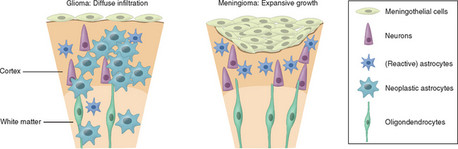
The use of each therapeutic modality therefore should be dictated by the location of the tumor within the brain and within the skull, specifically, whether it is intra-axial (e.g., glioma) or extra-axial (e.g., meningioma), the likely histology, and the patient’s age and general condition. The distinction between intra- and extra-axial tumors is important, as the effect on surrounding brain tissue and the likely plane of dissection are critical to the success of radical surgery (Fig. 98-2). When considering surgery for intrinsic tumors, patient factors, particularly age and performance status, are far more important in terms of prognosis than any specific treatments used, and this needs to be borne in mind when evaluating the efficacy of any treatment and claims that survival is significantly improved.

Surgery

There are three principal indications for surgery in the management of brain tumors: tissue diagnosis (via stereotactic or open biopsy), relief of raised intracranial pressure (via tumor debulking, lobectomy, aspiration of tumor-associated cysts, insertion of shunts for tumor-associated hydrocephalus), and prolongation of survival. The complete removal of an intracranial tumor is the goal of radical tumor surgery and is routinely achieved in the treatment of convexity meningiomas and other extra-axial tumors such as nonsecreting pituitary adenomas and vestibular schwannomas. Surgery may also be regarded as curative for a limited number of intrinsic tumors, particularly WHO grade I gliomas, such as juvenile pilocytic astrocytomas. In other malignant intrinsic tumors, such as medulloblastomas and solitary brain metastases, the extent of resection plays a vital prognostic role in determining the success of adjuvant treatment but is not in itself curative.

The main complications of surgery are neurological deficit, postoperative hydrocephalus, cerebrospinal fluid leak, and seizures, as well as the general complications of any surgery, such as wound infection, deep vein thrombosis, and pulmonary embolism.

The role of surgery in the management of the majority of primary intrinsic tumors, specifically gliomas, is more controversial as even radical removal is unlikely to be curative given the diffusely infiltrative nature of these tumors and their tendency to spread far beyond the core of the tumor along extracellular pathways and white matter tracts. Furthermore, as there are no prospective randomized studies correlating extent of resection with survival and it is highly unlikely that such studies will ever be carried out, controversy will continue to exist with regard to the impact that extent of resection has on outcome.

There have been a number of recent advances in tumor neurosugery including computerized neuronavigation techniques, improved preoperative mapping of eloquent brain areas using functional magnetic resonance imaging, and assessment of white matter pathways by diffusion tractography. Frameless stereotaxy now enables the surgeon to delineate the tumor boundaries seen on preoperative magnetic resonance imaging with the surface markings of the brain in three dimensions. However, this may be affected by brain shift as the cranial cavity is opened and therefore becomes less accurate at deeper levels of the brain. In this respect, intraoperative magnetic resonance imaging now allows the surgeon to view the results of his or her resection while the patient is still on the table and remove further tissue as necessary according to the scan appearances of the tumor. These have all contributed to improving the morbidity and mortality of neurosurgery, but an effect on overall survival has yet to be demonstrated. Nevertheless, the ability to remove more and more abnormal tissue with less and less risk to the patient and eventually to resect almost an entire intrinsic tumor remains the Holy Grail of oncological neurosurgery.

Radiotherapy

Radiotherapy has become the most commonly used tumor-specific therapy in neuro-oncology. Therapeutic radiation for brain tumors is most commonly produced from high-energy linear accelerators that generate x-rays and electrons, which have much higher energy than those used in diagnostic radiology. Protons, neutrons, and gamma rays from cobalt 60 decay are used particularly for extrinsic tumors. Radiotherapy may also be delivered directly into the tumor using radioisotopes such as iodine 125.

Radiation can be either directly (e.g., protons and other charged particles) or indirectly (e.g., x-rays and gamma rays) ionizing. Indirect ionizing radiation targets DNA by producing short-lived fast-moving charged particles when absorbed into water-rich tissue, leading to single- and double-strand breaks. Single-strand breaks are easily reparable, but double-strand breaks lead to difficulties with subsequent mitoses and chromosomal aberrations, some of which are lethal to the cell.

Traditionally, external beam radiotherapy is delivered in multiple daily fractions over several weeks, in order to spare normal tissue, which has a greater capacity than tumor tissue to repair sublethal damage and to repopulate between fractions.

Recent advances in the technology of diagnostic imaging and computerized treatment planning systems have allowed greater accuracy of radiotherapy delivery. The brain lends itself to high precision techniques due to the lack of internal motion within the skull and the ease of immobilization of the skull itself. Conformal radiotherapy allows the profile of the radiation beam to be shaped around the tumor (to conform to the tumor edges), and this reduces both the short- and long-term toxicity of the treatment. Highly focused radiation can be given either in a single high dose (stereotactic radiosurgery) or in smaller fractions (stereotactic radiotherapy) and is predominantly indicated for lesions less than 3 cm in diameter that are well circumscribed, extra-axial, and more than 5 mm from vital structures. This has led to more widespread use of stereotactic radiotherapy in the treatment of benign extra-axial tumors such as vestibular schwannomas, skull-based meningiomas, and small intrinsic tumors in eloquent locations. However, this approach is not appropriate for infiltrative tumors such as gliomas.

The main side effects of radiotherapy include hair loss, which is usually permanent and tiredness, particularly toward the end of a long course of treatment. Early delayed radiation toxicity is characterized by severe lethargy and occasionally worsening of a preexisting neurological deficit, giving rise to concerns about the possibility of tumor recurrence. This usually responds to steroids. In contrast, late delayed radiation toxicity is irreversible and includes vasculopathy, dementia, brain necrosis, and secondary tumors.

Chemotherapy

Chemotherapy has traditionally been the “poor relative” of brain tumor treatments, but over the past decade the role of chemotherapy has expanded and many options are now available to treat a wide variety of tumors. Tumors that had been previously regarded as chemoresistant are now being treated with new chemotherapy schedules and used in combination with other therapeutic modalities, particularly radiotherapy. There are a number of specific difficulties in evaluating new drugs for central nervous system tumors, specifically the determination of treatment response by magnetic resonance imaging alone, particularly when patients have been heavily pretreated, and the problems of drug delivery into tumors that may be shielded from the systemic circulation by the blood-brain barrier.

Chemotherapy is usually administered as multimodality therapy, concurrently with radiotherapy, either as single agents or as combinations of drugs. Combining agents with different mechanisms of action and different toxicities has been found to be effective for many different tumor types. Cytotoxic chemotherapy impairs DNA synthesis and, as tumor cells divide more rapidly than most normal cells, achieve an acceptable therapeutic index.

Chemotherapy may be delivered before surgery or radiotherapy, known as neoadjuvant treatment, simultaneously with radiotherapy, known as concomitant treatment, or after surgery or radiotherapy, known as adjuvant treatment. It may given as part of the primary treatment or at progression, particularly for malignant gliomas.

The main cytotoxic agents used in the treatment of brain tumors are temozolomide, procarbazine, lomustine (CCNU), carmustine (BCNU), vincristine, carboplatin, and cisplatin. Temozolomide, procarbazine, and lomustine are alkylating agents causing alkyl groups to bind to DNA, producing DNA cross-links or single- or double-strand breaks. In contrast, vincristine is a microtubule poison and disrupts the mitotic machinery, particularly in the G1 phase.

Temozolomide is increasingly being used for brain tumor treatment. It is an imidazotetrazine derivative of dacarbazine with good oral bioavailablity and is rapidly metabolized to an active derivative. It works by methylating the O6 position on guanidine and depletes the drug resistance enzyme methylyguanine methyltransferase (MGMT) enzyme. It is relatively well tolerated and so has become the chemotherapeutic agent of choice for many patients with gliomas, although it has yet to be compared head-to-head with the conventional combination regimen of procarbazine, CCNU, and vincristine (PCV).

There is extensive research interest in novel compounds known as small molecules that block signaling pathways mediated by various growth factors. In some cases, they produce tumor shrinkage when used as monotherapy, although in the majority of instances they are cytostatic rather than cytotoxic. Examples of compounds under investigation include EGFR antagonists such as OSI-7740–erlotinib and PDGFR antagonists such as STI-571. It is likely that these will find a place in the chemotherapy armamentarium in association with either radiotherapy or with more conventional cytotoxic drugs.

The main dose-limiting effects of chemotherapy are bone marrow suppression, which may be cumulative or noncumulative. This can be managed by either increasing the interval between cycles until bone marrow recovery has occurred or reducing the dosage in subsequent cycles. Other effects specific to brain tumor chemotherapy include constipation and headache (temozolomide), rash and jaundice (procarbazine), neuropathy (vincristine, cisplatin), and nephropathy (cisplatin, methotrexate). Most chemotherapy-induced toxicity is reversible on cessation of the drug.

In order to avoid systemic administration and hence toxicity altogether, various new methods of drug delivery are under investigation. In particular, the implantation of drug directly into the tumor resection cavity using biodegradable wafers containing carmustine (Gliadel) has now been shown in phase 3 trials to prolong survival in malignant gliomas. Convection-enhanced delivery uses catheters placed into the brain parenchyma around the resection cavity to deliver intratumoral chemotherapy by infusion over several days. The use of convection rather than diffusion alone results in larger volumes of distribution, an important consideration when treating gliomas which are known to grow for many centimeters around the tumor cavity.

SUPPORTIVE TREATMENT

Steroids are used extensively in neuro-oncology to reduce peritumoral edema, to improve symptoms and quality of life, and to reduce the morbidity of brain tumor surgery. Dexamethasone is the most frequently used steroid and is 10 times more potent than prednisolone. The main side effects include weight gain, fluid retention, impaired glucose tolerance, skin changes, proximal myopathy, and increased susceptibility to infections. Not uncommonly, the patient becomes cushingoid leading to dramatic changes in body habitus and facial appearance (Fig. 98-4). As with all steroids, the aim is to use the lowest dose possible that controls symptoms. Steroids have a lympholytic effect and are therefore cytotoxic to primary central nervous system lymphomas. If there is a clinical suspicion of lymphoma based on the results of a scan, then steroids should be avoided prior to biopsy as they may cause temporary disappearance of the tumor.

Patients with brain tumors are prone to seizures and are frequently treated with anticonvulsants. The management of tumor-associated epilepsy is beyond the scope of this chapter but, as a general rule, the principles are similar to those for the treatment of patients with epilepsy patients, although seizures are more likely to be resistant to monotherapy with antiepileptic drugs. Specific prescribing considerations for these patients include interaction with anticancer drugs and steroids and the greater potential for cognitive impairment due to the underlying tumor. For this reason, many neuro-oncologists are using non–enzyme-inducing antiepileptic drugs in preference, such as lamotrigine and levetiracetam.

INTRA-AXIAL TUMORS

Low-Grade Gliomas

The management of low-grade gliomas is one of the most controversial issues in neuro-oncology. These are typically diffusely infiltrating tumors, often invading but not destroying eloquent regions of the brain, and cannot be removed completely in the majority of cases by surgical resection alone. Although some patients may survive for decades, the majority of these tumors eventually become high-grade gliomas and these are usually fatal. There are a large number of surgical series advocating the benefits of complete resection over partial resection but these are all retrospective and include patients with a variety of tumor histologies, specifically pilocytic and non-pilocytic astrocytomas. They are by definition subject to selection bias, and therefore reports of better outcomes for patients with larger resections need to be interpreted in light of the known prognostic factors which include tumor size.12 Despite these reservations, surgery is the only certain way of removing a large volume of tumor tissue and is widely used in young, fit patients with tumors in noneloquent regions (e.g., nondominant frontal or temporal lobes). Some surgeons will resect tumors in dominant frontal temporal lobes, using the techniques of awake craniotomy to monitor language perioperatively, but there are no comparative data published comparing this approach with a more conservative policy. This lack of clear benefit is supported in part by a population-based study of nearly 1000 patients with low-grade glioma that showed a relatively small and nonsignificant difference in overall survival between patients who had had a biopsy only (6.4 years), a subtotal tumor resection (6.8 years), or a gross-total tumor resection (7.6 years).13

The benefit of early radiation in low-grade gliomas is similarly unclear. The European Organisation for Research and Treatment of Cancer (EORTC) reported interim results from a trial comparing early radiotherapy (at diagnosis) with radiotherapy at progression. This was a prospective trial and followed patients for a median of 5 years. Patients who had early radiotherapy showed a significant improvement in time to progression compared with patients irradiated at tumor progression but there was no difference in overall survival. The 5-year estimate was 63% versus 66% (overall survival) and 44% versus 37% (time to progression) for the treated and control arms respectively.14 On the basis of these data, the majority of oncologists irradiate patients with low-grade glioma only at progression or rarely for control of intractable seizures. The standard dosage schedule is 54 Gy in 33 fractions over 6.5 weeks.

Chemotherapy is being increasingly used for low-grade gliomas, particularly those with oligodendroglial elements. There are strong indications that a significant percentage of low-grade oligodendrogliomas respond favorably to PCV chemotherapy with improvements in seizure control and cognitive function more readily apparent than shrinkage of the tumors in magnetic resonance images.15 Temozolomide has also been found to be useful in oligodendrogliomas both as primary treatment and in recurrent disease after radiotherapy, particularly in those tumors with loss of chromosome 1p and 19q.

Because of the relatively indolent growth of these tumors, combined with their comparatively long survivals and treatment associated morbidity, low-grade gliomas are often managed conservatively with symptomatic treatment (antiepileptic drugs) alone. Regular surveillance imaging is used to detect early signs of tumor progression, such as the development of new areas of gadolinium enhancement, which frequently predates clinical deterioration. Patients need to be carefully informed of the pros and cons of active treatment versus no treatment at all. Ultimately, this is a highly individualized decision process that depends on the age of the patient, the tumor type and location, and the philosophy of the treating clinicians. As a result, there is a wide discrepancy in the way that patients with low-grade gliomas are managed from center to center, giving rise to considerable uncertainty over the most appropriate form of intervention for that individual patient. At our center, we are in the process of developing magnetic resonance imaging parameters as surrogate markers for biological behavior in order to determine whether it is possible to select patients at high risk of tumor progression while the tumor is still in the “premalignant” phase of its natural history.

Malignant Gliomas

In contrast to the situation with low-grade gliomas, the general principles of management of malignant gliomas are much more clear-cut. These are fast growing aggressive tumors, which, untreated, are associated with a median survival of 3 to 4 months. Initial management consists of dexamethasone to reduce vasogenic edema and to prepare the patient for surgery (biopsy or resection). Once surgery has been performed, the dose of dexamethasone should be rapidly reduced to the lowest dose possible to minimize the side effects of prolonged high-dose steroids. Phenytoin is often given prophylactically around the time of surgery and should be weaned 1 week after operation in patients who have not had seizures.

Surgery is the primary treatment modality for tumors where resection is believed to be technically feasible and in the best interest of the patient. Resection has the advantage over biopsy of providing more tissue for diagnosis as well as reduction of tumor bulk and possible prolongation of time to tumor progression. It also allows the surgeon to implant interstitial chemotherapy in the form of carmustine wafers and therefore start adjuvant tumor treatment immediately and hence slow down the inevitable regrowth, prior to radiotherapy. Despite these advantages, there is still wide variation in surgical practice with “aggressive” surgeons arguing the case for gross total resections as a means of achieving significant tumor “cytoreduction,” thereby improving the chances of successful treatment with adjuvant radiotherapy and chemotherapy and reducing the need for steroids. More conservative surgeons will often only perform a biopsy on a high-grade tumor, particularly tumors that are in the dominant hemisphere or deeply seated, arguing that there is limited evidence for the survival benefits of radical surgery in the absence of prospective randomized trials. A trial comparing resection with biopsy alone for patients in whom the surgeon is uncertain about the benefits of one over the other is unlikely ever to be carried out because of the perceived ethical problems and difficulties recruiting patients to a nontreatment arm.

Relative contraindications to surgery include poor performance status, significant medical comorbidity, and tumors in eloquent or inaccessible locations. For these patients, the risks of surgery may be outweighed by the potential benefits. The overall morbidity rate for untreated malignant gliomas is 24% with a mortality rate of 1.5%. The chances of neurological improvement with surgery are just over 20% with less than 10% of patients deteriorating.16

In contrast, there is unanimity of opinion about the benefits of radiotherapy for malignant gliomas as it is the only treatment that has been proved to extend survival in this patient group. Radiotherapy is indicated for the treatment of grades III and IV astrocytomas and oligodendrogliomas, and treatment with radical intent is given to patients under the age of 70 years who have a good performance status, that is, a Karnofsky performance scale of at least 70, implying the ability to self-care. The radiation field encompasses the contrast-enhanced T1-weighted target with a margin of between 2 and 3 cm to sterilize “satellite” tumor cells. The total dose is usually 60 Gy, delivered over 6 weeks in 30 fractions. In a landmark study, the median survival of patients with malignant gliomas increased from 14 weeks with supportive treatment alone to 36 weeks with radical radiotherapy.17 Lower-dose schedules, such as 30 Gy in 10 fractions, are used mainly in the palliative setting, particularly for patients over the age of 70 in poor general condition.

Chemotherapy

Chemotherapy for malignant gliomas has traditionally been used as adjuvant treatment following radiotherapy (mainly in the United States) or at recurrence. The common drugs used are nitrosoureas such as BCNU (as adjuvant treatment) and PCV or temozolomide at recurrence. There is no clear benefit for the use of adjuvant chemotherapy over radiotherapy alone, although a meta-analysis based on 12 randomized trials suggested a small benefit of chemotherapy compared with radiotherapy alone (a 5% increase in 2-year survival).18 A trial is under way comparing PCV with temozolomide in recurrent disease, and the results will not be available until 2008.

A trial has shown that the use of concomitant temozolomide with radical radiotherapy followed by six cycles of adjuvant temozolomide in patients with glioblastoma multiforme offered a significant survival advantage over radiotherapy alone with minimal additional toxicity. Although the increase in median survival from 12.1 months with radiotherapy alone to 14.6 months in the concomitant temozolomide group was relatively modest, the 2-year survival rate increased from 10.4% to 26.5%.19 These results represent a significant improvement in the outlook of patients with glioblastoma multiforme, although it remains to be seen whether these data can be extrapolated to patients with anaplastic astrocytomas. Further trials to confirm these findings are unlikely to occur given that the sample size was large (573 patients from 85 centers), that prognostic factors were well matched between the two groups, and that 85% of patients in the concomitant arm completed both radiotherapy and temozolomide as planned. Furthermore, an exploratory subgroup analysis defined according to known prognostic factors demonstrated a survival benefit in nearly all subgroups. However, in a parallel study on the same patient group investigating the role of genetic silencing of the MGMT (O6-methylguanine-DNA methyltransferase) DNA-repair gene by promoter methylation, there was a striking survival benefit in those patients who received temozolomide and whose tumors contained a methylated MGMT promoter compared with those who did not have a methylated MGMT promoter.20

Chemotherapy is being increasingly used for patients with anaplastic oligodendrogliomas following a landmark study from the National Cancer Institute of Canada study reporting a 75% response rate in patients with anaplastic oligodendrogliomas treated with PCV.11 Subsequently, temozolomide (TMZ) has also been found to have activity with high response rates and durable responses. Because of the increasing interest in chemotherapy for anaplastic oligodendrogliomas, trials are being carried out investigating the role of neoadjuvant and adjuvant chemotherapy. Rather surprisingly, a randomized controlled clinical trial of neoadjuvant intensive PCV chemotherapy followed by radiotherapy versus radiotherapy alone in patients with pure and mixed anaplastic oligodendrogliomas showed no benefit in terms of overall survival between the two groups. Although there was a slight prolongation of progression-free survival in the combined treatment group, this was at the expense of considerable acute toxicity in the PCV group. As predicted, patients with 1p and 19q loss lived longer than other patients irrespective of treatment.21 An abstract, just presented at the time of writing, has also failed to demonstrate a survival benefit for PCV therapy when used as adjuvant treatment to radiotherapy in patients with anaplastic oligodendrogliomas.

PROGNOSIS

Neither earlier diagnosis of tumors nor advances in treatment over the last decade have significantly changed the overall prognosis of primary brain tumors. The median survival of glioblastoma multiforme without treatment is 3 months, and with radiotherapy treatment, 9 months. Anaplastic astrocytomas are associated with a median survival of 18 months. Young age and good performance status are the most important prognostic factors.

The outlook for patients with low-grade gliomas is considerably better with a median survival of 5 to 10 years depending on age, performance status, and histology. Oligodendrogliomas are more chemosensitive than astrocytomas, have a more indolent course, and so their prognosis is correspondingly better with patients surviving 10 to 15 years after diagnosis.

As discussed, loss of chromosomes 1p/19q is an independent good prognostic factor in both newly diagnosed and recurrent oligodendrogliomal tumors. Similarly, hypermethylation of the MGMT promoter sequence seems to be a good prognostic factor for glioblastoma.

Primary Central Nervous System Lymphomas

Primary central nervous system lymphomas are rarer tumors than gliomas, accounting for 4% of primary brain tumors in a recent survey. Their incidence has increased significantly over the last two decades since the advent of acquired immunodeficiency syndrome, but they are also becoming more common in immunocompetent patients, particularly in the sixth and seventh decades. Over 50% of cases occur in the cerebral hemispheres, and one third have multifocal disease. The tumor presents frequently with behavioral cognitive and focal neurological dysfunction, particularly visual field defects, reflecting a predilection for the peritrigonal area interrupting the posterior visual pathways. On magnetic resonance imaging, these tumors are typically periventricular with a diffuse and homogeneous pattern of enhancement and, unlike malignant gliomas, rarely show central necrosis. Almost all primary central nervous system lymphomas are high-grade B-cell lymphomas, predominantly of the diffuse large-cell subtype. Cerebrospinal fluid spread occurs in about 25% of patients and ocular spread in 20% so all patients should be screened with a slitlamp and cerebrospinal fluid examination prior to treatment. Human immunodeficiency virus testing should be a routine part of the screening work-up. Systemic staging with computed tomography scanning of the chest, thorax, pelvis, and bone marrow biopsy is of little use, as less than 4% of patients with primary central nervous system lymphomas have extracerebral disease.

There is no benefit for surgical resection, and so where there is a high clinical and radiological index of suspicion, a stereotactic biopsy should be undertaken, preferably without steroid cover. There is some debate over the relative benefits of chemotherapy (based around high-dose systemic methotrexate), radiotherapy, and the two treatments combined. Standard combination chemotherapy for systemic lymphoma (e.g., CHOP) is ineffective in primary central nervous system lymphoma. Chemotherapy is associated with a complete response rate of between 50% and 80%, whereas radiotherapy alone is associated with a median survival of 12 to 18 months. The combination of the two increases median survival to 40 months, but there is a high risk of delayed neurotoxicity, particularly in older patients manifesting as a severe and rapidly progressive dementia.

Brain Metastases

These are very common tumors; 10% to 15% of all patients with cancer develop brain metastases during the course of their disease. Most metastases arise from cancers of the lung, breast, kidney, or melanomas, and the majority will be multiple. Diagnosis is established by enhanced computed tomography or magnetic resonance imaging, the latter being mandatory for all patients with a solitary metastasis on computed tomography scanning, to determine whether there are other multiple tumors that are not seen on computed tomography.

Initial management includes corticosteroids to reduce peritumoral edema and assessment of underlying tumor control. Metastases cause significant disruption of the blood-brain barrier and are often surrounded by considerable edema, frequently out of proportion to the size of the tumor. In patients with no known cancer, further investigations should be carried out to determine the site of a possible primary and should include chest x-ray, computed tomography scan of the thorax, abdomen, and pelvis, and mammography or, in certain centers, whole body positron emission tomography scanning that highlights areas of increased metabolic activity. If all these investigations are negative, biopsy of the brain lesion is necessary to rule out other potentially treatable causes.

Subsequent treatment depends on the age and condition of the patient, the stage of the underlying tumor, and the number and location of metastases.

Whole brain radiotherapy, at a dose of 30 Gy in 10 fractions, is used to treat patients with multiple metastases, although the benefit in terms of survival and quality of life compared with best supportive care has never been compared in a randomized controlled trial. These patients generally have a poor prognosis of between 3 and 7 months’ life expectancy depending on age, performance status, and presence of other metastatic disease.

Patients who present with solitary metastases and controlled or overt systemic disease should be considered for either surgical resection or stereotactic radiotherapy/radiosurgery. Two of three trials have shown that surgery followed by radiotherapy is associated with a superior local control rate and a significant improvement in overall median survival compared with radiotherapy alone. For patients with tumors in eloquent locations, stereotactic radiosurgery is an alternative to surgery provided that the tumor is less than 3.5 cm in diameter. This technique delivers highly focused radiation to the tumor. The efficacy is similar to surgery in uncontrolled studies but no head-to-head studies have been carried out. Side effects include headache, seizures, and radiation necrosis. The role of whole brain radiotherapy following radiosurgery or complete resection of solitary metastases is unclear and is being investigated in a randomized controlled study. The definition of “multiple” varies from center to center. Some oncologists advocate stereotactic radiosurgery for as many as seven tumors—most will treat a maximum of three.

In patients with multiple metastases who are relatively asymptomatic and who are about to undergo chemotherapy for their primary disease, it may be reasonable to defer whole brain radiotherapy and assess response after chemotherapy.

Extra-axial Tumors

Meningiomas

These are the second most common intracranial tumors, accounting for 15% to 20% of all primary brain tumors. They are more frequent in women, and some are hormonally sensitive, presenting in pregnancy. Risk factors include neurofibromatosis types I and II and prior cranial irradiation, particularly for childhood leukemia or brain tumors. They arise from the meningeal lining of the cerebral hemispheres (convexity meningiomas), the falx cerebri (parafalcine), the roof of the anterior cranial fossa (orbital prefrontal meningiomas), the sphenoid wing (sphenoid ridge meningiomas), the base of the skull, the cerebellopontine angle, and the foramen magnum. Rarely, they may grow out of the optic nerve sheath. They can also present in the spine, almost always in women. Increasingly they are picked up as an incidental finding discovered after a scan for an unrelated problem. The diagnosis is established by computed tomography scanning/magnetic resonance imaging that shows an extra-axial homogeneously enhancing tumor, often with a dural tail. There may be a central area of necrosis, which does not necessarily imply a worse prognosis. Brain invasion and perifocal edema are often seen in more aggressive tumors. The majority of tumors are histologically benign (90% are WHO grade I, 8% are WHO grade II [atypical], and 2% are WHO grade III [malignant or anaplastic]).

Surgery is the only curative treatment, but for tumors around the base of the skull and foramen magnum, a policy of watch and wait or stereotactic radiotherapy may be more appropriate. The recurrence rate depends on the extent of tumor resection and the histological grade. In order to achieve complete excision, the whole tumor and associated dura must be exposed and removed or coagulated. Even for seemingly completely excised tumors, the recurrence rate is 3% at 5 years and 10% at 10 years. Tumors with atypical or malignant histology are associated with 5-year recurrence rates of 40% and 80%, respectively. For this reason, all malignant meningiomas should have adjuvant radiotherapy. For atypical meningiomas, the decision to irradiate postoperatively will depend on the completeness of resection, the accessibility of the tumor, and the age of the patient. Fractionated radiotherapy at doses of around 54 Gy offers control rates between 80% and 95%. Stereotactic radiotherapy can be given to small tumors where the meningioma is in an inaccessible location, such as the skull base, and close to vital structures, such as the optic nerve. Radiosurgery with doses of approximately 15 Gy is probably equally effective for tumors less than 3 cm in diameter. Stereotactic radiotherapy is now the treatment of choice for optic nerve sheath meningiomas.

For recurrent tumors, a second resection followed by external beam radiotherapy is usually offered to delay the inevitable regrowth of the tumor. Some tumors continue to recur despite radiotherapy, and in these patients, chemotherapy with hydroxyurea or hormonal treatment with antiprogestogens is sometimes offered. The response rate, however, is generally poor. Malignant meningiomas have been treated with adjuvant combination therapy using cyclosphosphamide, adriamycin, and vincristine with limited success.

ACKNOWLEDGMENTS

The author would like to acknowledge the contribution of Professor Sebastian Brandner for Figures 98-2 and 98-3 and for the constructive suggestions about the pathology and genetics of brain tumors.

KEY POINTS

References

1 Pobereskin LH, Chadduck JB. Incidence of brain tumors in two English counties: a population based study. J Neurol Neurosurg Psychiatry. 2000;69:464-471.

2 Ries LAG, Eisner MP, Kosary CL, et al, editors. SEER cancer statistics review 1975–2001. Bethesda: National Cancer Institute, 2004.

3 Werner MH, Phuphanich S, Lyman GH. The increasing incidence of malignant gliomas and primary central nervous system lymphomas in the elderly. Cancer. 1995;76:1634-1642.

4 Ron E, Modan B, Boice J, et al. Tumors of the brain and nervous system following radiotherapy in childhood. N Engl J Med. 1988;319:1033-1039.

5 Inskip PD, Tarone RE, Hatch EE, et al. Cellular-telephone use and brain tumors. N Engl J Med. 2001;344:79-86.

6 Grant R. Overview: brain tumor diagnosis and management/Royal College of Physicians guidelines. J Neurol Neurosurg Psychiatry. 2004;75(Suppl II):ii37-ii42.

7 Forsyth PA, Posner JB. Headaches in patients with brain tumors: a study of 111 patients. Neurology. 1993;43:1678-1683.

8 van Veelen MLC, Avezaat CJJ, Kros JM, et al. Supratentorial low grade astrocytoma: prognostic factors, dedifferentiation, and the issue of early versus late surgery. J Neurol Neurosurg Psychiatry. 1998;64:581-587.

9 Rees JH. Advances in MR imaging of brain tumors. Curr Opinion Neurol. 2003;16:643-650.

10 Kleihues P, Cavenee WK, editors. Pathology and genetics of tumors of the nervous system. In World Health Organisation Classification of Tumors. Lyon, France: IARC Press, 2000.

11 Cairncross JG, Ueki K, Zlatescu MC, et al. Specific genetic predictors of chemotherapeutic response and survival in patients with anaplastic oligodendrogliomas. J Natl Cancer Inst. 1998;90:1473-1479.

12 Keles GE, Lamborn KR, Berger MS. Low-grade hemispheric gliomas in adults: a critical review of extent of resection as a factor influencing outcome. J Neurosurg. 2001;95:735-745.

13 Johannesen TB, Langmark F, Lote K. Progress in long-term survival in adult patients with supratentorial low-grade gliomas: a population-based study of 993 patients in whom tumors were diagnosed between 1970 and 1993. J Neurosurg. 2003;99:854-862.

14 Karim ABMF, Afra D, Cornu P, et al. Randomized trial on the efficacy of radiotherapy for cerebral low-grade glioma in the adult: European Organization for Research and Treatment of Cancer Study 22845 with the Medial Research Council BR04: an interim analysis. Int J Radiat Oncol Biol Phys. 2002;52:316-324.

15 Streffer J, Schabet M, Bamberg M, et al. A role for preirradiation PCV chemotherapy for oligodendroglial brain tumors. J Neurol. 2000;247:297-302.

16 Chang SM, Parney IF, McDermott M, et al. Perioperative complications and neurological outcomes of first and second craniotomies among patients enrolled in the Glioma Outcome Project. J Neurosurg. 2003;98:1175-1181.

17 Walker MD, Alexander EJr, Hunt WE, et al. Evaluation of BCNU and/or radiotherapy in the treatment of anaplastic gliomas: a co-operative clinical trial. J Neurosurg. 1978;49:333-343.

18 Stewart LA. Chemotherapy in adult high-grade glioma: a systematic review and meta-analysis of individual patient data from 12 randomised trials. Lancet. 2002;359:1011-1018.

19 Stupp R, Mason WP, van den Bent MJ, et al. Radiotherapy plus concomitant and adjuvant temozolomide for glioblastoma. N Engl J Med. 2005;352:987-996.

20 Hegi ME, Diserens AC, Gorlia T, et al. MGMT gene silencing and benefit from temozolomide in glioblastoma. N Engl J Med. 2005;352:997-1003.

21 Cairncross G, Seiferheld W, Shaw E, et al: An Intergroup randomized controlled clinical trial of chemotherapy plus radiation (RT) versus RT alone for pure and mixed anaplastic oligodendrogliomas: Initial report of RTOG 94–02 [abstract]. Presented at the 2004 ASCO annual meeting, New Orleans, La.