Metastatic Tumor to the Ovary

Published on 10/03/2015 by admin

Filed under Obstetrics & Gynecology

Last modified 22/04/2025

Print this page

rate 1 star rate 2 star rate 3 star rate 4 star rate 5 star
Your rating: none, Average: 0 (0 votes)

This article have been viewed 1504 times

Metastatic Tumor to the Ovary

Synonyms/Description

Secondary ovarian tumor
Krukenberg tumor (originating mostly from the gastrointestinal tract)

Etiology

Metastatic tumors to the ovary account for approximately 20% of ovarian malignancies. The most common primary origins for metastatic disease to the ovary include colon, stomach, breast, and the genitourinary tract, and less commonly, lymphoma and leukemia. Krukenberg tumor is a specific term, characteristically used to describe metastatic colon or stomach adenocarcinoma to the ovary, although breast and other sites may be the primary. Spread of the primary tumor to the ovaries may occur from direct seeding or, more likely, via lymphatics.

Ultrasound Findings

Metastases to the ovaries are bilateral in 60% to 80% of cases, especially when the primary malignancy originates from the stomach, colon, rectum, or breast.
Krukenberg tumors are bilateral in more than 70% of cases. They are typically large masses containing solid and cystic components. Frequently the mass is multiloculated with a multitude of small cystic compartments intermixed with solid areas, giving a frothy sonographic appearance. More than 50% of Krukenberg tumors have associated ascites, especially when there is bilateral disease. It is not uncommon to have the Krukenberg tumor be the initial symptom or finding, while the primary has not yet been discovered. Reportedly, up to 7% of ovarian lesions presenting as primary ovarian malignancies are actually metastatic in origin.
Metastatic tumors from nongastrointestinal origin tend to be more solid and smaller than those from a gastrointestinal origin.

Differential Diagnosis

Metastatic tumors to the ovary can have a similar appearance to primary ovarian malignancies. Both can be complex cystic and solid masses associated with ascites. The bubbly or frothy appearance of the mass, often large and bilateral, is a feature that should raise suspicion for metastatic adenocarcinoma of the gastrointestinal tract rather than a primary ovarian cancer.
Often the smaller unilateral masses are nonspecific-appearing and cannot be accurately diagnosed as a primary or secondary malignancy. Abundant color flow is usually present in these malignant tumors, whether they are primary or secondary. The presence of abundant color flow would make a benign etiology unlikely.

Clinical Aspects and Recommendations
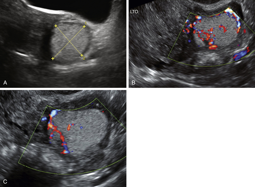
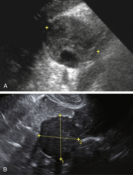
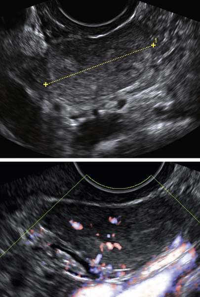
The prognosis of secondary ovarian tumors is typically poor; patients with gastric cancer metastatic to the ovaries usually succumb to the disease within 1 year. The treatment for malignancies that have metastasized to the ovary varies according to the primary tumor. Management may include a combination of oncologic and surgical approaches.

Figures

image

Figure M1-1 A and B, Metastatic colon cancer to the ovary—Krukenberg tumor. Note the multiple tiny cystic areas and septations, giving a frothy appearance. B shows that part of the mass is solid.

image

Figure M1-2 A and B, Adenocarcinoma of the stomach metastatic to the ovary—Krukenberg tumor. Note the large size of the tumor with multiple small cystic areas within a solid matrix.

image

Figure M1-3 Colon cancer metastatic to the ovary. Note the large size of the mass, which is typical.


image

Figure M1-4 A and B, Largely solid-appearing ovarian mass with some cystic areas as well as small punctate calcifications. This tumor proved to be metastatic colon adenocarcinoma.

image

Figure M1-5 A, B, and C Small echogenic mass with internal blood flow shown on Doppler examination (B and C). This proved to be an adenocarcinoma originating from the appendix and metastatic to the ovary, first presenting as an adnexal mass.


image

Figure M1-6 Two different cases of metastatic breast cancer to the ovary. Both are small tumors that are largely solid, although A shows a small cystic portion.

image

Figure M1-7 Small solid ovarian mass with internal blood flow. This was found to be metastatic angiosarcoma of the ovary at surgery. The appearance is nonspecific sonographically, although suspected to be a malignancy.


Suggested Reading

Guzel A.B., Gulec U.K., Paydas S., Khatib G., Gumurdulu D., Vardar M.A., Altintas A. Preoperative evaluation, clinical characteristics, and prognostic factors of nongenital metastatic ovarian tumors: review of 48 patients. Eur J Gynaecol Oncol. 2012;33(5):493–497.

Jain V., Guptay K., Kudvay R., Rodrigues G.S. A case of ovarian metastasis of gall bladder carcinoma simulating primary ovarian neoplasm: diagnostic pitfalls and review of literature. Int J Gynecol Cancer. 2006;16(suppl. 1):319–321.

Koyama T., Mikami Y., Saga T., Tamai K., Togashi K. Secondary ovarian tumors: spectrum of CT and MR features with pathologic correlation. Abdom Imaging. 2007;32:784–795.

Loke T.K.L., Lo S.S., Chan C.S. Case report: Krukenberg tumours arising from a primary duodenojejunal adenocarcinoma. Clin Radiol. 1997;52:154–155.

Yada-Hashimoto N., Yamamoto T., Kamiura S., Seino H., Ohira H., Sawai K., Kimura T., Saji F. Metastatic ovarian tumors: a review of 64 cases. Gynecol Oncol. 2003;89:314–317.