Brenner Tumor
Synonyms/Description
Etiology
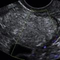
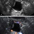
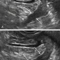
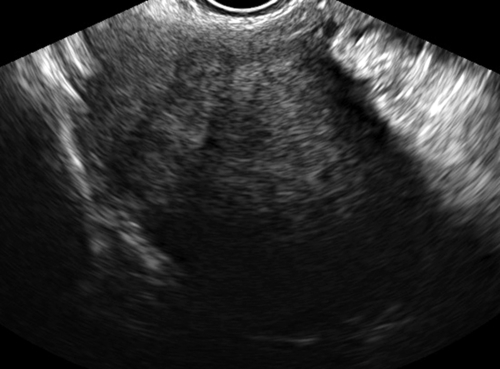
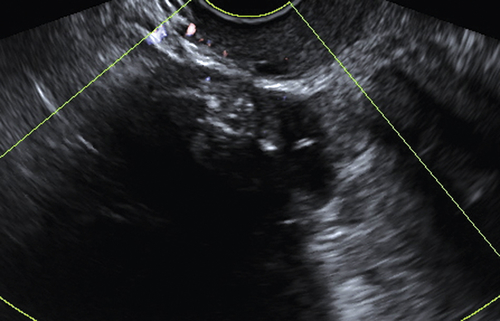
Ultrasound Findings
Differential Diagnosis
Clinical Aspects and Recommendations
Suggested Reading
Athey P.A., Siegel M.F. Sonographic features of Brenner tumor of the ovary. J Ultrasound Med. 1987;6:367–372.
Dierickx I., Valentin L., Van Holsbeke C., Jacomen G., Lissoni A.A., Licameli A., Testa A., Bourne T., Timmerman D. Imaging in gynecological disease (7): clinical and ultrasound features of Brenner tumors of the ovary. Ultrasound Obstet Gynecol. 2012;40:706–713.
Green G.E., Mortele K.J., Glickman J.N., Benson C.B. Brenner tumors of the ovary: sonographic and computed tomographic imaging features. J Ultrasound Med. 2006;25:1245–1251.
Hermanns B., Faridi A., Rath W., Füzesi L., Schröder W. Differential diagnosis, prognostic factors, and clinical treatment of proliferative Brenner tumor of the ovary. Ultrastruct Pathol. 2000;24:191–196.
Hiroi H., Osuga Y., Tarumoto Y., Shimokama T., Yano T., Yokota H., Taketani Y. A case of estrogen-producing Brenner tumor with a stromal component as a potential source for estrogen. Oncology. 2002;63:201–204.