162
Tuberous sclerosis
The shagreen patch. There is usually one lesion, but several may be present. They are soft, flesh-colored to yellow plaques with an irregular surface that has been likened to pig skin.
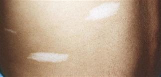
Hypomelanotic macules (oval or ash leaf shaped) are randomly distributed, with a concentration on the arms, legs, and trunk.
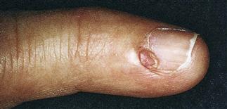
Periungual fibromas are conical, pink, firm projections from posterior nail folds of the fingers and toes. They appear around the time of puberty and persist indefinitely.
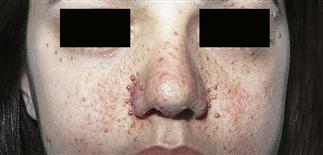
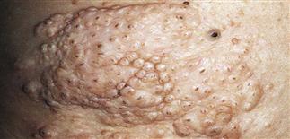
Adenoma sebaceum is the most common cutaneous manifestation of tuberous sclerosis. The lesions consist of smooth and firm 1- to 5-mm, yellow-pink papules with fine telangiectasia.
DESCRIPTION
An uncommon genodermatosis with characteristic features of the skin and central nervous system, as well as multiple other organs. Also called Bourneville disease and epiloia.
HISTORY
• Incidence estimated at 1 in 10 000. • Equal gender distribution. • Spontaneous mutations account for 75% of cases; 25% are autosomal dominant. • Two separate genes have been implicated. • About 40% of affected individuals have normal intelligence; the remainder have subtle to mild mental retardation. • Cutaneous manifestations may not correlate with mental ability. • Premature death occurs rarely, most often from status epilepticus or malignant brain tumor.
PHYSICAL FINDINGS
• Typically presents at or just after birth. • Earliest signs are ‘ash leaf’ hypopigmented macules, usually found on trunk or extremities. These should be examined with a Wood’s lamp. Polygonal, hypopigmented ‘confetti’ macules are also common, especially in the pretibial area. • Facial angiofibromas, also called adenoma sebaceum, appear in early childhood and increase in number throughout adolescence. These benign hamartomas are smooth, firm, pink, 1- to 5-mm papules appearing on nasolabial folds, cheeks, and chin. They may be misdiagnosed as acne. • The shagreen patch is a connective tissue nevus seen in roughly 80% of patients. Typically located in the lumbosacral area, it is a 1- to 5-cm, white to yellow plaque with a pebbled surface. • Periungual fibromas or angiofibromas are conical, pink, firm projections from the posterior nail folds of the fingers and toes. They appear around the time of puberty and persist indefinitely. • Associated non-skin findings include cortical tubers, paraventricular calcification, subependymal hamartomas, and astrocytomas. • Seizures occur in 75% of patients with central nervous system lesions. • Infantile spasms and mental retardation are also part of the syndrome. • Retinal hamartomas (phakomas) may be seen. • Angiomyolipoma, multiple renal cysts, cardiac rhabdomyoma, enamel pits, gingival fibromas, phalangeal cysts, periosteal thickening, and pulmonary cysts may be present.
TREATMENT
• Complete physical examination with routine follow-up by the primary care physician is important. • Imaging studies should be performed to look for cardiac, renal, and central nervous system tumors. • Referral to a pediatric neurologist, including long-term follow-up, should be considered for seizure management. • Baseline ophthalmologic evaluation should be performed. • Carbon dioxide laser ablation or shave removal of facial angiofibromas can significantly improve cosmetic appearance and self-image. • If needed, special educational planning should help an individual reach maximal potential. • Careful cutaneous and general examination of first-degree relatives, as well as genetic counseling, is recommended.


