Tropical infections and infestations
Infections constitute one of the biggest problems in dermatology in tropical countries of the developing world. Leprosy, for example, despite being a treatable disease, continues to ravage in many parts of the globe.
Leprosy
Clinical presentation
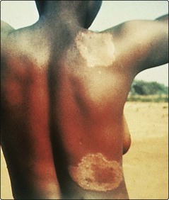
Tuberculoid leprosy affects the nerves and the skin. Nerves may be thickened, and anaesthesia or muscle atrophy is found. Skin lesions often occur on the face, and as few as one or two may be seen. They usually take the form of raised red plaques with a hypopigmented centre, which is typically dry and hairless (Fig. 1). Sensation may be impaired within the plaque.
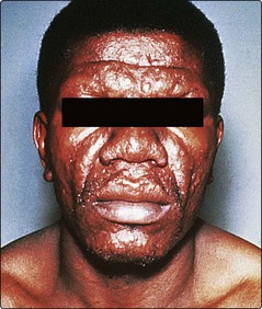
The skin lesions of lepromatous leprosy are multiple and take the form of macules, papules, nodules and plaques. They are symmetrical and tend to involve the face, arms, legs and buttocks. Sensation is not impaired. Untreated, the condition is infectious from the nasal involvement. Progression gives a thickened furrowed appearance to the face (leonine facies) with loss of eyebrows (Fig. 2). Borderline leprosy shows features intermediate between lepromatous and tuberculoid.
Leprosy must be distinguished from a variety of other dermatological conditions (Table 1).
| Type of leprosy | Differential diagnosis |
|---|---|
| Tuberculoid | Vitiligo, pityriasis versicolor, pityriasis alba, sarcoidosis, lupus vulgaris, granuloma annulare, post-inflammatory hypopigmentation |
| Lepromatous | Disseminated cutaneous leishmaniasis, yaws, guttate psoriasis, discoid lupus erythematosus, mycosis fungoides |
Leishmaniasis
1. Leishmania tropica causes the cutaneous ‘oriental sore’ and is seen around the Mediterranean coast, in the Middle East and in Asia.
2. L. braziliensis, endemic in Central and South America, leads to cutaneous and mucosal disease.
3. L. donovani is widely distributed in Asia, Africa and South America, and causes visceral disease (kala-azar) with associated skin lesions.
Clinical presentation
Oriental sore is a common infection in endemic areas normally affecting children, who subsequently develop immunity. In non-endemic regions, it is not infrequently seen in travellers after a Mediterranean holiday. The face, neck or arms are usually affected. At the site of inoculation, a red or brown nodule appears, which either ulcerates or spreads slowly to form a crust-topped plaque (Fig. 3). Untreated, the lesion will heal in 6–12 months, although a chronic form is seen. In mucocutaneous leishmaniasis, the skin lesion resembles an oriental sore but, subsequently, necrotic ulcers affect the nose, lips and palate with deformity. Kala-azar principally affects children and has a significant mortality. It causes hepatomegaly, splenomegaly, anaemia and debility. The cutaneous signs are patchy pigmentation on the face, hands and abdomen.
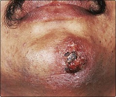
Fig. 3 The oriental sore of cutaneous leishmaniasis.
Small lesions may respond to cryotherapy. Otherwise, intravenous sodium stibogluconate may be given.
Leishmaniasis must be distinguished from some other disorders (Table 2).
| Variant | Differential diagnosis |
|---|---|
| Cutaneous | Lupus vulgaris, leprosy, discoid lupus erythematosus |
| Mucocutaneous | Syphilis, yaws, leprosy, blastomycosis |
| Kala-azar | Leprosy |
Larva migrans
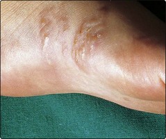
Larva migrans is a ‘creeping’ eruption due to penetration of the skin by the larval stage of animal hookworms. Larva migrans is often acquired from tropical beaches where ova from the hookworms of dogs and cats have hatched into larvae that are able to penetrate the human skin. Penetration usually occurs on the feet. The larvae advance at the rate of a few millimetres a day in a serpiginous route causing red intensely itchy tracks to appear (Fig. 4). They eventually die spontaneously after a few weeks, as they cannot complete their life cycle in humans.
Deep mycoses
Deep mycoses are defined as the invasion of living tissue by fungi, causing systemic disease. Brief details are given in Table 3.
| Mycosis | Clinical features | Management |
|---|---|---|
| Actinomycosis (filamentous bacteria) | A chronic suppurating granulomatous infection with multiple sinuses discharging yellow granules, particularly around the jaw, chest and abdomen | Long-term high-dose penicillin, surgical excision |
| Blastomycosis | Ulcerated discharging nodules that show central clearing with scarring; may spread from pulmonary infection | Oral itraconazole, systemic amphotericin B or ketoconazole |
| Histoplasmosis | Seen in immunosuppressed patients who develop lung disease with granulomatous skin lesions | Oral itraconazole or ketoconazole, or systemic amphotericin B |
| Mycetoma | A chronic granulomatous infection usually of the foot, involving skin, subcutaneous tissue and bones, due to several types of fungi or actinomycetes; nodules with abscesses, sinuses, ulceration and tissue necrosis result | Depends on the organism; surgical excision, dapsone with co-trimoxazole, and itraconazole may help |
| Sporotrichosis | An abscess forms with nodules subsequently occurring proximally along the line of lymphatic drainage | Potassium iodide, itraconazole or terbinafine |
Onchocerciasis
Tropical infections
 Leprosy: tuberculoid and lepromatous forms mainly affect the skin and nerves; treatment is with dapsone, rifampicin and clofazimine.
 Leishmaniasis: cutaneous, mucocutaneous and visceral types; treatment is with sodium stibogluconate.

 Deep mycoses: serious infections that may be difficult to eradicate.


