Toxoplasmosis

Published on 09/02/2015 by admin

Filed under Allergy and Immunology

Last modified 22/04/2025

Print this page

rate 1 star rate 2 star rate 3 star rate 4 star rate 5 star
Your rating: none, Average: 0 (0 votes)

This article have been viewed 1843 times

Toxoplasmosis

Epidemiology

T. gondii was first discovered in a North African rodent and has been observed in numerous birds and mammals worldwide, including human beings. It is a parasite of cosmopolitan distribution able to develop in a wide variety of vertebrate hosts.

Human infections are common in many parts of the world. The prevalence of infection in adults ranges from less than 10% to more than 90%; higher prevalences tend to occur at lower elevations and in latitudes closer to the equator. The highest recorded rate (93%) was in Parisian women who preferred undercooked or raw meat; a 50% rate of occurrence was documented in the children of these women. Toxoplasma infection rates vary around the world. In the United States, it’s about 10% to 15%, while rates in Europe and Brazil are much higher, around 50% to 80%. These are only estimates. Calculating exact rates is difficult because most infected people don’t have any symptoms.

Toxoplasmosis is not passed from person-to-person, except in cases of mother to child (congenital) transmission and blood transfusion or organ transplantation. People typically become infected by three principal routes of transmission:

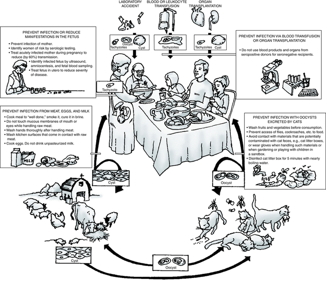
The definitive host is the house cat and other members of the Felidae family (Fig. 20-1). Domestic cats are a source of the disease because oocysts are often present in their feces. Accidental ingestion of oocysts by human beings and animals, including the cat, produces a proliferative infection in the body tissues. Fecal contamination of food or water, soiled hands, inadequately cooked or infected meat, and raw milk can be major sources of human infection. The risk for infection is higher in many developing and tropical countries, especially when people eat undercooked meat, drink untreated water, or are extensively exposed to soil.

Organ transplant recipients can become infected by receiving an organ from a Toxoplasma-positive donor. Transfusion-transmitted toxoplasmosis has been associated with the use of leukocyte concentrates. Patients at risk are those receiving immunosuppressive agents or corticosteroids. Laboratory workers who handle infected blood can also acquire infection through accidental inoculation.

Transplacental Transmission

All mammals, including human beings, can transmit the infection transplacentally. Transplacental transmission usually takes place in the course of an acute but inapparent or undiagnosed maternal infection. Evidence has shown that the number of infants born in the United States each year with congenital T. gondii infection is considerably higher than the 3000 previously estimated. It is estimated that 6 of 1000 pregnant women in the United States will acquire primary infection with Toxoplasma during a 9-month gestation. Approximately 45% of women who acquire the infection for the first time and who are not treated will give birth to congenitally infected infants. Consequently, the expected incidence of congenital toxoplasmosis is 2.7/1000 live births.

It is recommended that all pregnant women be tested for toxoplasmosis immunity. If a patient is susceptible, screening should be repeated during pregnancy and at delivery. Prevention of infection in pregnant women should be practiced to avert congenital toxoplasmosis (Box 20-1). To further prevent infection of the fetus, women at risk should be identified by serologic testing and pregnant women with primary infection should receive drug therapy.

Signs and Symptoms

In adults and children other than newborns, toxoplasmosis is usually asymptomatic. A generalized infection probably occurs. Although spontaneous recovery follows acute febrile disease, the organism can localize and multiply in any organ of the body or the circulatory system.

Toxoplasma can be harmful to individuals with suppressed immune systems. Toxoplasmic encephalitis in AIDS patients may result in death, even when treated (Fig. 20-2). Persons at risk can be identified by screening patients positive for human immunodeficiency virus (HIV) for antibody to T. gondii.
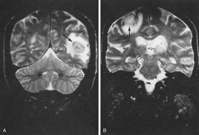
Acquired Infection

When seen, symptoms are frequently mild. Toxoplasmosis can simulate infectious mononucleosis, with chills, fever, headache, lymphadenopathy, and extreme fatigue. Primary infection may be promoted by immunosuppression. A chronic form of toxoplasmic lymphadenopathy exists. T. gondii presents a special problem in immunosuppressed or otherwise compromised hosts. Some of these patients have experienced reactivation of a latent toxoplasmosis. These patients have included those with Hodgkin’s and non-Hodgkin’s lymphoma, as well as recipients of organ transplants.

Reactivation of cerebral toxoplasmosis is not uncommon in patients with AIDS, in whom toxoplasmic meningoencephalitis is almost always a reactivation of a preexisting latent infection, most often occurring when the total CD4 count falls below 100 × 109/L. T. gondii–seropositive, HIV-infected patients may develop toxoplasmic encephalitis because of the following: (1) genetic susceptibility in the human immune response to T. gondii; (2) subtle differences in patients’ immunocompromised status; (3) differences in the virulence of individual strains of T. gondii; (4) possible recurrent infections with different strains; and (5) variable coinfections with other opportunistic pathogens.

Immunologic Manifestations

Both clinical and laboratory findings in toxoplasmosis resemble those of infectious mononucleosis. An increased number of variant lymphocytes can be seen on a peripheral blood smear.

The diagnosis can be established serologically by detecting a marked elevation of Toxoplasma antibodies. Antibodies are demonstrable within the first 2 weeks after infection, rising to high levels early in the infection and then falling slightly, but persisting at an elevated level for many months before declining to low levels after many years. The best evidence for current infection is a significant change on two appropriately timed specimens (paired acute and convalescent specimens), in which both tests are done in the same laboratory at the same time.

If a significant level of T. gondii immunoglobulin M (IgM) antibody is detected, it may indicate a current or recent infection. The presence of IgM to T. gondii in an adult indicates an infection, but low levels of IgM antibodies occasionally may persist for more than 12 months after infection. The Centers for Disease Control and Prevention (CDC) recommends that any equivocal or positive result should be retested using a different assay from another reference laboratory specializing in toxoplasmosis testing.

Diagnostic Evaluation

The diagnosis of toxoplasmosis is typically made by serologic testing. A test that measures immunoglobulin G (IgG) is used to determine whether a person has been infected. If it is necessary to try to estimate the time of infection, which is of particular importance for pregnant women, a test that measures IgM is also used along with other tests, such as an avidity test.

Diagnosis can be made by direct observation of the parasite in stained tissue sections, cerebrospinal fluid (CSF), or other biopsy material. These techniques are used less frequently because of the difficulty of obtaining these specimens.

Parasites can also be isolated from blood or other body fluids (e.g., CSF) but this process can be difficult and requires considerable time. Molecular techniques that can detect the parasite’s DNA in the amniotic fluid can be useful in cases of possible mother to child (congenital) transmission.

The diagnosis of toxoplasmosis can be established by the following:

• Serologic tests (Table 20-1)

Table 20-1

Serologic Evaluation of Toxoplasmosis

Test Method Recommended Use
T. gondii antibodies, IgG and IgM Chemiluminescent immunoassay First-line test in endemic areas for identifying T. gondii infection in pregnant women; diagnosis of opportunistic infections in immunocompromised hosts
T. gondii by PCR   Confirmation of toxoplasmosis infection in immunocompromised hosts

Note: The CDC suggests that equivocal or positive results be retested using a different assay from another reference laboratory specializing in toxoplasmosis testing (IgG Sabin dye test, IgM ELISA, reflex to avidity and/or other tests).

Adapted from Associated Regional and University Pathologists (ARUP): Reference test guide, 2012 (http://www. aruplab.com).

• Polymerase chain reaction (PCR)

• Indirect fluorescent antibody (IFA)

• Isolation of the organism

Serologic Tests

The mainstay of diagnosis of T. gondii infection is serologic testing. A relatively high proportion of people have antibodies to T. gondii, which makes interpretation of serologic test results difficult. Assays for different isotypes of antibodies have been developed to support the diagnosis of an acute or chronic T. gondii infection.

For the detection of IgM antibodies to T. gondii, a variety of procedures are available—IFA, automated enzyme-linked immunosorbent assay (ELISA), and chemiluminescent immunoassay for IgM and IgG antibodies.

IgM Antibodies

The IgM assay was widely used in the past, but this is not recommended for routine use in adults because it may yield frequent false-positive or false-negative results, particularly in pregnant women, immunocompromised patients, and patients from areas in which Toxoplasma infection is highly endemic. IgM antibodies tend to appear earlier and decline more rapidly than IgG antibodies. Persistently elevated IgM-specific antibody titers after the initial infection can lead to false-positive results and difficulty in interpreting these tests.

In patients with recently acquired infection, IgM T. gondii antibodies are detected initially and, in most cases, these titers become negative within a few months. In some patients, however, positive IgM T. gondii–specific titers can be observed during the chronic stage of the infection. IgM antibodies have been reported to persist as long as 12 years after the acute infection. However, their persistence does not seem to be clinically relevant and these patients should be considered chronically infected.

Clinicians should be cautious when using IgM antibody levels in prenatal screening. Any positive result in a pregnant patient confirmed positive by a second reference laboratory should be evaluated by amniocentesis and PCR testing for T. gondii. A negative result does not rule out the presence of PCR inhibitors in the patient specimen or T. gondii DNA concentrations below the level of detection of the assay.

The U.S. Food and Drug Administration (FDA) has recommended that sera with positive IgM test results obtained at nonreference laboratories should be sent to a Toxoplasma reference laboratory. After IgM-positive sera undergo confirmatory testing, the results are interpreted as the following: (1) a recently acquired infection; (2) an infection acquired in the past; or (3) a false-positive result.

IgG Antibodies

IgG antibodies appear 1 to 2 weeks after the initial infection, peak after about 6 to 8 weeks, decline gradually over the next 1 to 2 years and, in some cases, persist for life.

Sabin-Feldman Dye Test.

IgG antibodies are primarily measured by the Sabin-Feldman dye test (DT), considered the gold standard. The DT is a sensitive and specific neutralization test in which live organisms are lysed in the presence of complement and the patient’s IgG T. gondii–specific antibody. IgG antibodies usually appear within 1 to 2 weeks of the infection, peak within 1 to 2 months, fall at variable rates, and usually persist for life. The titer does not correlate with the severity of illness. This test is available mainly in reference laboratories.

A negative test result practically rules out prior T. gondii exposure—unless the patient is hypogammaglobulinemic. In a small number of patients, IgG antibodies might not be detected within 2 to 3 weeks after initial exposure to the parasite. Rare cases of toxoplasmic chorioretinitis and toxoplasmic encephalitis have been documented in immunocompromised patients negative for T. gondii–specific IgG antibodies.

Avidity Test.

The functional affinity of specific IgG antibodies is initially low after primary antigenic challenge and increases during subsequent weeks and months. Protein-denaturing reagents are used to dissociate the antibody-antigen complex. The avidity result is determined using the ratios of antibody titration curves of urea-treated and untreated serum.

The avidity test can be used as an additional confirmatory diagnostic tool in patients with a positive or equivocal IgM test or with an acute or equivocal pattern in the differential agglutination test (AC/HS test). Its highest value is observed when laboratory test results reveal high–IgG avidity antibodies and the serum is obtained during the time window of exclusion of acute infection for a particular method (range, 12 to 16 weeks). Low– or equivocal–IgG avidity antibody results should not be interpreted as diagnostic of recently acquired infection. These low- or equivocal-avidity antibodies can persist for months to 1 year or longer.

Studies of the avidity of IgG in pregnant women who have seroconverted during gestation have shown that women with high-avidity test results are infected with T. gondii at least 3 to 5 months earlier (time to conversion from low- to high-avidity antibodies varies with the method used). Because low-avidity antibodies may persist for many months, their presence does not necessarily indicate recently acquired infection.

Cell Culture

Detection of T. gondii in the blood may represent a major advance in the diagnosis of toxoplasmosis in patients with AIDS. A cell culture method for the growth of T. gondii has been developed using monocytes. After 4 days, parasites in the culture are revealed by immunofluorescence with an anti-P30 monoclonal antibody. A quantitative and qualitative analysis by cytofluorometry can then be performed on the cultured cells.

CASE STUDY

image Rapid TORCH Procedure

Principle

The ImmunoDOT TORCH test (GenBio, San Diego, Calif) uses an enzyme immunoassay (EIA) dot technique for the detection of antibodies. The antigens are dispensed as discrete dots onto a solid membrane. After adding specimen to a reaction vessel, an assay strip is inserted, allowing patient antibodies reactive with the test antigen to bind to the strip’s solid support membrane. In the second stage, the reaction is enhanced by the removal of nonspecifically bound materials. During the third stage, alkaline phosphatase–conjugated antihuman antibodies are allowed to react with bound patient antibodies. Finally, the strip is transferred to an enzyme substrate reagent that reacts with bound alkaline phosphatase to produce an easily seen distinct dot.

Refer to the image website for the procedural protocol.

Limitations

If testing of a sample occurs less than 5 days following primary infection, detectable specific antibody may not yet be present.

This test is a qualitative screening procedure and cannot be used to detect increases in antibody titer or to diagnose active infection. The assay is not intended for final selection of cytomegalovirus (CMV)–negative donors for blood transfusion or organ transplantation. The test may be used to screen potential recipients. Antibody screening in the compromised host must be interpreted with caution. The antibody response of an immunosuppressed individual may differ from that of the immunocompetent host. Because maternal antibody will be detected in infants younger than 1 year, the assessment of previous infections or of the immune status of infants is inappropriate using this test alone.

Chapter Highlights

• Toxoplasmosis is a widespread disease in human beings and animals caused by Toxoplasma gondii, often found in cat feces.

• Fecal contamination of food or water, soiled hands, inadequately cooked or infected meat, and raw milk are sources of human infection. All mammals, including human beings, can transmit the infection transplacentally.

• In adults and children other than newborns, the disease is usually asymptomatic. A generalized infection probably occurs.

• Although spontaneous recovery follows acute febrile disease, the organism can multiply in any organ of the body or circulatory system.

• Congenital toxoplasmosis can result in CNS malformation or prenatal mortality.

• T. gondii is difficult to culture and diagnosis must be supported by serologic methods to determine levels of IgM and IgG antibodies to T. gondii. The presence of IgM antibodies to T. gondii in an adult indicates active infection. Detection of IgM also suggests active infection in the newborn.

• Serologic tests include IFA, chemiluminescent immunoassay, and PCR.