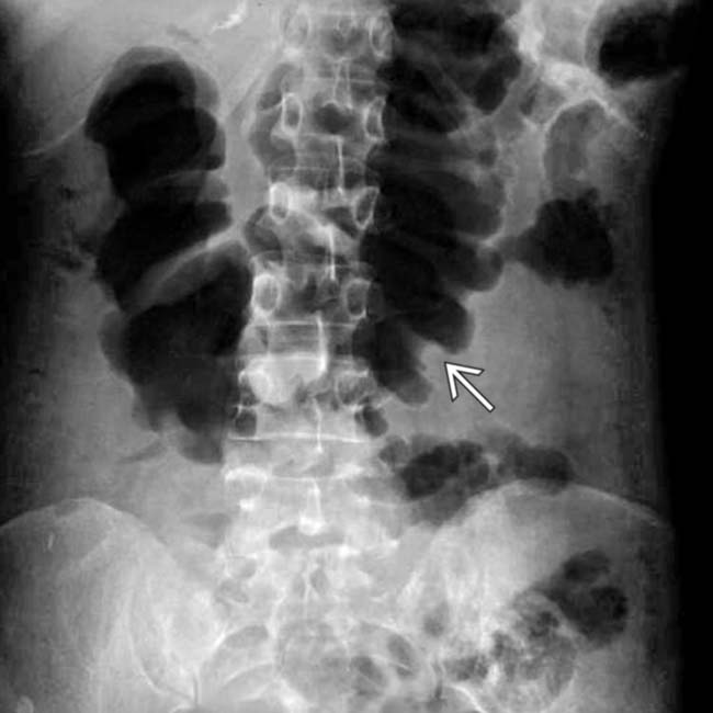
Transverse colonic folds may be thickened (edema or hemorrhage), or lost (sloughed mucosa and submucosa)


 .
.
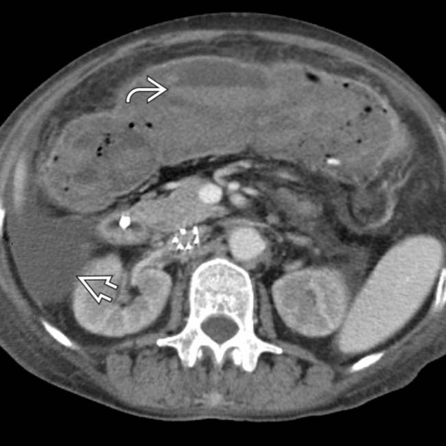
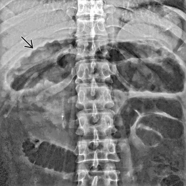
 , marked dilation of the colon with loss of transverse folds, and intraluminal high-density material
  representing hemorrhage and sloughed mucosa.
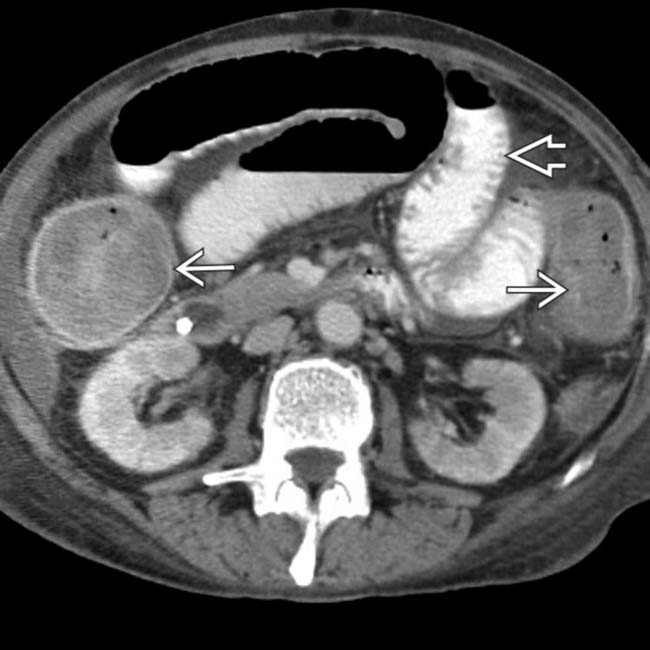
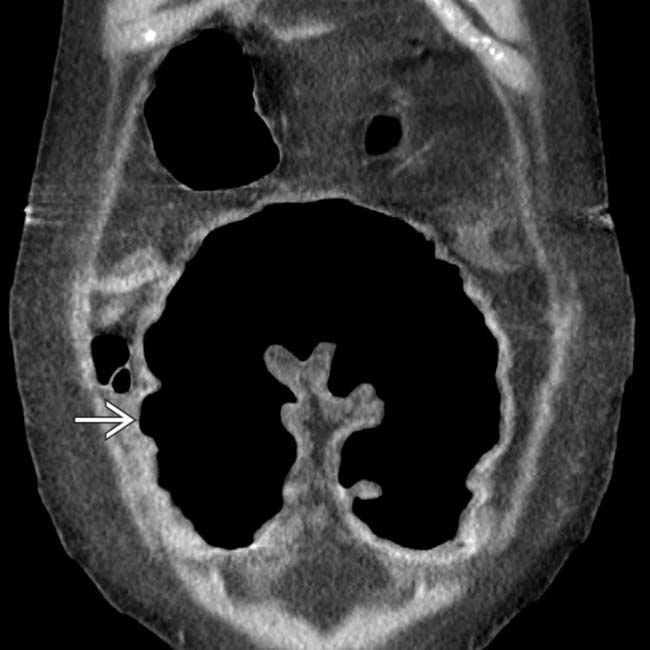
 . The colon
  is massively distended with blood and debris and its wall is relatively thin. Soon after this scan, the colon perforated and a total colectomy was required.


 , suggesting mucosal sloughing or pseudopolyps.
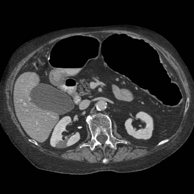
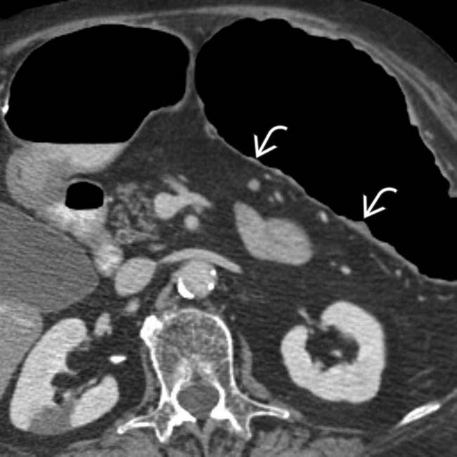
 within the dilated, thin-walled transverse colon.
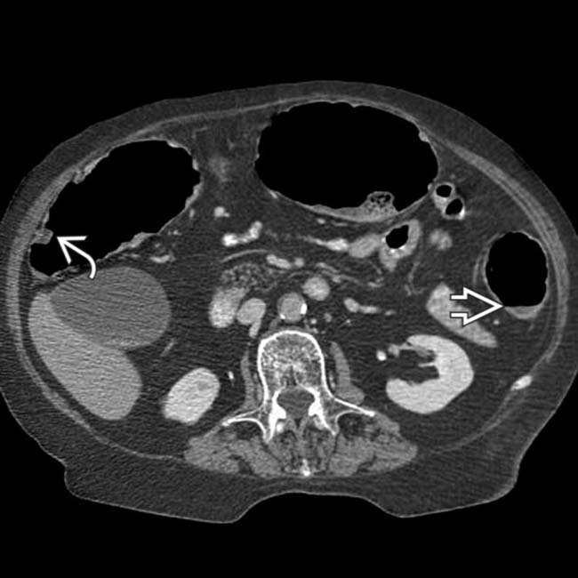
 , distention and thinning of the walls of the colon, as well as more mucosal pseudopolyps
  .
.
 . Because of symptoms that were refractory to medical management, as well as these CT findings, the patient had a total colectomy.
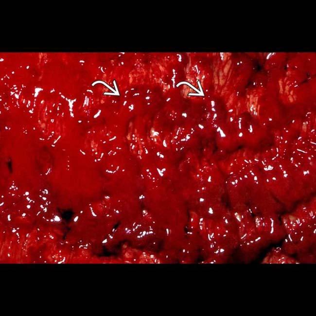
 in a case of toxic megacolon.
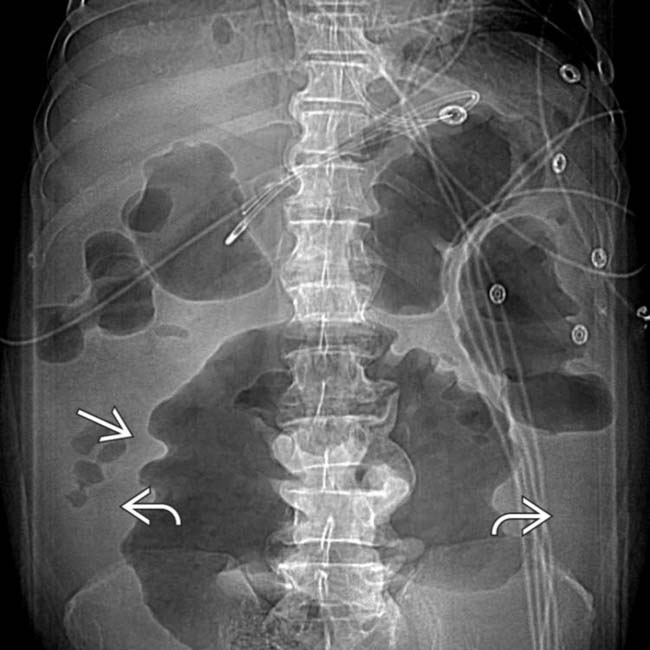
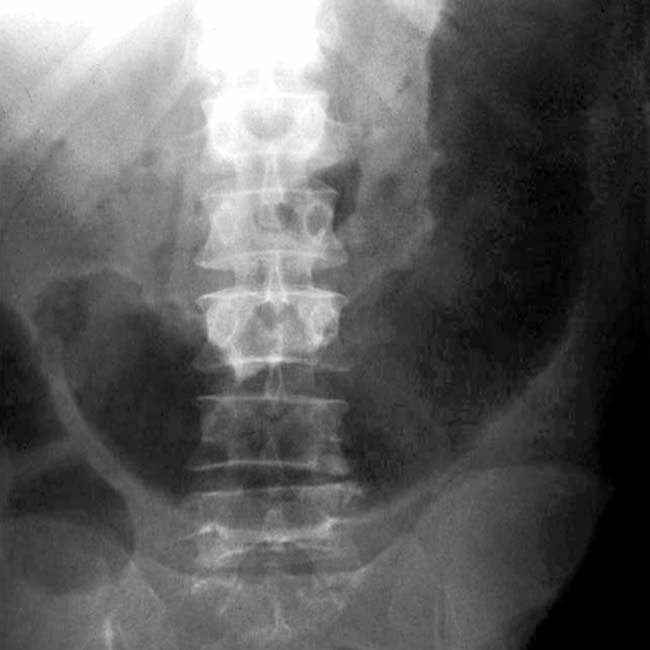
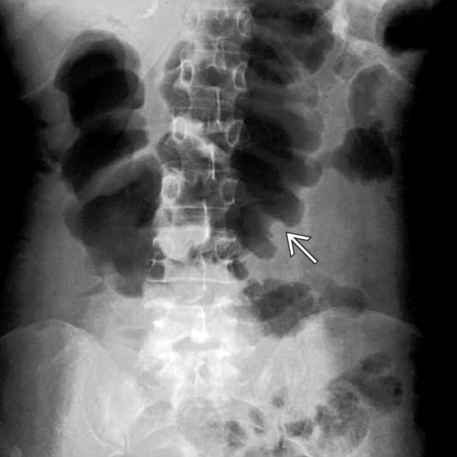
 due to ascites. The colon is diffusely dilated with a loss of the normal transverse fold pattern. The folds that are seen are grossly thickened and have a “thumbprint” appearance
  .
.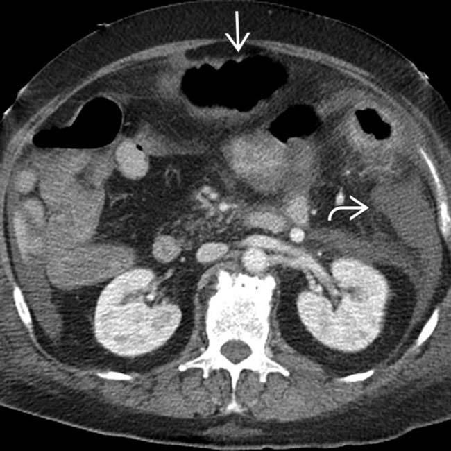
 and colonic distension
  .
.
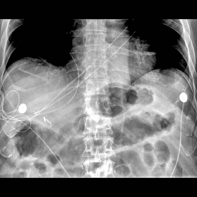
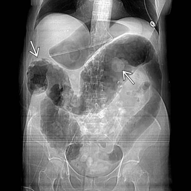
 . No normal transverse folds are evident. Ascites fills the paracolic gutters
  , accounting for the bulging flanks seen on plain films.
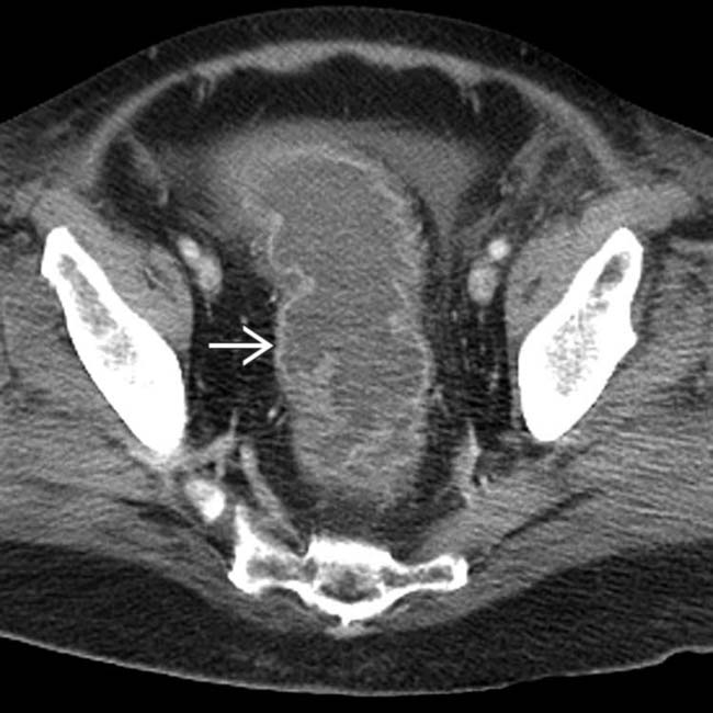
 .
.
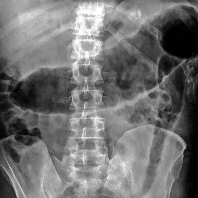
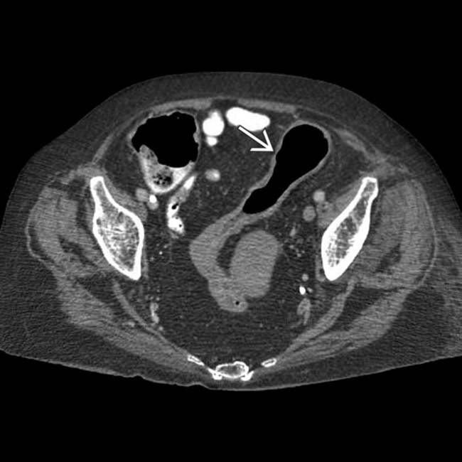
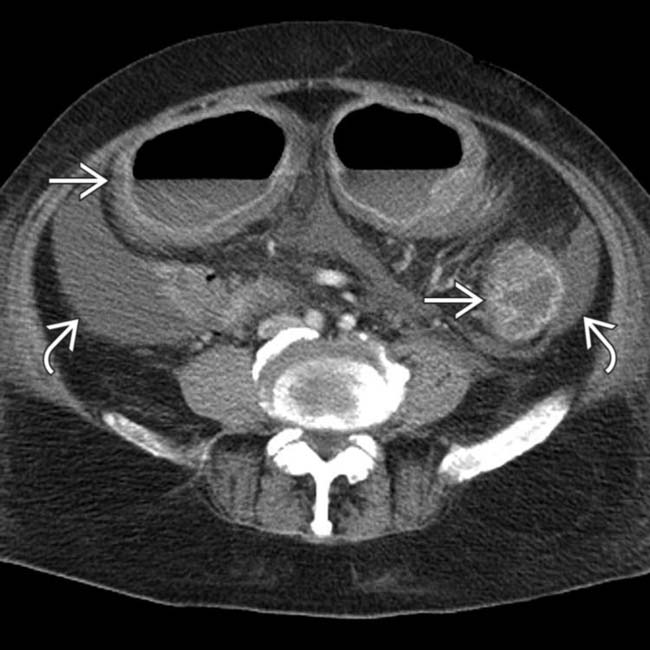
 with a complete loss of its normal transverse fold pattern.
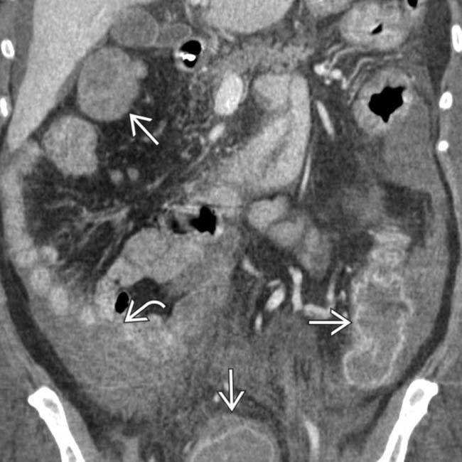
 and ascites
  . These CT and clinical features are classic for toxic megacolon due to infectious colitis (C. difficile), confirmed at urgent colectomy. The colonic mucosa showed extensive necrosis and sloughing, but no frank perforation was found.
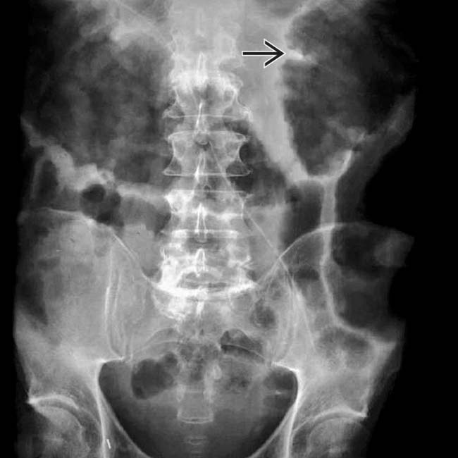
 due to subserosal and omental edema.
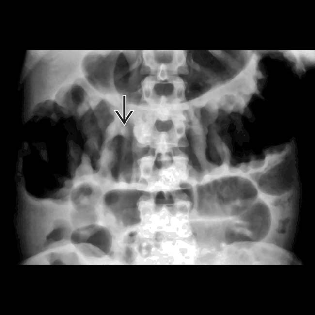
 .
.
 .
.
 indicating edematous mucosa.