71
Tinea versicolor
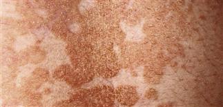
Lesions begin as multiple small circular macules in various colors (white, pink-brown) that enlarge radially. Lesions may be inconspicuous in fair-complexioned individuals during winter.
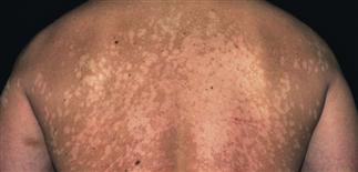
Extensive eruption in a dark-skinned person. Lesions are lighter than normal skin; accentuated when uninfected skin tans. Demonstrating the fine scale helps differentiate from vitiligo.
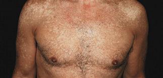
Lesions may be pink or white in fair skinned patients. This extensive eruption has spread down onto the abdomen.
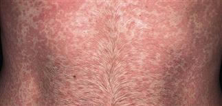
Lesion may be light to dark brown when scale accumulates. Demonstrating scale by scraping with a blade helps confirm the diagnosis.
DESCRIPTION
Common infection caused by the lipophilic yeast Pityrosporum orbiculare (Malassezia furfur). The organism is part of the normal skin flora.
HISTORY
• More common during years of higher sebaceous activity (adolescence, young adulthood); very common especially in tropical, semitropical regions. Potentially transmissible; people with oily skin may be more susceptible. Excess heat and humidity predispose to infection. • May itch but usually asymptomatic; appearance often major concern. • Recurrences common (40–60%) after successful treatment. • Adrenalectomy, Cushing disease, pregnancy, malnutrition, burns, corticosteroid therapy, immunosuppression, oral contraceptives may lower resistance, allowing this normally non-pathogenic resident yeast to proliferate.
PHYSICAL FINDINGS
• Numerous small, circular, scaling papules on upper trunk extending to the upper arms, neck, abdomen. Facial involvement more common in children and in persons of African descent. • Lesions hypopigmented in tanned skin and pink or fawn-colored in untanned skin. Lesions may be inconspicuous in fair-complexioned people during winter. • Dyspigmentation persists several weeks after yeast eliminated. • Potassium hydroxide examination of scale: numerous hyphae that tend to break into short, rod-shaped fragments intermixed with round spores in grape-like clusters, giving ‘spaghetti and meatballs’ pattern.
TREATMENT
• Topical. For limited disease. Apply selenium sulfide lotion 2.5% to entire skin surface from lower posterior scalp area down to thighs; wash off in 10 min. This can be repeated every day for 7 consecutive days. Ketoconazole 2% shampoo is applied to dampened skin, lathered, left on for 5 min, then rinsed. This has clinical response rate of about 70% when used as a single application or daily for 3 days. Zinc pyrithione soap (ZNP bar) is applied in shower, lathered, left on for 5 min, then rinsed off. Miconazole, clotrimazole, econazole, ketoconazole applied to entire affected area at bedtime for 2–4 weeks is effective. Patient should not bathe for at least 12 h after treatment. • Oral. For extensive disease and patients who do not respond to topical treatment or who have frequent recurrences. Itraconazole 200 mg q.d. for 5 days, taken with food to enhance absorption. Ketoconazole 400 mg in single dose or 200 mg q.d. for 5 days, taken at breakfast with fruit juice. Fluconazole 150 mg (two capsules per week for 4 weeks or two capsules as an initial dose to be repeated after 2 weeks). Sweating may improve transfer of oral ketoconazole and fluconazole to the skin surface. Oral Lamisil not effective.


