80
Tinea of the scalp
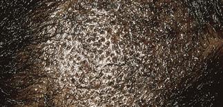
Seborrheic dermatitis pattern. Fine white diffuse or patchy scale-like dandruff. Hair shaft infection then occurs; hairs break at surface, leaving alopecia areas. Broken hairs look like black dots.
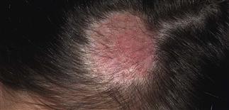
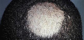
Black dot pattern. Well-demarcated areas of hair loss appear. Infected hairs break, leaving black dots on scalp surface. Little or no inflammation. Occipital adenopathy may be present.
Inflammatory tinea capitis. The lesion near the vertex is boggy, with a scaling and crusted surface.
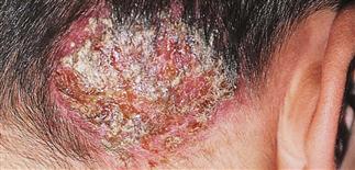
Inflammatory tinea capitis (kerion). A deep boggy mass with pustules and exudate occurs in some patients. The painful lesion heals with scarring and permanent localized hair loss.
DESCRIPTION
Caused by invasion of stratum corneum and hair shaft with fungal hyphae. Most patients prepubertal children. Localized scalp scale in children is a common presentation for seborrheic dermatitis. This is frequently diagnosed as tinea capitis.
HISTORY
• Most US cases caused by Trichophyton tonsurans. • Microsporum canis infection acquired from pets (particularly cats). M. canis-, M. audouinii-infected hair fluoresces blue-green under Wood’s light. • Poverty, crowded city living risk factors. Fungal spores remain on clothing, combs, brushes, telephones for long periods.
PHYSICAL FINDINGS
Four clinical infection patterns for T. tonsurans. • Seborrheic dermatitis type. Diffuse, patchy scale on scalp. Adenopathy may be present. Potassium hydroxide examination often negative. Culture scale. • Black dot pattern. Fungal hyphae invade hair shaft. Hair then breaks at scalp surface. Broken shafts like black dots (red if hair red). Round areas of alopecia with scale present without inflammation. Occipital adenopathy may be present. • Inflammatory tinea capitis (kerion). Deep boggy round masses with pustules, surface exudate. Fever, occipital adenopathy, leukocytosis may occur. Intense inflammation destroys hyphae; potassium hydroxide wet mounts, fungal cultures often negative. May initiate treatment based on clinical appearance without laboratory proof of infection. • Pustular. Pustules or scabbed areas without scaling, significant hair loss may be present; look like scalp bacterial infection. Cultures, potassium hydroxide wet mounts may be negative.
TREATMENT
Examine siblings and adult household members. Clean or discard contaminated objects such as combs. Daily dosing with following drugs is effective. • Griseofulvin. Taken with a fatty meal. Used for 2 weeks beyond the time that cultures and potassium hydroxide preparations become negative (usually 6–12 weeks). • Terbinafine, itraconazole, or fluconazole. Taken for 4–8 weeks. Itraconazole and fluconazole are available as a suspension. A single dose of 150 mg once weekly for 4 weeks may also be effective for older children.
Consider suppressing kerion inflammation with prednisone 1–2 mg/kg q.d. or intralesional triamcinolone 10 mg/cc.
Shampoo with selenium sulfide 1% (Selsun Blue) or ketoconazole (Nizoral) every other day for the first 2 weeks, then twice weekly throughout course of rest of oral therapy. Shampoo is left on for 5 min. Other family members should shampoo two or three times weekly.


