72
Tinea of the nails
Distal subungual onychomycosis. Various patterns are produced as the fungus grows proximally.
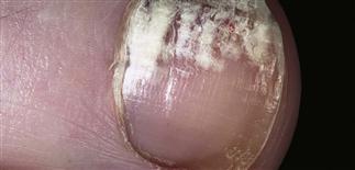
White superficial onychomycosis. The surface of the nail is soft, dry, and powdery, and can easily be scraped away.
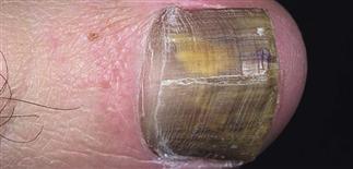
Proximal subungual onychomycosis. Hyperkeratotic debris accumulates and causes the nail to separate from the nail bed.
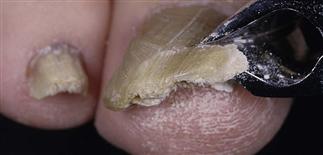
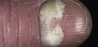
Distal subungual onychomycosis. A yellow to brown color extends the full thickness of the nail.
DESCRIPTION
Onychomycosis: fungal infection of fingernail or toenail plate. Cause: many different species of fungus.
HISTORY
• Trauma from tight-fitting shoes that are too short predisposes to infection. • A large mass composed of a thick nail plate and underlying debris may cause discomfort with footwear.
PHYSICAL FINDINGS
There are four distinct patterns. Several patterns may occur simultaneously. • Distal subungual onychomycosis. Most common pattern. Fungi invade distal area of nail bed. Distal plate turns yellow or white as an accumulation of hyperkeratotic debris causes nail to rise and separate from underlying bed. • White superficial onychomycosis. Caused by surface invasion of nail plate, most often by Trichophyton mentagrophytes. Nail surface is soft, dry, powdery, and can easily be scraped away. Nail plate is not thickened. • Proximal subungual onychomycosis. Microorganisms enter the area of posterior nail fold cuticle, invade nail plate from below. Surface remains intact. Hyperkeratotic debris causes nail to separate. Trichophyton rubrum is most common cause. • Candidal onychomycosis. Nail plate infection caused by Candida albicans is seen almost exclusively in chronic mucocutaneous candidiasis, a rare disease. Generally involves all the fingernails. Nail plate thickens and turns yellow-brown.
All nails and skin are examined to rule out other diseases that mimic onychomycosis.
Aspergillus, Cephalosporium, Fusarium, and Scopulariopsis species, considered contaminants or non-pathogens, can also infect nail plate. Multiple pathogens may be present in a single nail.
In a potassium hydroxide wet mount, subungual debris and nail plate are examined for hyphae.
Culture to establish presence of dermatophytes—organisms susceptible to itraconazole (Sporanox), terbinafine (Lamisil), and fluconazole (Diflucan).
TREATMENT
• Topical antifungal creams. Little value. • Oral therapy. Highest success rate in fingernail and toenail infections in young persons. • Systemic therapy. From 50% to over 80% effective. Relapse rate approximately 15–20% in 1 year. Indications for treatment include pain with thick nails, functional limitations, secondary bacterial infection, and appearance. • Terbinafine (Lamisil) 250 mg q.d. Administered for 6 weeks for fingernail infection, 12 weeks for toenail infection. Not effective for some candidal species. • Itraconazole (Sporanox) 200 mg q.d. Prescribed for 6 weeks for fingernail infection, 12 weeks for toenail infection. Pulse dosing with 200 mg b.i.d. for 1 week on then 3 weeks off another option. Fingernail infection: two or three pulses. Toenail infection: three or four pulses.