75
Tinea of the foot
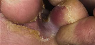
Maceration of the fourth web is a very common finding and is asymptomatic in most cases. Here, the fungal infection has progressed out of the web space and onto the sole.
The acute onset of erythema surrounding the web indicates the acute inflammatory stage of the fungal infection or a secondary bacterial infection.
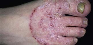
Classic ‘ringworm’ pattern on the dorsal foot with concentric active borders and intervening dry powdery scale. Cracks and painful fissures may eventually appear.
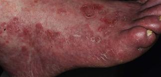
Tinea of the soles is sometimes red and inflamed. It progresses on to the heels and dorsum of the foot. Itching is usually moderate to intense.
DESCRIPTION
Tinea pedis (‘athlete’s foot’) is epidermal infection with dermatophyte fungi. On webs, soles, dorsum. Different inflammation patterns. Psoriasis, eczema, bacterial infections appear similar.
HISTORY
• Occurs in genetically predisposed persons. Common in adults, especially men. Uncommon in children. • Infection persists, recurs. • Wet locker room floor a possible infection source. • Acute, chronic phases. • Interdigital infections often asymptomatic. • Extension out of web on to dorsum may result in acute erythema, secondary bacterial infection. • Infection of soles mildly itchy, persistent. Difficult to produce long remissions.
PHYSICAL FINDINGS
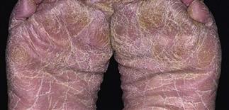
Potassium hydroxide culture sometimes necessary for diagnosis. Toe web infection (especially third, fourth) common. Shoes compress toes, create warm, moist environment. Infection produces white, dry or macerated, dense, persistent scale. Bacterial secondary infection sometimes occurs. Three infection patterns. • Ringworm pattern and acute vesicular infections. On sides and dorsum of foot or extend from webs. Itching common. • Dermatophytid or ‘id reactions.’ Allergic reactions to fungal infection, sometimes misinterpreted as drug rash. Itchy vesicles erupt at distant sites (palms, trunk, along sides of fingers). Prompts examination of webs, sides of feet. Chronic scaly appearance of entire sole occurs with dry skin but often unrecognized tinea infection. • ‘Moccasin’-type pattern. Difficult to treat, recurs. Orange-brown soles are thick, scaly, tender, sometimes itchy. Same pattern on palms. Classic presentation is infection of two feet, one hand, or two hands, one foot. Toenail or fingernail fungal infection may eventually occur.
TREATMENT
• Over-the-counter terbinafine (Lamisil) and butenafine (Lotrimin Ultra) cream may be more effective than miconazole, clotrimazole; b.i.d., 2–6 weeks. Chronic sole infections require at least 3–4 weeks. Antifungal-corticosteroid combination creams, lotions (e.g. Lotrisone) minimally effective. Econazole active against tinea, some bacteria. Useful for macerated, secondarily infected web spaces. • Treat acute vesicular infections with cool Burow’s or tap water dressings, 30 min several times daily. May apply immediately after cream. • Treat acute, extensive infections orally: terbinafine (Lamisil) 250 mg q.d. (2 weeks), itraconazole (Sporanox) 200 mg b.i.d. (1 week), Diflucan 150 mg once a week (3–4 weeks). • Treat secondary bacterial infection with oral antibiotics. Treat id reactions at distant sites with cool wet dressings, group V topical steroids, and (if intense) short course of prednisone (1 mg/kg per day q.d.), 5–10 days. • Wear wide shoes. Open web space with small strand of lamb’s wool (Dr Scholl’s Lamb’s Wool), cotton ball. • Powders, not necessarily medicated, absorb moisture (Zeasorb). Apply to feet rather than shoes. Change wet socks.


