77
Tinea of the body
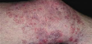
Classic ringworm infection pattern. Round to oval plaque, sharp scaling border. Single lesion or many. Eczema, psoriasis, impetigo may be similar. Patients call anything round ‘ringworm.’
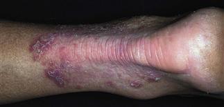
Initial plaque appeared eczematous; treated with topical steroids. Acute inflammation occurred when steroids were stopped. Potassium hydroxide examination of border scale proved the diagnosis.
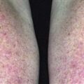
Tinea can progress for years with few symptoms. Topical steroids had no effect. Follicular papules indicate extension of infection into follicle depths. Cleared by terbinafine 250 mg q.d. for 2 weeks.
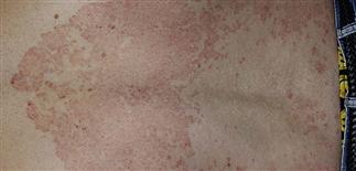
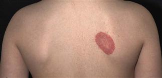
The sharp scaling annular border in this geographic plaque on the back is an important clue to the diagnosis. Psoriatic and eczematous plaques are more homogeneous and symmetric.
DESCRIPTION
Tinea corporis: dermatophyte infection of face, body, trunk, limbs. Other diseases present with round lesions that look like ‘ringworm.’ Nummular eczema and psoriasis lack scaly advancing border of tinea. Pityriasis rosea has collarette of scale that does not reach edge of red border as it does in tinea.
HISTORY
• Infections common in warm, humid climates. • Lesions grow slowly for months or years, may eventually cover very wide areas. • Itching minimal to moderate for chronic infections, intense with inflammatory lesions. Very large lesions tend to be mildly itchy or asymptomatic. Patients may be unaware of their presence. • Wrestlers unknowingly transmit disease to opponents. • Infections can be acquired from animals (cows and cats).
PHYSICAL FINDINGS
• Most common presentation: classic ringworm pattern of infection. Lesions appear as round, flat papules with a scaly raised border. Lesion slowly expands in diameter. Plaques vary from a few centimeters to huge lesions that may cover half of skin surface. Slowly advancing, scaly border is highly characteristic. May have red raised papules or vesicles. Central area becomes red, purple, brown, or hypopigmented and less scaly. Fungal hyphae invade deep into follicle, produce red papules. With treatment, scaling resolves before erythema fades. • Deep inflammatory lesions usually occur from infection with fungi from animals, such as Trichophyton verrucosum from cattle. Localized, intensely inflamed lesions have elevated, red, boggy, pustular surface. Secondary staphylococcal infection possible. Destructive inflammatory process causes brown hyperpigmentation, scarring. Postinflammatory dyspigmentation blends or disappears over several months. • Potassium hydroxide examination shows abundant hyphae. Perform fungal cultures of inflamed lesions to establish the animal of origin.
TREATMENT
• Treat localized superficial infections with clotrimazole, miconazole, ketoconazole, econazole, butenafine, or terbinafine applied twice each day for at least 2 weeks. Continue treatment for at least 1 week after infection resolves. • Extensive lesions and those with red papules require oral therapy. Red papules indicate extension into hair follicle. Topical medication may not penetrate deeply enough to be effective. Treat with terbinafine (Lamisil) 250 mg q.d. for 1–2 weeks, itraconazole (Sporanox) 100 mg q.d. for 1–2 weeks or 200 mg q.d. for 1 week, or fluconazole (Diflucan) 150 mg once a week for 2–4 weeks. • Griseofulvin (ultramicrosize) adult 333 mg q.d. or b.i.d. for 2–4 weeks also effective. • Treat secondary bacterial infection with oral antibiotics. • Consider short course of prednisone for highly inflamed lesions to minimize scarring.