79
Tinea incognito
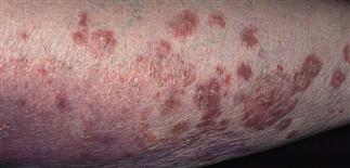
This fungal infection was misdiagnosed as psoriasis. Group I topical steroids were prescribed. The inflammation became confluent and spread over much of the lower leg.
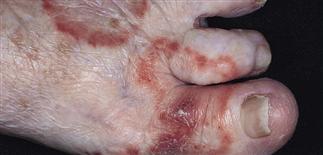
A diagnosis of eczema of the dorsum of the foot was made and treated with a group IV topical steroid. The misdiagnosed fungal infection spread in a ringworm-like pattern.
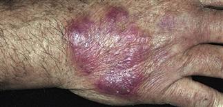
Initial plaque appeared eczematous; treated with topical steroids. Acute inflammation occurred when steroids stopped. Potassium hydroxide examination of border scale showed fungal hyphae.
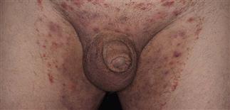
A group V topical steroid was applied intermittently after an incorrect diagnosis of intertrigo. The dermatophyte fungal infection flared with a yeast-like pattern with satellite pustules. Note sparing of the scrotum.
DESCRIPTION
Fungal infection whose clinical presentation has been altered by application of topical corticosteroids. Bizarre patterns of inflammation are produced by the inappropriate use of these topical anti-inflammatory agents. The fungal infection is most often misdiagnosed as eczema, psoriasis, pityriasis rosea, folliculitis, or rosacea.
HISTORY
• A cutaneous fungal infection is misdiagnosed as an inflammatory skin rash. Topical steroids are prescribed. These anti-inflammatory agents decrease inflammation, and the eruption appears to be improving. The fungus, however, flourishes with cortisone-induced local immunosuppression. The topical steroid is stopped and the infection rapidly flares. The cycle of treatment and flaring continues until infections with bizarre patterns spread over a wide area. • The altered clinical picture is called tinea incognito. • The intensity of itching varies. • Perform a potassium hydroxide examination when a rash does not respond as expected to topical steroids.
PHYSICAL FINDINGS
• Fungal infections of the dorsum of the hand or foot, lower leg, face, and groin may not present with a typical ringworm pattern of infection. The typical sharp scaling border is not always present. • Infections become much worse in the presence of immunosuppressive anti-inflammatory agents. • A well-defined border may not be present, and a once-localized process may expand over wide areas. • Diffuse scale and erythema make the eruption look eczematous and encourage further use of topical steroids. • Papules and pustules appear inside and outside the border and may represent a bacterial infection. • Brown postinflammatory hyperpigmentation appears in time.
TREATMENT
Stop topical steroids. Superficial lesions respond to antifungal creams (e.g. butenafine, clotrimazole, miconazole, econazole, oxiconazole, terbinafine) applied twice each day. Continue treatment for at least 1 week after resolution of infection. Extensive lesions or those with papules or pustules are treated with oral therapy. • Lamisil: 250 mg q.d. for 2 weeks. • Sporanox: 100 mg, two tablets q.d. for 2 weeks. • Diflucan: 150 mg once a week for 2 weeks. • Griseofulvin (ultramicrosize): Adult 333 mg q.d. or b.i.d. for 2–6 weeks.
Secondary bacterial infection is treated with oral antibiotics such as cephalexin 500 mg b.i.d.
Cool water wet dressings suppress inflammation and control itching.


