Thyroid pathophysiology

Published on 01/03/2015 by admin

Filed under Basic Science

Last modified 22/04/2025

Print this page

rate 1 star rate 2 star rate 3 star rate 4 star rate 5 star
Your rating: none, Average: 0 (0 votes)

This article have been viewed 1304 times

44

Thyroid pathophysiology

Introduction

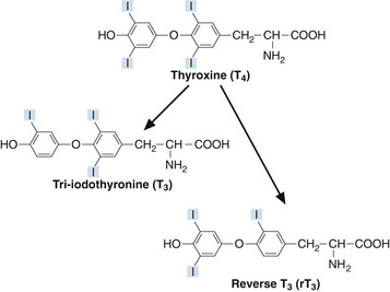
Thyroxine (T4) and tri-iodothyronine (T3) are together known as the ‘thyroid hormones’. They are synthesized in the thyroid gland by iodination and coupling of two tyrosine molecules whilst attached to a complex protein called thyroglobulin. T4 has four iodine atoms while T3 has three (Fig 44.1).

The thyroid gland secretes mostly T4 whose concentration in plasma is around 100 nmol/L. The peripheral tissues, especially the liver and kidney, deiodinate T4 to produce approximately two-thirds of the circulating T3, present at a lower concentration of around 2 nmol/L. Most cells are capable of taking up T4 and deiodinating it to the more biologically active T3. It is T3 that binds to receptors and triggers the end-organ effects of the thyroid hormones. Alternatively, T4 can be metabolized to reverse T3 (rT3), which is biologically inactive. By modulating the relative production of T3 and rT3, tissues can ‘fine tune’ their local thyroid status. Exactly how this is accomplished is not yet fully understood.

Goitre

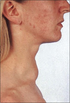
A goitre is an enlarged thyroid gland (Fig 44.2). This may be associated with hypothyroidism, hyperthyroidism or a euthyroid state. Globally, iodine deficiency is the commonest cause of goitre. The WHO estimates that approximately 2 billion people have an inadequate iodine intake making it the commonest preventable cause of neuro-developmental problems. In many developed countries this problem has been overcome by the addition of iodine to a staple food such as iodised salt.

Thyroid hormone action

Thyroid hormones are essential for the normal maturation and metabolism of all the tissues in the body. Their effects on tissue maturation are most dramatically seen in congenital hypothyroidism, a condition which, unless treated within 3 months of birth, results in permanent brain damage. Hypothyroid children have delayed skeletal maturation, short stature and delayed puberty.

Thyroid hormone effects on metabolism are diverse. The rates of protein and carbohydrate synthesis and catabolism are influenced. An example of the effect of thyroid hormones on lipid metabolism is the observation of a high serum cholesterol in some hypothyroid patients. This is a consequence of a reduction in cholesterol metabolism due to down regulation of low-density lipoprotein (LDL) receptors on liver cell membranes, with a subsequent failure of sterol excretion via the gut.

Regulation of thyroid hormone secretion

The components of the hypothalamic–pituitary–thyroid axis are TRH, TSH and the thyroid hormones. TRH, a tripeptide, is secreted by the hypothalamus and in turn causes the synthesis of a large glycoprotein hormone, TSH, from the anterior pituitary. This drives the synthesis of thyroid hormones by the thyroid. Production of TSH is regulated by feedback from circulating unbound thyroid hormones. A knowledge of these basics is essential for the correct interpretation of results in the investigation of primary thyroid disease. Remember:

Thyroid function tests

Biochemical measurements in the diagnosis of thyroid disease have traditionally been known as ‘thyroid function tests’. TSH and some estimate of T4 status (either total T4 or free T4) are the first-line tests.

image TSH. Measurement of TSH is a good example of how better technology has helped in the diagnosis and monitoring of disease. Early assays for TSH were unable to measure low concentrations of the hormone – the detection limits of the radioimmunoassays overlapped significantly with concentrations of the low end of the reference interval in healthy subjects. Now, very sensitive TSH assays can detect much lower concentrations and it is possible to tell with a greater degree of certainty whether TSH secretion really is lower than normal.

    Because of its log-linear relationship with TRH, TSH is very sensitive to derangements in thyroid control, and many laboratories use TSH alone as the first-line thyroid function test. There is, however, one situation in which TSH cannot be used to diagnose primary thyroid disease, or to monitor replacement, namely hypopituitarism. For example, TSH is undetectable post hypophysectomy and an estimate of T4 status must be used instead to monitor the adequacy of replacement.

image Total T4 or free T4. Following the introduction of replacement thyroxine or of anti-thyroid treatment, e.g. carbimazole, or indeed following any alteration in dosage, TSH may take many weeks to unsuppress and adjust to its new level. During this time, it is essential to have some estimate of T4 status. This applies particularly to the monitoring of anti-thyroid treatment; patients can become profoundly hypothyroid quite quickly.

image Total T3 or free T3. Occasionally it may be useful to have an estimate of T3 status in addition to T4. In hyperthyroidism, the rise in T3 is almost always disproportionate to the rise in T4; an estimate of T3 status may permit earlier identification of thyrotoxicosis. In some patients only the T3 rises – the T4 remains within the reference interval (T3 toxicosis).

image Antibodies. The titre of autoantibodies to thyroid tissue antigens may be helpful in the diagnosis and monitoring of autoimmune thyroid disease. Anti-thyroid peroxidase (anti-TPO) may be useful in hypothyroidism and stimulating thyroid receptor antibodies in thyrotoxicosis.

Drugs and the thyroid

Various drugs affect thyroid function tests. The effects of some of these are summarized in Table 44.1.

Table 44.1

Drugs affecting thyroid function tests

Drug Mechanism Major effects
Amiodarone Reduced peripheral deiodination
Amiodarone can also stimulate or inhibit release of thyroid hormones from thyroid
↑T4,↓T3, transient ↑ in TSH
Hyperthyroidism
Hypothyroidism
Lithium Reduced thyroid uptake of iodine
Reduced release of thyroid hormone
Goitre
Hypothyroidism
Anticonvulsants (phenytoin, carbamazepine, phenobarbital) Displace T4 and T3 from binding proteins ↑ free T4, ↑ free T3
Heparin Releases lipoprotein lipase into plasma with resultant increase in free fatty acids. These displace T4 and T3 from binding proteins ↑ free T4, ↑ free T3
Aspirin In high concentrations displaces T4 from transthyretin ↑ free T4